https://doi.org/10.36253/phyto-15955
Accepted: March 11, 2025; Published: May 15, 2025
Research Papers
Alternaria and Curvularia leaf spot pathogens show high aggressivity on watermelon, and are emerging pathogens in cucurbit production
1 Department of Fruit Growing, Institute of Horticulture, Hungarian University of Agriculture and Life Sciences, 2100 Budapest, Hungary
2 Department of Botany, Institute of Agronomy, Hungarian University of Agriculture and Life Sciences, H-1118 Budapest, Hungary
3 Department of Vegetable and Mushroom Growing, Institute of Horticulture, Hungarian University of Agriculture and Life Sciences, 2100 Budapest, Hungary
*Corresponding author. E-mail: Papp.David@uni-mate.hu
Summary. Fungal leaf spot pathogens of cucurbits cause significant yield losses. They cause extensive leaf necroses and defoliation, reducing host photosynthesis. They increase risks of fruit sunscald, and can cause substantial crop damage. Alternaria cucumerina has been recognized as the causal agent of leaf spot disease of cucurbits, and recent studies have identified other Alternaria species, and other emerging pathogens such as Curvularia. This study characterized 25 isolates obtained from infected watermelon and cucumber leaves from Hungary, Spain, and Kosovo. Morphological characterization and molecular analyses using TEF1-α, HIS3, and ITS gene regions identified Alternaria alternata and A. arborescens, and for the first time on this host, the genus Curvularia. Detached leaf assays of ten isolates on 73 watermelon accessions showed variation in isolate pathogenicity. The tested Curvularia isolate was the most aggressive, followed by the A. arborescens and A. alternata isolates, although A. alternata was the most frequently identified species. These results highlight the potential for emerging fungal pathogens causing cucurbit leaf spot, such as Curvularia sp., and show that these fungi can cause damage on economically important plants. This study also showed differing resistance within the watermelon collection, indicating potential for the plant introduction (PI) accessions as sources of resistance breeding.
Keywords. Alternaria alternata, Alternaria arborescens, bar coding, leaf spot, Citrullus spp.
Contents
Collection and culturing of fungal isolates
Culture and conidium morphology
Phylogenetic analyses of fungal isolates
Introduction
Watermelon (Citrullus lanatus, Cucurbitaceae) is an important food crop which has been cultivated for at least 5000 years since domestication in northeastern Africa (Paris, 2015; Chomicki et al., 2020). In 2022, world watermelon production was approx. 100 million tons, while the total area of watermelon cultivation was approx. 2.9 million hectares, highlighting the extensive land use dedicated to this crop (FAO, 2022).
Watermelon plants are susceptible to several important fungal diseases, such as powdery mildew (Podosphaera xanthii, Erysiphe cichoracearum), downy mildew (Pseudoperonospora cubensis), anthracnose (Colletotrichum orbiculare), Fusarium wilt (Fusarium oxysporum) and gummy stem blight (Didymella bryoniae) (Egel et al., 2022). Fungal leaf spot of cucurbits has also been a growing concern during the last decade, resulting in significant economic losses for farmers due to reduced yields and increased production costs (Abu-Nasser and Abu-Naser, 2018; Ma et al., 2021; Shanthi Avinash et al., 2021).
Alternaria cucumerina has been identified as the causal agent of the Alternaria leaf spot of cucurbits, including watermelon (AA.VV., 2021; Kucharek, 1985). In recent decades, new Alternaria species have been reported as pathogens of cucurbits, including A. alternata f. sp. cucurbitae (Vakalounakis, 1990; Zhou and Everts, 2008; Ahmed et al., 2021), A. tenuissima, A. infectoria, and A. gaisen (Kwon et al., 2021; Ma et al., 2021).
The importance of these pathogens is indicated by Alternaria being among the ten most cited pathogenic fungal genera. This diverse genus includes more than 600 species, many of which are major plant pathogens causing leaf spots, blights, rots, and seed infections of a wide range of agricultural plants, including cereals, fruit crops, vegetables, and ornamentals (Bhunjun et al., 2022, 2024). New cucurbit host species and outbreaks of Alternaria diseases are being reported, highlighting the increasing threats of these pathogens to cucurbit production, especially in regions with extended periods of high humidity (Abu-Nasser and Abu-Naser, 2018; Matić et al., 2020; Shanthi Avinash et al., 2021). Alongside Alternaria leaf spot, more recent reports have shown that members of the related Curvularia genus can also affect cucurbits, including Trichosanthes dioica (pointed gourd), Cucurbita argyrosperma (kershaw, or silver-seed gourd), and Cucumis melo (melon) (Sarkar et al., 2018; Ayvar-Serna et al., 2022; Vanitha et al., 2024).
The Pleosporaceae includes 23 genera with Alternaria and Curvularia as two of the most important genera (Torres-Garcia et al., 2022; Hyde et al. 2024). Curvularia spp. are ubiquitous as pathogens and saprobes of plants, animals, and humans (Sivanesan, 1987; Marin-Felix et al., 2017), and many are known as causes of diseases of grasses and staple food crops, including rice, maize, wheat, and sorghum (Khan et al., 2023). More than 200 Curvularia species are recognized (Hyde et al. 2024). Curvularia and Alternaria have been described as “sister genera” within Pleosporaceae, due to their close evolutionary relationship (Bao and Roossinck, 2013). Therefore, they share morphological traits (Torres-Garcia et al., 2022), and cause similar symptoms on host plants (Rabaaoui et al., 2022), making species identification difficult. Results showing different capabilities of Alternaria species to cause disease on a given host have highlighted the importance of accurate species level diagnoses, for plant protection and resistance breeding (Fontaine et al., 2021).
Phylogenetic analyses utilizing barcoding markers are important for identification of fungi. In the last decade, Alternaria (Peever et al., 2004; Woudenberg et al., 2015; Dettman and Eggertson, 2021) and Curvularia (Manamgoda et al., 2012a; 2015; Marin-Felix et al., 2020; Connally et al., 2022) have undergone substantial taxonomic changes, with reclassification of several species and identification of new species based primarily on molecular genetic analyses. Considerable progress has been made in systematic revision of both genera through multi-locus sequence analyses, particularly using the genetic regions of Internal Transcribed Spacer (ITS), Translation Elongation Factor 1- α (TEF1-α), Glyceraldehyde-3-phosphate dehydrogenase (GAPDH), RNA polymerase II second largest subunit (RPB2), Histone H3 (HIS3), Small Subunit ribosomal RNA (SSU, 18S rRNA), and Large Subunit ribosomal RNA (LSU, 28S rRNA) (Dettman and Eggertson, 2021; Aung et al., 2024). Those phylogenetic analyses have often been refined, by using endopolygalacturonase gene (endoPG) and two anonymous genome regions (OPA1-3 and OPA2-1), which are suitable for functional studies and species delimitations (Woudenberg et al., 2015; Aung et al., 2024). Species delimitation within Alternaria has challenges, and is often considered to be difficult, emphasizing the need for integrated approaches combining morphological, ecological, and molecular data for species identifications.
Selection of host plants that are tolerant or resistant gene sources for the breeding requires accurate identification of the respective pathogens. Among germplasm collections, plant introductions (PIs, USDA) of watermelon offer a valuable source of genetic diversity for resistance breeding programmes, that contribute to reducing the reliance on fungicides, and can improve fruit quality and yield to increase crop profitability (Grumet et al., 2021). Currently, little is known about resistance watermelon towards leaf spot diseases caused by Alternaria or Curvularia spp.
The present study reports occurrence of a Curvularia sp. on watermelon. Furthermore, A. alternata, A. arborescens, and Curvularia isolates were characterized as causal agents of leaf spot disease in cucurbits, combining morphological and phylogenetic analyses, and evaluation of their pathogenicity on a diverse collection of watermelon accessions.
Materials and methods
Collection and culturing of fungal isolates
Diseased watermelon and cucumber (Cucumis sativus) leaves were collected from plants grown in open fields and greenhouses in Hungary, Spain, and Kosovo, between June and September of 2024. A total of 25 single conidium isolates were obtained from collected leaves. Isolations were carried out according to Venkatesagowda et al. (2012), with some modifications. Fungal lesions were cut out from leaves, and were surface-sterilized in 4% sodium hypochlorite solution for 60 sec, then rinsed with distilled water for 10 sec. Leaf fragments were then dried on sterile paper, and placed on Potato Dextrose Agar (PDA) in Petri plates. After 24 h, hyphal tips resembling Alternaria sp. were aseptically cut under a stereo microscope, and placed on new PDA plates. From the developing cultures, after 4–5 d, conidia were harvested by mixing a small piece of sporulating mycelium with sterile distilled water containing 0.03% Tween 20, and filtering the suspension through double cheesecloth. Resulting conidium suspensions were then spread on the surface of 1.6% water agar, (and individual conidia were each picked up with a needle and transferred to a new PDA plate. Single conidium isolates were maintained on 25°C, and were subcultured every 10 d.
To obtain conidium suspensions for morphological characterization and for host inoculations, isolates were grown on a calcium carbonate-based sporulation medium as described by Shahin (1979), with sucrose omitted from the medium formulation. Seven-day-old conidia were collected from the surfaces of abundantly-sporulating isolates, by washing with sterile distilled water containing Tween 20, or using the tape touch method for microscope slide preparations (Harris, 2000).
Culture and conidium morphology
Photographs of 1-week-old PDA cultures of the isolates were taken to evaluate culture appearance, including colour according to RAL code system (RAL GmbH), and texture following the description of Nobles (1948). Colony growth rates (cm d-1) were calculated from the age of cultures, and their final colony diameters measured with a vernier caliper.
Conidium length (µm), width (µm), and numbers of conidia in chains or clusters were assessed using a Zeiss Axio Imager A2 (Carl Zeiss Microscopy). Conidium characteristics were measured for at least 20 replicates per species, with isolates selected randomly, using the software ImageJ2 (Rueden et al., 2017).
Molecular analyses
To identify species of the collected isolates, their genetic sequences were analysed and a phylogenetic tree was constructed. To extract DNA, mycelia of each monospore isolate grown on PDA were cut and stored at -80 °C. Frozen samples were then ground in a mortar, and DNA was extracted using the E.Z.N.A. plant DNA kit (Norcoss), according to the manufacturer’s recommendations. DNA concentrations were verified by staining and running the extracted DNA on 1% agarose gels (BioReagent, Sigma-Aldrich).
Three barcoding markers were used for the amplifications: the Internal Transcribed Spacer (ITS) region was amplified using the primers ITS1-F (5’-TCCGTAGGTGAACCTGCGG-3’) and ITS4-R (3’-TCCTCCGCTTATTGATATGC-5’) (White, 1990), Histone 3 (HIS3), using HIS3-F (5’-ACTAAGCAGACCGCCGCGAAG-3’), and (HIS3-R (3’-GCGGGCGAGCTGGATGTCCTT-5’) (Steenkamp et al., 2000), and the Translation Elongation Factor 1-alpha (TEF1-α) using TEF1-F (5’-CATCGAGAAGTTCGAGAAGG-3’) and TEF1-R (5′-TACTTGAAGGAACCCTTACC-3′) (Wu et al., 2013).
The polymerase chain reaction (PCR) was carried out for DNA samples and a negative control (without DNA), using a Thermal Cycler 2720 PCR machine (Applied Biosystems). The PCR mixture (final volume of 16 μL) was prepared with the DreamTaq Green PCR Master Mix (Fermentas). The PCR program was set according to the specified temperatures, times, and number of cycles as described in Table S1. Integrity of amplicons was evaluated by running them on 1% agarose gel (BioReagent, Sigma-Aldrich). Amplicons were sequenced using an ABI 3100 automatic sequencer. All generated sequences were uploaded to GenBank (Table S2).
The quality of the resulting chromatograms was checked using CodonCode Aligner 7.0.1 (CodonCode Corporation). Reference sequences for Alternaria and Curvularia from the GeneBank database were included in the analyses (Table S2). HIS3 was only used for Blast analyses. Sequences of ITS and TEF1-α, together with sequences of related species downloaded from GenBank, were aligned separately with the online MAFFT v. 7.0, using the L-INS-i strategies (Katoh and Standley, 2013). The alignments were checked and edited by manual adjustment in Multiple sequence alignment, and manual corrections (cutting of low-quality edges, indels) were achieved using AliView (Larsson, 2014). Maximum Likelihood (ML) analysis was performed using RAxML in raxmlGUI2.0 (Stamatakis, 2014; Edler et al., 2021), with 1,000 rapid bootstraps and the GTRGAMMA substitution model. The resulting phylogenetic tree was visualized in MEGA11 (Tamura et al., 2021).
Pathogenicity assays
Healthy watermelon leaf samples were collected from an open greenhouse located in Soroksár, Hungary (47°23′49′′ N, 19°09′10′′ E), containing a collection of 73 watermelon varieties. The collection included 71 plant introduction (PI) accessions from the United States of America, and the commercial C. lanatus cultivars ‘Black Diamond’ and ‘Calhoun Grey’. The plant accessions originated from a wide geographic range, including Turkey, Spain, Zimbabwe, Democratic Republic of Congo, Senegal, Iran, Japan, Syria, Ghana, Zambia, South Africa, and the United States of America.
The leaf samples were put between wet sterile tissue for transport to the laboratory. Leaves were then washed gently in 1% sodium hypochlorite solution for 60 sec and immediately placed in sterile water for a further 60 sec. Young undamaged leaf sections (each of 6 mm) were cut with a cork borer and surface water was removed by placing these on sterile paper towels. the leaf sections were then placed adaxial surface upwards on 1% water agar.
Following identification of isolates to genus or species, pathogenicity tests were carried out using six representative isolates from watermelon in Spain, identified as A. arborescens, one isolate from watermelon in Hungary, identified as Curvularia, and three isolates from watermelon in Hungary, identified as A. alternata. These assays were each conducted using a conidium suspension (1 × 10⁶ conidia mL-1, with 5 µL of the suspension applied on each leaf disc. Each plate contained three inoculated leaf discs per plant accession, for biological replicates and one leaf disc mock-inoculated with 5 µL of sterile water. The plates were incubated at 25°C in dark conditions. Relative disease severity (DS) was estimated visually at 5 d post-inoculation (DPI), using a 0 to 9 scale (Singh et al., 2020) and an Olympus BX41 stereomicroscope (Olympus Corporation). Development of new conidium chains, hyphae, and the presence of necrotic tissues were considered compatible reactions (Figure S1).
Statistical analyses
Morphological features of conidia were evaluated using the Wilks’s lambda MANOVA model to compare the mean lengths, widths, and conidium counts per chain, among the different species. This model had the assumptions of homogeneity of covariance matrices, multivariate normality, and homogeneous variances. Assumption of homogeneity of covariance matrices was tested by Box’s M-test and multivariate normality with the Shapiro-Wilk test, while homogeneity of variances was tested using Bartlett’s test. MANOVA was carried out with Wilk’s Lambda test. After identifying statistically significant overall differences among species using Wilks’ Lambda (P < 0.05), pairwise comparisons between species were subsequently made using estimated marginal means based on the fitted multivariate linear model. P-values were adjusted using the Bonferroni correction for multiple comparisons.
A Principal Component Analysis (PCA) was conducted on five morphological traits related for each culture (growth rate, appearance) and conidia (length, width, number per chain or cluster). Principal Components (PC) were computed, and proportion of variance explained by each function was assessed to determine their contributions to species discrimination. To visualize the relationship between the main two PC across species, a two dimension scatterplot was created.
For pathogenicity assessments, Disease Index (DI) was calculated from DS values using the formula described by Singh et al. (2020). A one-way ANOVA model was carried out to compare mean Disease Index (DI) across the different fungal species. This model had assumptions of independent samples, non-homogeneous variances, and normally distributed residuals. Normality was checked using the d’Agostino test, and graphically by histogram and QQ-plot, while homogeneity of variances was tested using Levene’s test. Pairwise comparisons were calculated by the Games-Howell post hoc test. All statistical tests used were two-sided, and the significance level was set at α = 0.05. All statistical analyses and visualizations were carried out using the software R (Version 4.4.1. R Foundation for Statistical Computing).
Results
Based on morphology and genetic barcoding, three species of fungi were isolated from watermelon leaf spots from the sampled plant material (Table 1).
| Isolate species | Isolate code | Location of origin | Host species | ||||||||||||||||
|---|---|---|---|---|---|---|---|---|---|---|---|---|---|---|---|---|---|---|---|
| Alternaria arborescens | cc_11 | Spain (La Puebla) | Citrullus lanatus | ||||||||||||||||
| Alternaria arborescens | cc_2 | Spain (La Puebla) | Citrullus lanatus | ||||||||||||||||
| Alternaria alternata | cc_31 | Spain (La Puebla) | Citrullus lanatus | ||||||||||||||||
| Alternaria arborescens | cc_41 | Spain (La Puebla) | Citrullus lanatus | ||||||||||||||||
| Alternaria alternata | cc_6 | Spain (La Puebla) | Citrullus lanatus | ||||||||||||||||
| Alternaria alternata | cc_71 | Spain (La Puebla) | Citrullus lanatus | ||||||||||||||||
| Alternaria arborescens | cc_81 | Spain (La Puebla) | Citrullus lanatus | ||||||||||||||||
| Alternaria arborescens | cc_9 | Spain (La Puebla) | Citrullus lanatus | ||||||||||||||||
| Alternaria arborescens | cc_10 | Spain (La Puebla) | Citrullus lanatus | ||||||||||||||||
| Alternaria alternata | cc_11 | Hungary (Buda) | Cucumis sativus | ||||||||||||||||
| Alternaria alternata | cc_12 | Hungary (Polgár) | Cucumis sativus | ||||||||||||||||
| Alternaria alternata | cc_13 | Hungary (Polgár) | Cucumis sativus | ||||||||||||||||
| Alternaria alternata | cc_14 | Hungary (Polgár) | Cucumis sativus | ||||||||||||||||
| Alternaria alternata | cc_15 | Hungary (Polgár) | Cucumis sativus | ||||||||||||||||
| Alternaria alternata | cc_16 | Hungary (Polgár) | Cucumis sativus | ||||||||||||||||
| Alternaria alternata | cc_17 | Hungary (Polgár) | Cucumis sativus | ||||||||||||||||
| Alternaria alternata | cc_20 | Hungary (Soroksár) | Citrullus lanatus | ||||||||||||||||
| Alternaria alternata | cc_21 | Hungary (Soroksár) | Citrullus lanatus | ||||||||||||||||
| Curvularia sp. | cc_221 | Hungary (Soroksár) | Citrullus lanatus | ||||||||||||||||
| Curvularia sp. | cc_24 | Hungary (Soroksár) | Citrullus lanatus | ||||||||||||||||
| Alternaria alternata | cc_25 | Hungary (Soroksár) | Citrullus lanatus | ||||||||||||||||
| Alternaria alternata | cc_36 | Kosovo (Drenas) | Citrullus lanatus | ||||||||||||||||
| Alternaria alternata | cc_411 | Hungary (Ócsa) | Citrullus lanatus | ||||||||||||||||
| Alternaria alternata | cc_421 | Hungary (Ócsa) | Citrullus lanatus | ||||||||||||||||
| Alternaria alternata | cc_451 | Hungary (Jászszentandrás) | Citrullus lanatus | ||||||||||||||||
| 1 Isolates used for leaf disc assays. | |||||||||||||||||||
Morphology of isolates
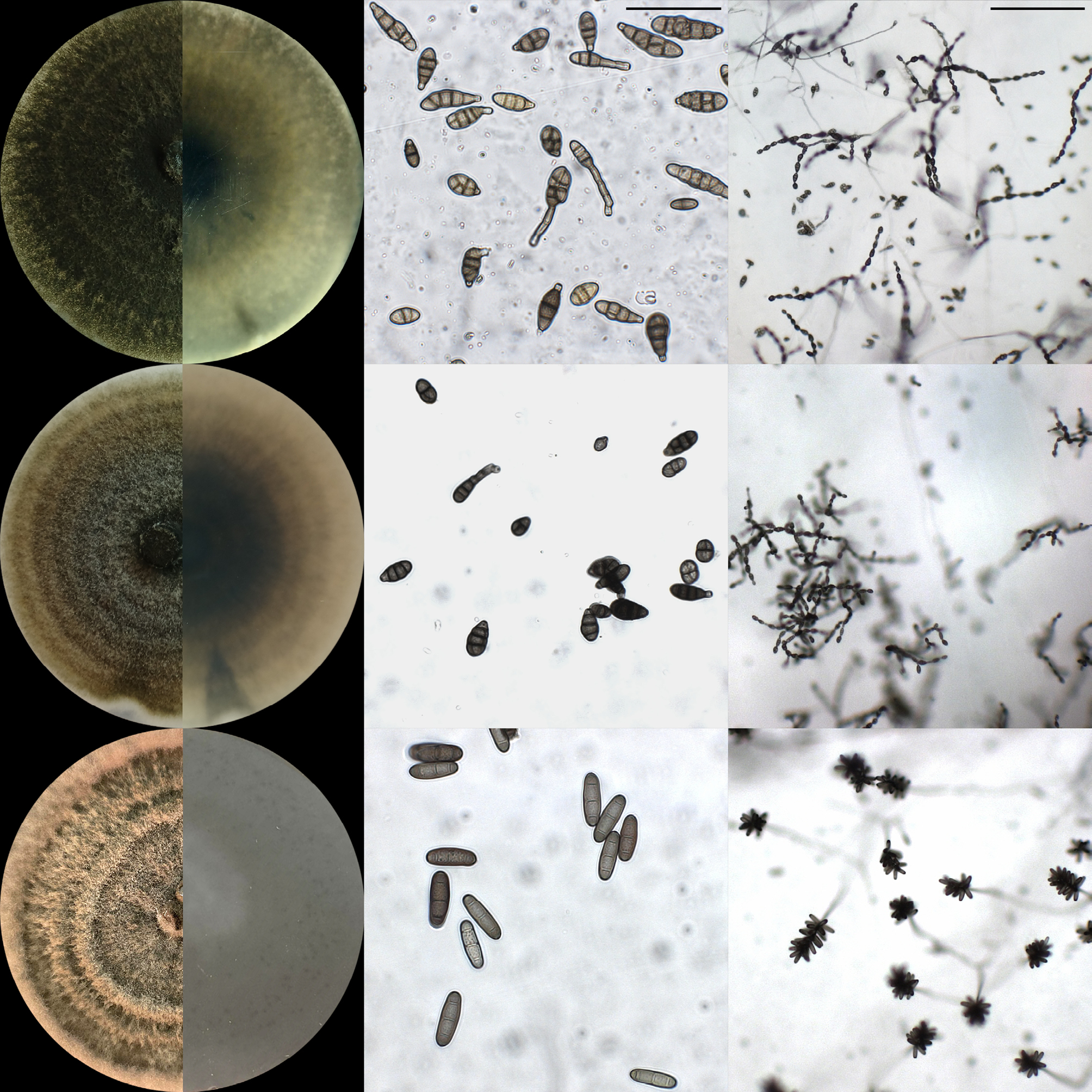
The morphological analyses showed variations in culture colour and texture between the studied species (Table 2). Alternaria alternata isolates had colony colours ranging from grey to black, with cottony textures, and generally smooth regular margins. The A. arborescens isolates had colony colours of various tones of grey, with a predominantly cottony appearance, and regular and irregular colony margins. Colonies of Curvularia isolates were green-brown in colour, of velvety texture, and had smooth margins. Differences between A. alternata, A. arborescens, and Curvularia sp. were further quantified based on conidium features.
| Species | Texturea | RAL colourb | Mean growth rate (cm d-1) | Mean conidium length (µm) | Mean conidium width (µm) | Number of conidia | |||||||||||||
|---|---|---|---|---|---|---|---|---|---|---|---|---|---|---|---|---|---|---|---|
| Alternaria alternata | Cottony | RAL 7013 | 1.14 ± 0.10 | 23.51 ± 3.55 | 9.76 ± 1.59 | 71 | |||||||||||||
| Alternaria arborescens | Cottony | RAL 7002 | 1.21 ± 0.04 | 21.83 ± 2.92 | 9.59 ± 1.16 | 51 | |||||||||||||
| Curvularia sp. | Velvety | RAL 8000 | 1.23 ± 0.07 | 28.03 ± 3.57 | 8.99 ± 1.12 | 82 | |||||||||||||
| a Predominant texture among the studied strains. | |||||||||||||||||||
| b Predominant RAL colour among the studied strains. | |||||||||||||||||||
| 1 Mean number (± standard deviation) of conidia per chain. | |||||||||||||||||||
| 2 Mean number (± standard deviation) of conidia per cluster. | |||||||||||||||||||
Alternaria alternata and A. arborescens had dark-coloured conidia, that were multicellular, ovoid or obclavate, and with short conical or cylindrical beaks. The average conidium dimensions of A. alternata were 23.5 ± 3.6 µm × 9.8 ± 1.6 µm, while those of A. arborescens were 21.8 ± 2.9 µm × 9.6 ± 1.2 µm. Conidia were produced in chains on conidiophores. The Curvularia sp. conidia were dark, ellipsoid, and each had three transverse septa. The average conidium dimensions of Curvularia sp. were 28.0 ± 3.6 µm × 9.0 ± 1.1 µm. These conidia developed close together in chains, appearing as clusters (Figure 1).
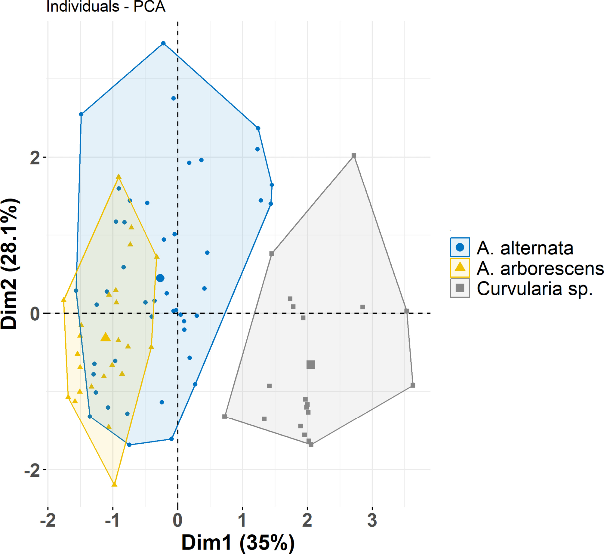
Evaluation of morphological traits of conidia using MANOVA Wilk’s Lambda test showed statistically significant differences in mean lengths, widths, and numbers of conidia per chain across the three fungal species (Wilks’ F(6,168) = 14.60; P < 0.0001). Pairwise comparisons using estimated marginal means, based on the multilinear model, showed significant differences in length, width, and number of conidia per chain between all pairs Curvularia sp. and A. alternata (P < 0.01), Curvularia sp. and A. arborescens (P < 0.0001), and A. alternata and A. arborescens (P < 0.001).
Morphological variations among the three species, A. alternata, A. arborescens, and Curvularia sp., based on the first two principal components (35.0% Dim. 1 and 28.1% Dim.2) are shown in Figure 2. Curvularia sp. was clearly distinct from the other two species, forming a separate cluster along the first principal component. In contrast, A. alternata and A. arborescens had overlapping clusters, indicating some morphological similarities and shared traits. Also, A. alternata had greater variability across both dimensions, reflecting high morphological diversity, while A. arborescens had a narrow cluster that indicated less variability within the species.
Phylogenetic analyses of fungal isolates
The phylogenetic analyses based on ITS sequences placed the studied isolates into two well-supported and clearly differentiated clades (Figure 3). Isolates cc_22 and cc_24 clustered with the type strains of Curvularia buchloes, C. hawaiiensis, C. rouhanii, and C. spicifera. BLAST analysis of TEF1-α sequences from the Hungarian Curvularia isolates (cc_24 and cc_24) identified the closest match with a C. hawaiiensis strain (GenBank no. OR841149.1), showing 96% sequence similarity. In contrast, no relevant Curvularia sequences for HIS3 were available in GenBank. Consequently, the Hungarian Curvularia isolates (cc_22 and cc_24) were reliably identified only to genus level.
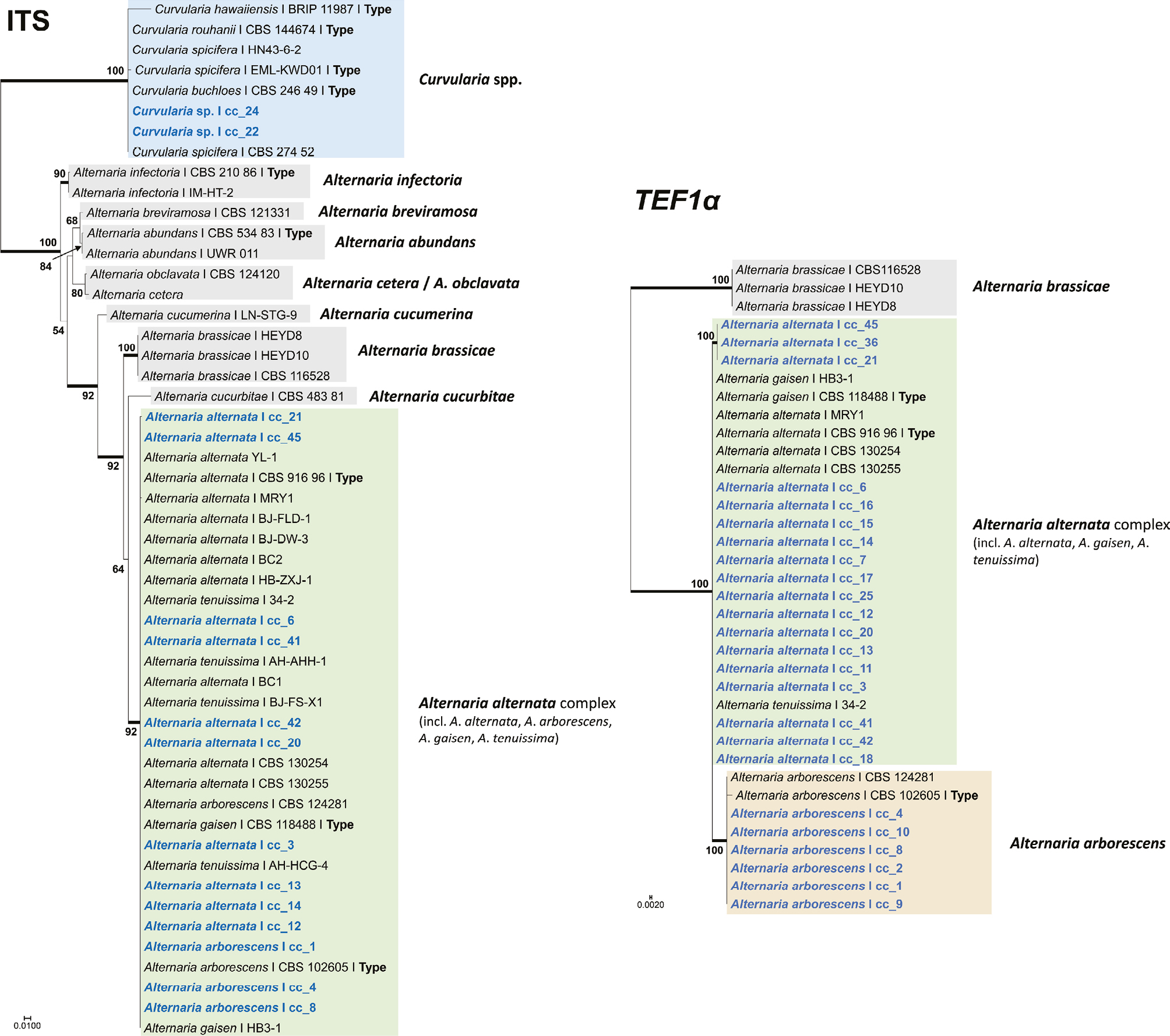
In addition to the two Curvularia isolates, all the other isolates examined in this study were classified within the Alternaria alternata species complex, based on ITS sequence analysis (Figure 3). To achieve more precise species-level identifications within the complex, TEF sequence analysis was conducted. Six isolates (cc_1, cc_2, cc_4, cc_8, cc_9, and cc_10) formed a strongly supported clade (ML = 100%) with the type strain of A. arborescens, clearly separating them from other members of the A. alternata species complex (Figure 3). To further differentiate A. alternata s. str. from A. tenuissima, HIS3 sequences were also analysed. However, none of the examined isolates showed sequence identity with A. tenuissima, indicating their affiliation with A. alternata s. str.
For species frequencies, 68% of the isolates were identified as A. alternata, 24% as A. arborescens, and 8% as Curvularia sp. The majority of A. alternata isolates were obtained from Hungary (77%), with smaller proportions from Spain (18%) and Kosovo (6%). In contrast, A. arborescens isolates were all (100%) collected in Spain, and Curvularia sp. isolates were all (100%) collected in Hungary.
Pathogenicity assays
Leaf disc assays showed variations in host resistance to the pathogens, expressed by the disease index (DI) for the 73 different plant accessions and the ten fungal isolates (Figure 4). A one-way ANOVA showed differences in mean DI between the investigated groups of fungal species (F(2,77.22) = 521.7; P < 0.0001). According to Welch’s ANOVA post hoc test, DI evaluated upon infection by A. alternata, Curvularia sp., and A. arborescens differed between all species (P < 0.0001). Curvularia sp. was the most aggressive with a meanDI of 63.69±26.0, followed by A. arborescens 50.75±26.04, and the least severe infection was caused by A. alternata 42.59±20.89 (Figure S2).
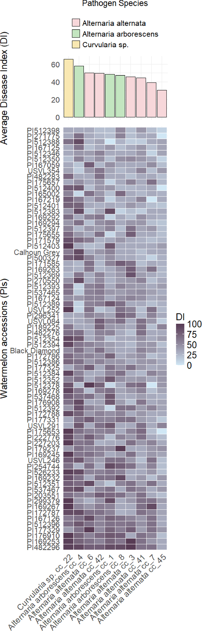
To focus on the pathogenicity of Alternaria and Curvularia species, and to draw conclusions about their aggressiveness to two reference cultivars with practical relevance, statistical analyses were narrowed to evaluate pathogenicity of Alternaria and Curvularia species using ‘Calhoun Grey’ and ‘Black Diamond’ as reference host cultivars. There was a statistically significant difference in mean DI between fungal species (F(2,5.5) = 21.368; P < 0.01). The Games- Howell post hoc test showed that Curvularia sp. (DI = 59.3 ± 6.4) and A. arborescens (DI 59.3 ± 26.1) were both more aggressive to watermelon than A. alternata (DI 33.3 ± 6.6) (P < 0.05). For ‘Black Diamond’, no statistically significant differences (P > 0.05) in mean DI were observed among the three fungal species. However, a consistent pattern was observed, with Curvularia sp. giving the greatest mean disease index (DI = 51.9, ± 12.8), followed by A. arborescens (DI = 46.9±16.4), and A. alternata (DI = 46.3 ± 16.7). Resistant plant accessions were identified with lower DI values than their commercial counterparts.
Based on the lowest recorded mean DI, which falls within the first quartile of the assessed plant accessions, the most resistant watermelon PIs were identified with a mean DI of 37.3 ± 2.9. These included accession PI512398 (from Spain), PI271775 (South Africa), PI512388 (Spain), PI167125 (Turkey), PI512346 (Spain), PI512350 (Spain), PI167059 (Turkey), PI482283 (Zimbabwe), PI512400 (Spain), PI175657 (Turkey), PI165002 (Turkey), PI167219 (Turkey), PI512401 (Spain), PI512383 (Spain), PI169292 (Turkey), PI169294 (Turkey), and accession PI512397 (from Spain).
No statistically significant differences (P > 0.05) in mean DIs were detected across the plant accessions originating from different countries , indicating that country of origin did not affect resistance to leaf spots caused by Alternaria and Curvularia isolates among the studied watermelon collection.
Discussion
Fungal leaf spot diseases pose increasing threats to cucurbit production (Abu-Nasser and Abu-Naser, 2018; Ma et al., 2021; Shanthi Avinash et al., 2021). Theses diseases cause premature losses of green leaf area through necroses and defoliation, as the infections progress, reducing photosynthetic rates, increasing fruit susceptibility to sunscald, and ultimately leading to important yield losses (Singh et al., 2011; Kaniyassery et al., 2024). Identifying the causal agents of these diseases is often challenging, as it requires distinction between closely related pathogen taxa that cause similar host symptoms and share similar morphological features (Rabaaoui et al., 2022; Torres-Garcia et al., 2022). Consequently, understanding the differences in pathogenicity among these fungi has been limited. The present study aimed to evaluate the morphology, genetic backgrounds, and pathogenicity of Alternaria and Curvularia species associated with leaf spot diseases in cucurbits collected from different geographical regions during 2024.
The most frequently isolated fungal leaf spot pathogen from infected leaves in the sampled locations was A. alternata, followed by A. arborescens, and Curvularia sp. The majority of A. alternata isolates were obtained from Hungary, while most A. arborescens isolates were collected from Spain, and Curvularia sp. isolates were exclusively from Hungary. Detection of Curvularia sp. in Hungary is notable since this is the first report of Curvularia sp. as a causal agent of fungal leaf spot on watermelon.
In the last decade, there have been few reports of Curvularia species affecting the cucurbits pointed gourd (Sarkar et al., 2018), kershaw (Ayvar-Serna et al., 2022), and melon (Vanitha et al., 2024). This limited number of reports suggests that the impacts of Curvularia on cucurbits may not be widespread, or may not have been consistently studied. In the present study, the observed symptoms of leaf spot diseases caused by Curvularia sp. on watermelon were similar to those described by Ayvar-Serna et al. (2022) and Sarkar et al. (2018), rather than blight symptoms reported by Vanitha et al. (2024).
The general morphological description of Curvularia sp. in the present study fits with other descriptions, for culture appearance and conidium size, septum number, and shape, and colony growth pattern (Ayoubi et al., 2017; Cui et al., 2020; Garganese et al., 2015). Curvularia species have been previously described considering only morphology, including conidium hila, septa, ontogeny, and wall structure, as well as culture colony appearance (Sivanesan, 1987). More recent studies have documented Curvularia spp. features including colony growth rates and conidium size and shape, which can depend on the culture conditions (dos Santos et al., 2018; Sun et al., 2003), which might lead to inaccurate pathogen identification. In the present study, species identification based on morphological characterization was challenging. To overcome these limitations multi-locus sequence analyses targeting ITS, TEF1-α, GAPDH, RPB2, HIS3, SSU, LSU, endoPG, OPA1-3, and OPA2-1 have been recommended (Dettman and Eggertson, 2021; Woudenberg et al., 2015; Aung et al., 2024). ITS, as the main barcoding marker for fungi, has been frequently employed for identifying Curvularia species (Ayoubi et al., 2017). However, recent studies have highlighted limitations in the use of ITS for species-level identification within Pleosporales (Bhunjun et al., 2020), suggesting the need for additional markers for more accurate delimitation of Curvularia species. Therefore, both HIS3 and TEF1-α were included in the present study, based on previous research indicating their suitability to delineate Curvularia species (Manamgoda et al., 2011; 2012a; 2012b; 2015). However, in the present case, the two barcoding markers were not sufficient to determine the species identity of the isolates. BLAST results for ITS suggested C. hawaiensis, while TEF1-α suggested C. spicifera as the species, but with low support for both species. To confirm this identification or to describe our isolates as a new species, multilocus sequencing is recommended.
During sampling in Soroksár, Hungary, morphologically similar symptoms caused by Curvularia sp. and Alternaria spp. were observed co-occurring on the same leaves, raising the possibility that Curvularia may cause opportunistic infections. Recent studies have also reported the occurrence of Curvularia spp. along with other fungal pathogens suggesting that Curvularia spp. may be secondary pathogens infecting stressed plants, especially under environmental conditions of high temperatures and poor water quality (Katushova et al., 2021; Bessadat et al., 2023).
The present study fulfilled Koch’s postulates for Curvularia as a pathogen of watermelon, using assays with ‘Black Diamond’ leaf disk, indicating pathogenicity. Furthermore, pathogenicity assessments were carried out for 73 watermelon accessions originating from 12 different countries. These assays showed statistically significant differences in DIs among the studied fungi, with Curvularia sp. having greatest pathogenicity, followed by A. arborescens, and A. alternata being the least pathogenic.
Several new host species has been recently reported for Curvularia spp. causing leaf blight and leaf spot diseases, many of which are important crops including sorghum (Akram et al., 2014), strawberry (Ayoubi et al., 2017), Chinese fir (Cui et al., 2020), maize (Garcia-Aroca et al., 2018; Manzar et al., 2024), citrus (Garganese et al., 2015), tomato (Huang et al., 2023), coffee (Nam et al., 2024), lettuce (Pornsuriya et al., 2018), rice (Majeed et al., 2015; Ren et al., 2022), vetiver (Sari et al., 2023), and dates (Rabaaoui et al., 2022). Previous reports have reported that within Pleosporaceae, the pathogenicity profiles and host ranges often depend on the production of host-specific toxins (HSTs) and non-host-specific toxins (nHSTs) (Akimitsu et al., 2014; Gao et al., 2014; Meena et al., 2017). Alternaria isolates produce several nHSTs, and low molecular weight HSTs that have host specificity. In contrast, Curvularia isolates have only been associated with nHSTs, which affect broader ranges of hosts (Meena et al., 2017; Rabaaoui et al., 2022). Understanding these differences will provide insights into the pathogenicity mechanisms and host-pathogen interactions of these fungi.
The present study results highlight the different host resistances within watermelon germplasm accessions, which could be utilized to select promising PI accessions, such as the Spanish PI512398, PI512350, and PI512388 which showed greater resistance than other accessions to Alternaria and Curvularia. The accession PI512398 has been used as a source of resistance to gummy stem blight (caused by Stagonosporopsis cucurbitacearum), so could be utilized as having multiple disease resistance (Gusmini et al., 2005; Rivera-Burgos et al., 2021). Additionally, during field sampling in the present study, PI512350 and PI512388 (both from Spain) were strongly tolerant to gummy stem blight under greenhouse conditions in Hungary (unpublished data, 2024). This suggests that some watermelon accessions classified as resistant in the present study may also possess resistance to a broader range of pathogens.
Conclusions
This study investigated morphology, phylogeny, and pathogenicity of Alternaria and Curvularia isolates associated with fungal leaf spots of cucurbits. Alternaria alternata occurred most frequently in the sampled locations, but was less aggressive in leaf disc assays with a diverse watermelon collection than the other fungi assessed. In contrast, only a relatively low number of Curvularia sp. isolates were detected, but the assessed isolate was the most aggressive in the leaf disc assays. This study is the first report of Curvularia sp. as a causal agent of fungal leaf spot disease on watermelon. This highlights the importance of considering species diversity and pathogenicity when managing leaf spot diseases of cucurbits. The present study also identified promising watermelon germplasm accessions among the plant material studied, that had resistance against Alternaria and Curvularia pathogens. Therefore, future research should focus on screening the watermelon germplasm collection under controlled and field conditions, and to further select resistant plant accessions. This knowledge will aid development of sustainable management strategies aimed at protecting cucurbit production from these emerging pathogens.
Acknowledgements
Dr Maxim Byatets and Jeszenszky Szilvia, of the Watermelon Breeding Team, Syngenta Research Station, Ócsa, Hungary, donated samples and plant material, and strongly supported this research. Their expertise and guidance were instrumental in completion of this study.
Author Contributions
C.P.; writing (original draft), visualization, validation, software, methodology, investigation, formal analysis, data curation, and conceptualization; V.P.; software, investigation, and formal analysis; V.B.; methodology, investigation; G.B.; methodology, resources, D.P.; supervision, writing, visualization, validation, software, methodology, investigation, formal analysis, data curation, conceptualization, resources, project administration, and funding acquisition. All the authors have read and agreed to the published version of the manuscript.
Literature cited
Abu-Nasser B. S., Abu-Naser S. S., 2018. Rule-Based System for Watermelon Diseases and Treatment. International Journal of Academic Information Systems Research 2(7): 1–7. https://hal.science/hal-01855441
Ahmed M. Z., Saeed S., Hassan A., Ghuffar S., Abdullah A., … Shafique M. S., 2021. Alternaria alternata causing Alternaria Leaf Spot of Cucumis melo (Muskmelon) in Pakistan. Plant Disease 105(6): 1853. https://doi.org/10.1094/PDIS-05-20-0973-PDN
Akimitsu K., Tsuge T., Kodama M., Yamamoto M., Otani H., 2014. Alternaria host-selective toxins: Determinant factors of plant disease. Journal of General Plant Pathology 80(2): 109–122. https://doi.org/10.1007/S10327-013-0498-7
Akram W., Anjum T., Ahmad A., Moeen R., 2014. First Report of Curvularia lunata Causing Leaf Spots on Sorghum bicolor from Pakistan. Plant Disease 98(7): 1007. https://doi.org/10.1094/PDIS-12-13-1291-PDN
Aung S. L. L., Liu F. Y., Gou Y. N., Nwe Z. M., Yu Z. H., Deng J. X., 2024. Morphological and phylogenetic analyses reveal two new Alternaria species (Pleosporales, Pleosporaceae) in Alternaria section from Cucurbitaceae plants in China. MycoKeys 107: 125–139. https://doi.org/10.3897/MYCOKEYS.107.124814
Ayoubi N., Soleimani M. J., Zare R., Zafari D., 2017. First report of Curvularia inaequalis and C. spicifera causing leaf blight and fruit rot of strawberry in Iran. Nova Hedwigia 105(1–2): 75–85. https://doi.org/10.1127/nova_hedwigia/2017/0402
Ayvar-Serna S., Díaz-Nájera J. F., Vargas-Hernández M., Mena-Bahena A., Mora-Romero G. A., Leyva-Madrigal K. Y., Tovar-Pedraza J. M., 2022. Curvularia brachyspora causing leaf spot on Cucurbita argyrosperma in Mexico. Journal of General Plant Pathology 88(5): 331–335. https://doi.org/10.1007/S10327-022-01078-1
Bao X., Roossinck M. J., 2013. Multiplexed Interactions: Viruses of Endophytic Fungi. Advances in Virus Research 86: 37–58. https://doi.org/10.1016/B978-0-12-394315-6.00002-7
Bessadat N., Hamon B., Bataillé-Simoneau N., Hamini-Kadar N., Kihal M., Simoneau P., 2023. Identification and characterization of fungi associated with leaf spot/blight and melting-out of turfgrass in Algeria. Phytopathologia Mediterranea 62(1): 73–93. https://doi.org/10.36253/PHYTO-14169
Bhunjun C. S., Dong Y., Jayawardena R. S., Jeewon R., Phukhamsakda C., … Sheng J. (2020). A polyphasic approach to delineate species in Bipolaris. Fungal Diversity 102(1): 225–256. https://doi.org/10.1007/S13225-020-00446-6
Bhunjun C. S., Niskanen T., Suwannarach N., Wannathes N., Chen Y. J., … Lumyong S., 2022. The numbers of fungi: are the most speciose genera truly diverse? Fungal Diversity 114(1): 387–462. https://doi.org/10.1007/S13225-022-00501-4
Bhunjun C. S., Chen Y. J., Phukhamsakda C., Boekhout T., Groenewald J. Z., … Crous P. W., 2024. What are the 100 most cited fungal genera? Studies in Mycology 108: 1–411. https://doi.org/10.3114/sim.2024.108.01
Chomicki G., Schaefer H., Renner S. S., 2020. Origin and domestication of Cucurbitaceae crops: insights from phylogenies, genomics and archaeology. New Phytologist 226(5): 1240–1255. https://doi.org/10.1111/nph.16015
Connally A., Smith D., Marek S., Wu Y., Walker N., 2022. Phylogenetic evaluation of Bipolaris and Curvularia species collected from turfgrasses. International Turfgrass Society Research Journal 14(1): 916–930. https://doi.org/10.1002/ITS2.16
Cui W. L., Lu X. Q., Bian J. Y., Qi X. L., Li D. W., Huang L., 2020. Curvularia spicifera and Curvularia muehlenbeckiae causing leaf blight on Cunninghamia lanceolata. Plant Pathology 69(6): 1139–1147. https://doi.org/10.1111/PPA.13198
Dettman J. R., Eggertson Q., 2021. Phylogenomic analyses of Alternaria section Alternaria: A high-resolution, genome-wide study of lineage sorting and gene tree discordance. Mycologia 113(6): 1218–1232. https://doi.org/10.1080/00275514.2021.1950456
dos Santos P. R. R., Leão E. U., Aguiar R. W. de S., de Melo M. P., dos Santos G. R., 2018. Morphological and molecular characterization of Curvularia lunata pathogenic to andropogon grass. Bragantia 77(2): 326–332. https://doi.org/10.1590/1678-4499.2017258
Edler D., Klein J., Antonelli A., Silvestro D., 2021. RaxmlGUI 2.0: A graphical interface and toolkit for phylogenetic analyses using RAxML. Methods in Ecology and Evolution 12: 373–377. https://doi.org/10.1111/2041-210X.13512
Egel D. S., Adkins S. T., Wintermantel W. M., Keinath A. P., D’Arcangelo K. N., … Quesada-Ocampo L. M., 2022. Diseases of Cucumbers, Melons, Pumpkins, Squash, and Watermelons. In: Handbook of Vegetable and Herb Diseases. Handbook of Plant Disease Management (W. H. Elmer, M. McGrath, R. J. McGovern ed.), Springer, Cham, 1–105. https://doi.org/10.1007/978-3-030-35512-8_33-1
AA.VV., 2021. Alternaria leaf blight. Available at: http://ephytia.inra.fr/en/C/7941/Melon-Alternaria-cucumerina
FAOSTAT, 2022. FAOSTAT: Crops and livestock products. Available at: https://www.fao.org/faostat/en/#data/QCL. Accessed January 30, 2025.
Fontaine K., Fourrier-Jeandel C., Armitage A., Boutigny A. L., Crépet M., … Aguayo J., 2021. Identification and pathogenicity of Alternaria species associated with leaf blotch disease and premature defoliation in French apple orchards. PeerJ 9: e12496. https://doi.org/10.7717/PEERJ.12496
Gao S., Li Y., Gao J., Suo Y., Fu K., Li Y., Chen J., 2014. Genome sequence and virulence variation-related transcriptome profiles of Curvularia lunata, an important maize pathogenic fungus. BMC Genomics 15(1): 1–18. https://doi.org/10.1186/1471-2164-15-627
Garcia-Aroca T., Doyle V., Singh R., Price T., Collins K., 2018. First report of Curvularia leaf spot of corn, caused by Curvularia lunata, in the United States. Plant Health Progress 19(2): 140–142. https://doi.org/10.1094/PHP-02-18-0008-BR
Garganese F., Sanzani S. M., Mincuzzi A., Ippolito A., 2015. First report of Curvularia spicifera causing brown rot of citrus in Southern Italy. Journal of Plant Pathology 97(3): 543. https://doi.org/10.4454/JPP.V97I3.001
Grumet R., McCreight J. D., McGregor C., Weng Y., Mazourek M., … Fei Z., 2021. Genetic resources and vulnerabilities of major cucurbit crops. Genes 12(8): 1222. https://doi.org/10.3390/genes12081222
Gusmini G., Song R., Wehner T. C., 2005. New Sources of Resistance to Gummy Stem Blight in Watermelon. Crop Science 45(2): 582–588. https://doi.org/10.2135/CROPSCI2005.0582
Harris J. L., 2000. Safe, Low-Distortion Tape Touch Method for Fungal Slide Mounts. Journal of Clinical Microbiology 38(12): 4683. https://doi.org/10.1128/JCM.38.12.4683-4684.2000
Huang Y., Jones C., Urbina H., Zhang S., 2023. First Report of Leaf Blight Caused by Curvularia aeria and C. senegalensis on Tomato (Solanum lycopersicum) in Florida, U.S.A. Plant Disease 107(12): 4027. https://doi.org/10.1094/PDIS-06-23-1209-PDN
Hyde K.D., Noorabadi M.T., Thiyagaraja V., He M.Q., Johnston P.R., … Zvyagina E., 2024. The 2024 Outline of Fungi and fungus-like taxa. Mycosphere 15(1): 5146–6239. https://doi.org/10.5943/mycosphere/15/1/25
Kaniyassery A., Hegde M., Sathish S. B., Thorat S. A., Udupa S., … Muthusamy A., 2024. Leaf spot-associated pathogenic fungi alter photosynthetic, biochemical, and metabolic responses in eggplant during the early stages of infection. Physiological and Molecular Plant Pathology 133: 102320. https://doi.org/10.1016/j.pmpp.2024.102320.
Katoh K., Standley D.M., 2013. MAFFT multiple sequence alignment software version 7: improvements in performance and usability. Molecular Biology and Evolution 30(4): 772-80. https://doi.org/10.1093/molbev/mst010
Katushova M., Beloshapkina O., Tarakanov R., Shipulin A., Dzhalilov F., 2021. Fungi of the genus Curvularia sp. - new pathogens of turfgrass in Russia. IOP Conference Series: Earth and Environmental Science 663(1): 012007. https://doi.org/10.1088/1755-1315/663/1/012007
Khan N. A., Asaf S., Ahmad W., Jan R., Bilal S., … Al-Harrasi A., 2023. Diversity, lifestyle, genomics, and their functional role of Cochliobolus, Bipolaris, and Curvularia species in environmental remediation and plant growth promotion under biotic and abiotic stressors. Journal of Fungi 9(2): 254. https://doi.org/10.3390/JOF9020254
Kucharek T., 1985. Alternaria Leaf Spot of Cucurbits. University of Florida, Insitute of Food and Agricultural Sciences, Plant Pathology Department, Plant Pathology Fact Sheet. PP-32. https://original-ufdc.uflib.ufl.edu/UF00066896
Kwon O. K., Jeong A. R., Jeong Y. J., Kim Y. A., Shim J., … Park C. J., 2021. Incidence of Alternaria species associated with watermelon leaf blight in Korea. Plant Pathology Journal 37(4): 329–338. https://doi.org/10.5423/PPJ.OA.02.2021.0018
Larsson A., 2014. AliView: a fast and lightweight alignment viewer and editor for large datasets. Bioinformatics 30(22): 3276–3278. https://doi.org/10.1093/BIOINFORMATICS/BTU531
Ma G., Bao S., Zhao J., Sui Y., Wu X., 2021. Morphological and molecular characterization of Alternaria species causing leaf blight on watermelon in China. Plant Disease 105(1): 60–70. https://doi.org/10.1094/PDIS-01-20-0130-RE
Majeed R. A., Shahid A. A., Ashfaq M., Saleem M. Z., Haider M. S., 2015. First Report of Curvularia lunata Causing Brown Leaf Spots of Rice in Punjab, Pakistan. Plant Disease 100(1): 219. https://doi.org/10.1094/PDIS-05-15-0581-PDN
Manamgoda D. S., Cai L., Bahkali A. H., Chukeatirote E., Hyde K. D., 2011. Cochliobolus: An overview and current status of species. Fungal Diversity 51: 3–42. https://doi.org/10.1007/S13225-011-0139-4
Manamgoda D. S., Cai L., McKenzie E. H. C., Crous P. W., Madrid H., … Hyde K. D., 2012a. A phylogenetic and taxonomic re-evaluation of the Bipolaris - Cochliobolus - Curvularia Complex. Fungal Diversity 56(1): 131–144. https://doi.org/10.1007/S13225-012-0189-2
Manamgoda D. S., Cai L., McKenzie E. H. C., Chukeatirote E., Hyde K. D., 2012b. Two new Curvularia species from northern Thailand. Sydowia 2(64): 255–267.
Manamgoda D. S., Rossman A. Y., Castlebury L. A., Chukeatirote E., Hyde K. D., 2015. A taxonomic and phylogenetic re-appraisal of the genus Curvularia (Pleosporaceae): Human and plant pathogens. Phytotaxa 212(3): 175–198. https://doi.org/10.11646/PHYTOTAXA.212.3.1
Manzar N., Kashyap A. S., Sharma P. K., Srivastava A. K., 2024. First Report of Leaf Spot on Maize Caused by Curvularia verruculosa in India. Plant Disease 108(3): 793. https://doi.org/10.1094/PDIS-07-23-1410-PDN
Marin-Felix Y., Senwanna C., Cheewangkoon R., Crous P. W., 2017. New species and records of Bipolaris and Curvularia from Thailand. Mycosphere 8(9): 1555–1573. https://doi.org/10.5943/mycosphere/8/9/11
Marin-Felix Y., Hernández-Restrepo M., Crous P. W., 2020. Multi-locus phylogeny of the genus Curvularia and description of ten new species. Mycological Progress 19(6): 559–588. https://doi.org/10.1007/S11557-020-01576-6
Matić S., Tabone G., Garibaldi A., Gullino M. L., 2020. Alternaria leaf spot caused by Alternaria species: an emerging problem on ornamental plants in Italy. Plant Disease 104(8): 2275–2287. https://doi.org/10.1094/PDIS-02-20-0399-RE
Meena M., Gupta S. K., Swapnil P., Zehra A., Dubey M. K., Upadhyay R. S., 2017. Alternaria toxins: Potential virulence factors and genes related to pathogenesis. Frontiers in Microbiology 8(1451): 1-14. https://doi.org/10.3389/FMICB.2017.01451
Nam H. S., Park H. S., Kim Y. C., 2024. First report of coffee leaf spot caused by Curvularia geniculata. Journal of Phytopathology 172(1): e13245. https://doi.org/10.1111/JPH.13245
Nobles M. K., 1948. Studies in forest pathology: VI. Identification of cultures of wood-rotting fungi. Canadian Journal of Research 26(3): 281–431. https://doi.org/doi.org/10.1139/cjr48c-026
Paris H. S., 2015. Origin and emergence of the sweet dessert watermelon, Citrullus lanatus. Annals of Botany 116(2): 133–148. https://doi.org/10.1093/aob/mcv077
Peever T. L., Su G., Carpenter-Boggs L., Timmer L. W., 2004. Molecular systematics of citrus-associated Alternaria species. Mycologia 96(1): 119–134. https://doi.org/10.1080/15572536.2005.11833002
Pornsuriya C., Ito S., Sunpapao A., 2018. First report of leaf spot on lettuce caused by Curvularia aeria. Journal of General Plant Pathology 84(4): 296–299. https://doi.org/10.1007/S10327-018-0782-7
Rabaaoui A., Masiello M., Somma S., Crudo F., Dall’Asta C., … Moretti A., 2022. Phylogeny and mycotoxin profiles of pathogenic Alternaria and Curvularia species isolated from date palm in southern Tunisia. Frontiers in Microbiology 13: 1034658. https://doi.org/10.3389/FMICB.2022.1034658
Ren X., Chen S., Guo J., Wang M., Liu X., Wei Z. Z., 2022. First Report of Curvularia lunata Causing Leaf Spot on Oryza sativa in Sabah, Malaysian Borneo. Plant Disease 107(7): 2234. https://doi.org/10.1094/PDIS-08-22-1939-PDN
Rivera-Burgos L. A., Silverman E., Sari N., Wehner T. C., 2021. Evaluation of Resistance to Gummy Stem Blight in a Population of Recombinant Inbred Lines of Watermelon × Citron. HortScience 56(3): 380–388. https://doi.org/10.21273/HORTSCI15599-20
Rueden C. T., Schindelin J., Hiner M. C., DeZonia B. E., Walter A. E., Arena E. T., Eliceiri K. W., 2017. ImageJ2: ImageJ for the next generation of scientific image data. BMC Bioinformatics 18(1): 1–26. https://doi.org/10.1186/S12859-017-1934-Z
Sari M. P., Wahyuno D., Hardiyanti S., Miftakhurohmah., 2023. First report of Curvularia akaiiensis as a causal agent of leaf spot disease on Vetiver. Australasian Plant Disease Notes 18(1): 1–4. https://doi.org/10.1007/S13314-023-00516-Z
Sarkar T., Chakraborty P., Das S., Saha D., Saha A., 2018. Curvularia leaf spot of pointed gourd in India. Canadian Journal of Plant Pathology 40(4): 594–600. https://doi.org/10.1080/07060661.2018.1504822
Shahin E. A., 1979. An Efficient Technique for Inducing Profuse Sporulation of Alternaria Species. Phytopathology 69(6): 618. https://doi.org/10.1094/PHYTO-69-618
Shanthi Avinash T., Jai Shanker Pillai H. P., Biradar M., Shinde V. M., 2021. A Review on Fungal Diseases of Cucurbitaceae and their Management. International Journal of Current Microbiology and Applied Sciences 10(08): 653–672. https://doi.org/10.20546/ijcmas.2021.1008.075
Singh M. P., Erickson J. E., Boote K. J., Tillman B. L., Jones J. W., van Bruggen A. H. C., 2011. Late Leaf Spot Effects on Growth, Photosynthesis, and Yield in Peanut Cultivars of Differing Resistance. Agronomy Journal 103(1): 85-91 https://doi.org/10.2134/agronj2010.0322
Singh S. P., Khan N., Singh R., Singh H., Prasad S., Dwivedi D., 2020. Documentation variation for Alternaria blight resistance in varieties of Rapeseed mustard. International Journal of Chemical Studies 8(4): 1397–1400. https://doi.org/10.22271/CHEMI.2020.V8.I4M.9793
Sivanesan A., 1987. Graminicolous species of Bipolaris, Curvularia, Drechslera, Exserohilum and their teleomorphs. Mycological Papers 158: 154–185.
Stamatakis A., 2014. RAxML version 8: A tool for phylogenetic analysis and post-analysis of large phylogenies. Bioinformatics 30(9): 1312-1313. https://doi.org/10.1093/bioinformatics/btu033
Steenkamp E., Britz H., Coutinho T., Wingfield B., Marasas W., Wingfield M., 2000. Molecular characterization of Fusarium subglutinans associated with mango malformation. Molecular Plant Pathology 1(3): 187–193. https://doi.org/10.1046/J.1364-3703.2000.00024.X
Sun G., Oide S., Tanaka E., Shimizu K., Tanaka C., Tsuda M., 2003. Species separation in Curvularia “geniculate” group inferred from Brn1 gene sequences. Mycoscience 44(3): 239–244. https://doi.org/10.1007/S10267-003-0104-5
Tamura K., Stecher G., Kumar S., 2021. MEGA11: Molecular Evolutionary Genetics Analysis Version 11, Molecular Biology and Evolution 38(7): 3022–3027. https://doi.org/10.1093/molbev/msab120
Torres-Garcia D., García D., Cano-Lira J. F., Gené J., 2022. Two Novel Genera, Neostemphylium and Scleromyces (Pleosporaceae) from Freshwater Sediments and Their Global Biogeography. Journal of Fungi 8(8): 868. https://doi.org/10.3390/JOF8080868
Vakalounakis D. J., 1990. Alternaria alternata f. sp. cucurbitae, the cause of a new leaf spot disease of melon (Cucumis melo). Annals of Applied Biology 117(3): 507–513. https://doi.org/10.1111/J.1744-7348.1990.TB04817.X
Vanitha S., Kavitha M., Ragul S., Mohanapriya S., Angappan K., … Rani C. I., 2024. First report of Curvularia lunata causing leaf blight on muskmelon (Cucumis melo). New Disease Reports 50(2): e12310. https://doi.org/10.1002/NDR2.12310
Venkatesagowda B., Ponugupaty E., Barbosa A. M., Dekker R. F. H., 2012. Diversity of plant oil seed-associated fungi isolated from seven oil-bearing seeds and their potential for the production of lipolytic enzymes. World Journal of Microbiology and Biotechnology 28(1): 71–80. https://doi.org/10.1007/S11274-011-0793-4
White T., Bruns T., Lee S., Taylor J., Innis M., … Sninsky J., 1990. Amplification and Direct Sequencing of Fungal Ribosomal RNA Genes for Phylogenetics. In: PCR Protocols, Academic Press, 315–322.
Woudenberg J. H. C., Groenewald J. Z., Binder M., Crous P. W., 2013. Alternaria redefined. Studies in Mycology 75: 171–212. https://doi.org/10.3114/sim0015
Woudenberg J. H. C., Seidl M. F., Groenewald J. Z., de Vries M., Stielow J. B., Thomma B. P. H. J., Crous P. W., 2015. Alternaria section Alternaria: Species, formae speciales or pathotypes? Studies in Mycology 82: 1–21. https://doi.org/10.1016/j.simyco.2015.07.001
Wu Q., Li Y., Li Y., Gao S., Wang M., Zhang T., Chen J., 2013. Identification of a novel fungus, Leptosphaerulina chartarum SJTU59 and characterization of its xylanolytic enzymes. PLoS ONE 8(9): e73729. https://doi.org/10.1371/journal.pone.0073729
Zhou X. G., Everts K. L., 2008. First report of Alternaria alternata f. sp. cucurbitae causing Alternaria leaf spot of melon in the Mid-Atlantic region of the United States. Plant Disease 92(4): 652. https://doi.org/10.1094/PDIS-92-4-0652B