https://doi.org/10.36253/phyto-15662
Accepted: April 28, 2025; Published: May 15, 2025
Research Papers
Microbiota dynamic communities in sweet orange infected by “huanglongbing” in Iran
1 Department of Plant Protection, Islamic Azad University, Varamin, Pishva Branch, Varamin, Iran
2 Department of Plant Pathology, Islamic Azad University, Varamin, Pishva Branch, Varamin, Iran
3 Iranian Research Institute of Plant Protection, AREEO, Tehran, Iran
4 The National Institute of Genetic Engineering and Biotechnology, NIGEB, Tehran, Iran
5 Plant Protection Research Department, Fars Agricultural and Natural Resources Research and Education Center, AREEO, Zarghan, Iran
6 Plant Protection Research Department, Isfahan Agricultural and Natural Resources Research and Education Center, AREEO, Isfahan, Iran
*Corresponding authors. E-mail: mojdehmaleki@yahoo.com; aalizadeh1340@yahoo.com
Summary. “Huanglongbing”-(HLB) or citrus-greening is one of the most serious citrus diseases worldwide. This study aimed to investigate the bacterial-communities associated with HLB-symptomatic sweet orange trees (Citrus sinensis) from different geographical regions in southern Iran. The 16S rRNA gene amplicon metagenomics sequencing of DNA extracted from the midrib and petiole tissues of symptomatic plants confirmed that the ‘Candidatus Liberibacter asiaticus’, was spread along the citrus-plantation regions in southern Iran, including Kerman, Sistan and Baluchistan, Fars, Hormozgan, and Khuzestan Provinces. The frequency of Operational Taxonomic Units (OTUs) related to ‘Ca. L. asiaticus’ was remarkable in the HLB symptomatic tree in the Fars region. No OTUs of ‘Ca. Liberibacter’ or ‘Ca. Phytoplasma’ were detected in the asymptomatic samples in the Kerman region. However, in asymptomatic materials representatives of the class Bacilli, including Lactobacillus spp. and Bacillus spp., showed 12- and 4-fold presence compared to the symptomatic samples of Kerman groves. Furthermore, the presence of OTUs belonging to ‘Ca. L. europaeus’ and ‘Ca. Phytoplasma aurantifolia’ was detected in sweet oranges. The simultaneous occurrence of ‘Ca. L. asiaticus’, ‘Ca. L. europaeus’, and ‘Ca. P. aurantifolia’ in HLB symptomatic orange trees in the Fars groves provided worthwhile insights for further research, although their epidemiological role in co-infections remains unknown. Microbial dataset in relation to variables associated with the plant health, defense, and disease helps to understand how these variables shape the citrus microbial community and identify individual that play a role in HLB suppression or promotion.
Keywords. ‘Candidatus Liberibacter’ species, plant microbiota, Valencia sweet orange.
Contents
DNA extraction, PCR amplification, and microbiome sequencing
Phylogenetic analysis of identified ‘Ca. Liberibacter’ species
PCR detection of ‘Ca. L. asiaticus’
OTU clustering and beta diversity
Microbial community structures of individual samples
Introduction
Citrus fruits are economically significant crops for Iran. They are grown in two commercial citrus-growing areas, including the Caspian Sea belt (Mazandaran and Guilan provinces) and the southern inland belt scattered through the low valleys of the Southern Zagros mountain range, particularly in the provinces of Fars, Kerman, Hormozgan and Khuzestan. Statistics show that Iran is the world’s seventh-largest citrus fruit producer and ranks eighth in farmland under citrus fruit cultivation (Cochran and Samadi, 1976).
“Huanglongbing” (HLB), previously known as citrus greening, is the most devastating disease of citrus worldwide which is associated with three phloem limited Gram-negative α-proteobacteria, ‘Candidatus Liberibacter asiaticus’, ‘Ca. L. africanus’, and ‘Ca. L. americanus’ (Bove, 2006; Hu et al., 2011). Both ‘Ca. L. asiatisus’ and ‘Ca. L. americanum’ are transmitted by the Asian citrus psyllid, Diaphorina citri Kuwayama (Sternorrhyncha, Liviidae), while ‘Ca. L. africanus’ is transmitted by the African citrus psyllid Trioza erytreae Del Guercio (Sternorrhyncha, Triozidae). HLB-infected trees show different symptoms including blotchy mottle and corky vein in leaves, defoliation, yellowing and die back as well as small misshapen fruits, with color inversion (Bendix and Lewis, 2018; da Graça, 2008). The disease results in phloem malfunction, root decline, and altered plant source-sink relationships, leading to a deficient plant with minimal yield before it dies (Limayem et al., 2024). HLB disease, in Florida alone, has caused $7 billion in crop losses and over 8,000 jobs lost in a 7-year period (2007–2014) (Hodges et al., 2017). HLB disease was first observed in China in the late 19th century and was called “huanglongbing” (which means yellow dragon tail). Then after, in 1928, it was noticed in South Africa and called greening and was also reported from other Asian and African countries. Until 2004, it was reported in orange trees (Citrus sinensis) in Brazil and a year later on pomelo trees (Citrus maxima) in Florida (Filho et al., 2004; Halbert, 2005).
In Iran, D. citri was first observed in 1997 in an area close to the Pakistan border, and since then, high populations of the insect have been reported frequently in citrus plantations of Hormozgan and Kerman Provinces suggesting the possible presence of ‘Ca. L. asiaticus’ that was detected and confirmed in various locations of Sistan-Baluchistan and Hormozgan Provinces in Valencia sweet orange trees (C. sinensis) and over 50 psyllid samples (Faghihi et al., 2009). The Asian HLB bacterium has been detected in citrus growing areas in southern Iran in Sistan and Baluchistan, Kerman, Hormozgan, and Fars Provinces and its presence has been confirmed employing DNA-based methods (Faghihi et al., 2009; Mohkami et al., 2011; Salehi et al., 2012, Faghihi et al., 2016; Salehi and Rasoulpour, 2016). The disease is one of the agents associated with citrus decline in some areas of southern Iran, such as mandarin decline in Siyahoo in Hormozgan Province of Iran (Faghihi, 2018; Alizadeh et al., 2022). Since 2010, almost 10% of losses have emerged in citrus cultivated trees belonging to different citrus species in various groves of Kerman Province, Iran (Passera et al., 2018). Moreover, emergence and spread of the citrus HLB indicating that the disease expanded and spread in citrus cultivation areas in the south parts of Iran with a gradual slope in the recent years. The reduction of the disease-carrying psyllid population seems to be effective in slowing down the spread of the disease accordingly (Alizadeh et al., 2020).
So far, no definitive treatment for HLB has been identified. The only way to grow high-yielding citrus where the disease is prevalent is to manage it using integrated management strategies. These include obtaining healthy seedlings from infection-free nurseries to prevent the spread of infection to trees in uninfected areas; focusing on reducing the initial source of infection by removing infected trees or branches, and controlling vectors and keeping the pest population at the lowest possible level. However, one of the most effective ways to manage this disease is to find cultivars that have a high degree of resistance to HLB. Safarpour et al. (2022) evaluated the response of seedlings of ten Iranian commercial citrus cultivars inoculated with ‘Ca. L. asiaticus’ using conventional grafting. In this regard, the cultivar Mexican lime was the most tolerant, while Valencia orange and Orlando tangelo were the most susceptible ones to HLB.
The symptoms of HLB have been related by callose accumulation and depositions in the phloem sieve plates as a mechanism of defense against invasive tissue pathogens that inhibit nutrient uptake and transport (Bendix and Lewis, 2018). Phloem-limited pathogens represent a significant research challenge because they are difficult to detect within plants and induce disease symptoms to develop and emerge slowly in infected plants
Recent developments in high-throughput DNA sequencing technology have opened new ways to investigate plant-associated microbial communities in the ‘Ca. L. asiaticus’ infected trees (Zhang et al., 2017; Tedersoo et al., 2019). Plant phyllosphere hosts a variety of bacteria which can play a positive role in the performance of the host plant. These bacterial communities are influenced by both biotic (host species, genotype, pathogens, and leaf age) and abiotic (mainly related to geographical location) factors that can play significant roles in plant growth and disease resistance. The study of plant-associated microbial communities may result in identification of synergistic and antagonistic agents against pathogens from a similar niche (Bodenhausen et al., 2013; Monazzah et al., 2022).
Recent research on the citrus microbiome has established a foundation of data concerning the rhizosphere- or leaf-associated microbes in citrus plants affected by HLB (Blaustein et al., 2017; Trivedi et al., 2010; Zhang et al., 2017; Xu et al., 2018). Blaustein et al. (2017) proposed that the diversity of bacterial communities within leaf tissues diminishes as HLB symptoms progress and that the relative abundance of ‘Ca. Liberibacter’ species exhibited a negative correlation with the α-diversity of the bacterial community in leaf tissues. They also anticipated adverse interactions between ‘Ca. Liberibacter’ species and specific bacterial families found within the native leaf bacteriome of citrus, which could extend to the entire tree level.
Research on microbiota dynamics in sweet oranges infected with HLB (exhibiting decline symptoms) indicated that the families Micrococcaeae, Gemellaceae, and Streptococcaeae were significantly prevalent in asymptomatic trees compared to those displaying symptoms, while the families Coriobacteriaceae, Lachnospiraceae, Ruminococcaceae, Erysipelotrichaceae, and Desulfovibrionaceae showed a markedly higher abundance in symptomatic trees in comparison to the asymptomatic ones (Passera et al., 2018).
Today, a few studies have been conducted in terms of the microbial communities associated with citrus HLB for managing the disease in Iran. Therefore, given the importance of this disease in citrus orchards in Iran and its increasing spread, this study aimed to investigate the bacterial communities of midribs and petiole tissues of HLB symptomatic orange (C. sinensis) trees from five major groves located in different geographical regions in southern Iran including Kerman, Sistan and Baluchistan, Fars, Hormozgan, and Khuzestan Provinces, so that the management strategy and breeding programs can be planned accordingly. Moreover, there is a critical need to analyze and characterize the bacterial communities in HLB-affected citrus trees using 16S rRNA gene amplicon sequencing and to explore the presence of co-infecting and or co-existing bacterial taxa, including potential pathogenic or beneficial bacteria.
Materials and method
Plant samples collection
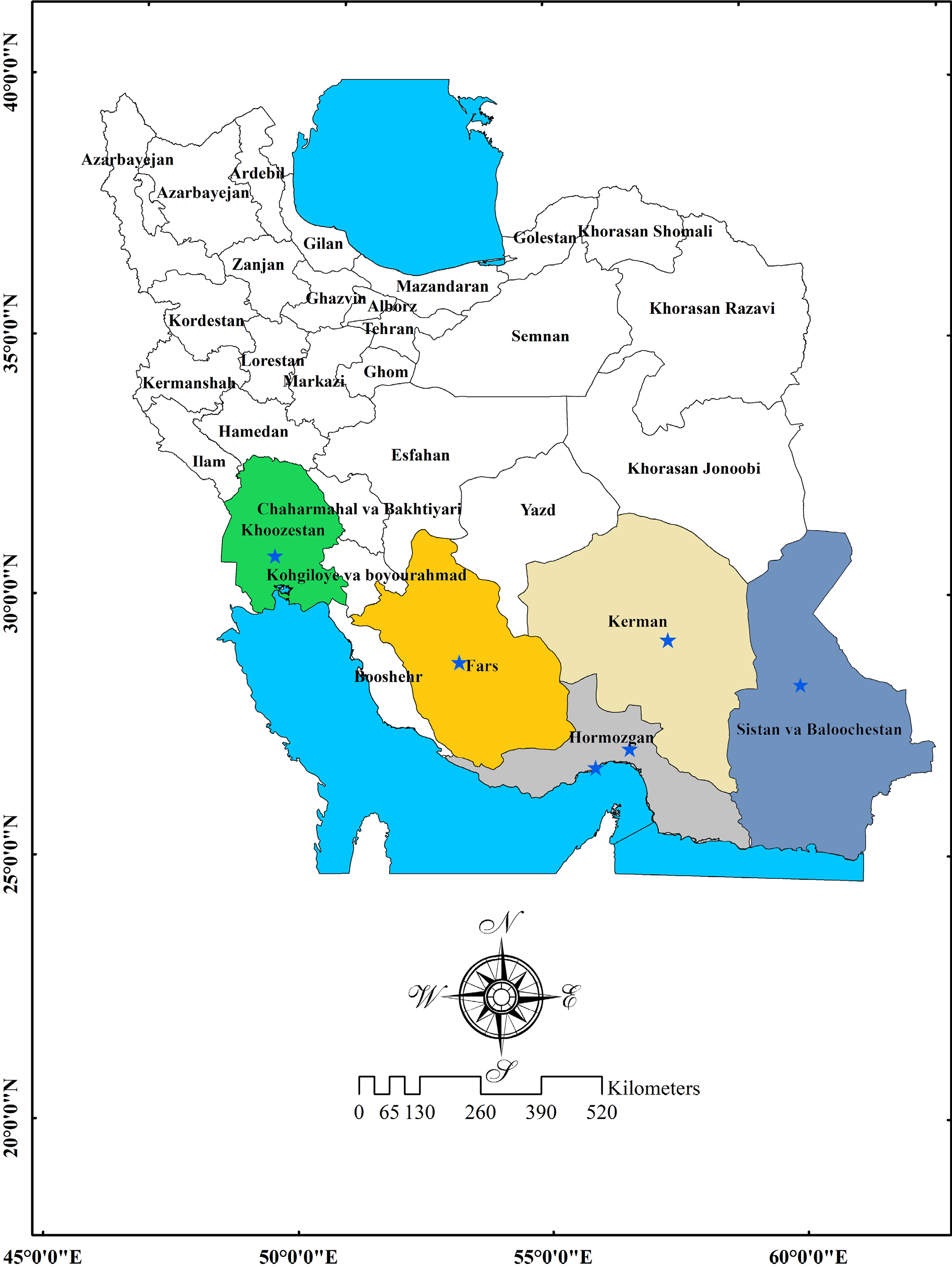
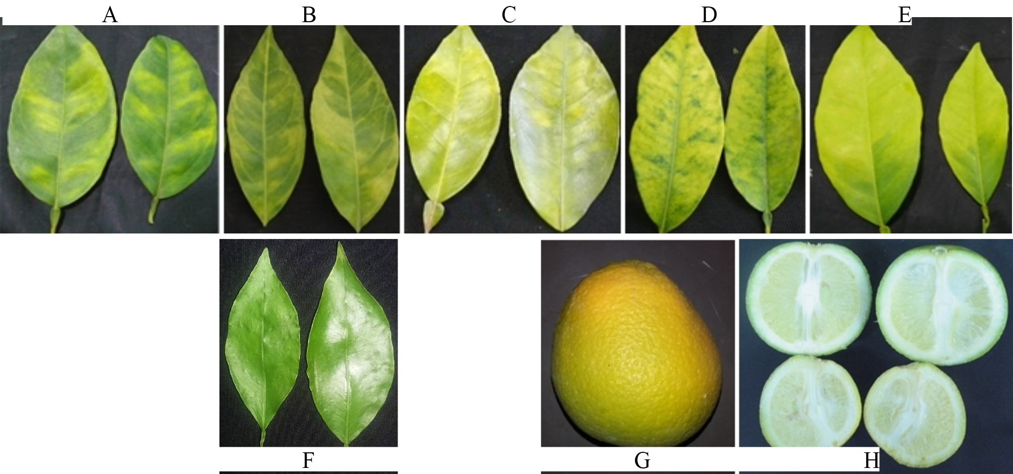
Sampling methods are critical for the detection, identification, and quantification of ‘Ca. Liberibacter’ species since their distribution in host plants can be irregular. Sweet orange trees infected may have symptomatic leaves only on some branches and others may remain free of symptoms or have low bacterial concentration. In the initial phase of the study, in October 2017, totally 75 leaf samples of Valencia sweet orange (C. sinensis) trees exhibiting symptoms of the HLB disease were collected from orchards located in the provinces of Fars (Darab), Hormozgan (Rudan), Kerman (Jiroft), Sistan and Baluchestan (Sarbaz), and Khuzestan (Dezful). Subsequently, the presence of infection was confirmed by PCR assay for ’Ca. L. asiaticus’, and the infected trees were identified in each region. Following the identification of the infected trees, composite leaf sampling was taken from three infected orange trees in each area in December 2017. Additionally, a composite leaf sample was obtained from three healthy orange trees (confirmed by PCR and quantitative PCR) located in Kerman Province. The trees targeted for sampling were almost at the same age and growing stage. In total, five composite leaf samples were collected from infected trees across the mentioned provinces (Suppl. Table 1), along with one composite leaf sample from healthy trees in Kerman Province (Figure 1). The leaf samples were placed in separated bags and transferred to the laboratory under cold conditions (Figure 2).
Plant samples preparation
The preparation of the midrib and petiole tissues was done after a proper surface sterilization to strictly ensure that only endophytic bacterial communities were present. Thus, the collected midrib and petiole tissues were surface sterilized separately. The related segments (approx. 60 mm long) were washed in mild liquid soap solution and rinsed thoroughly with running tap water. Surface sterilization was achieved by sequentially submerging each segment in 70% ethanol for 1 min and 0.625% sodium hypochlorite (10% Clorox, the Clorox Company, Oakland, CA, USA) solution for 4 min, followed by 3 sequential immersions in sterile distilled water. Surface sterilization was verified by pressing disinfected midrib and petiole segments along with aliquots of the sterile distilled water used in the final rinse. For more confidence, again the midrib and petiole tissues were rinsed five times in sterile distilled water and allowed to drain. Then, the related midribs and petioles were cut into small pieces and were flash-frozen accordingly.
DNA extraction, PCR amplification, and microbiome sequencing
The surface sterilized flash-frozen tissues of the collected symptomatic and asymptomatic tree samples were ground separately with sterile pestle and to ensure that the data only reflected endophytic or phloem-associated bacterial communities. Genomic DNA was extracted from 0.5 g of petioles and midribs using the DENAzist Plant DNA Isolation Kit (DENAzist, Iran) according to the manufacturer’s instructions. The quantity and purity of total DNA was evaluated using a NanoDrop™ 2000/2000c Spectrophotometers (Thermo Fisher Scientific).
The identity of the HLB-associated bacterium was confirmed through PCR amplification of a specific fragment of the ribosomal protein rplKAJL rpoBC gene of ‘Ca. L. asiticus’ using the primer pair A2/J5 (Villechanoux et al., 1993; Hocquellet et al., 1999). The DNA extracted from leaves of healthy sweet orange trees and sterile distilled water were used as negative controls. PCR was conducted in 20 μL of reaction mixture containing 10 μL 2X PCR Master Mix (Ampliqon, Denmark), 1 μL of each primer (10 μM), 1–2 μL of template DNA (ca. 100 ng) and 6 μL sterile distilled water. The thermocycling program included an initial denaturation step at 95°C for 4 min, succeeded by 35 cycles comprising denaturation at 94°C for 30 s, annealing at 59°C for 30 s and extension at 72°C for 60 s, with a final extension step at 72°C for 10 min. Two PCR products were directly sequenced in both directions. The sequences were compared with GenBank sequences using BLAST search (https://blast.ncbi.nlm.nih.gov/Blast.cgi) to identify and confirm the identity of ’Ca. L. asiaticus’.
It was designed a custom set of 10-base pair indices specifically tailored for the preparation of libraries from amplicons derived from the V3-V4 region of the 16S rRNA gene for the analysis of the microbial composition through sequencing on the Illumina MiSeq and HiSeq platforms. The 16S rRNA gene sequencing (V3-V4 region) was used to identify bacterial taxa to capture bacterial diversity.
Amplicon libraries were prepared for the V3–V4 region of 16S rRNA genes with PCRs that incorporated Illumina-compatible universal bacteria/archaeal primers, 341F and 805R (Klindworth et al., 2013). Amplicon libraries were sequenced using a MiSeq sequencer (Microsynth AG, Switzerland). This analysis was based on data obtained from 16S rRNA metagenomic sequencing conducted on the Illumina MiSeq and HiSeq sequencing platforms.
Data availability
Raw sequencing reads were deposited in the NCBI’s Sequence Read Archive under the accession number PRJNA578610 (SRX7033691-5).
Data analysis
The obtained forward and reverse DNA reads were analyzed using the QIIME v.1.8. pipeline (Bolyen et al., 2019). The reads were processed with Cutadapt (https://github.com/marcelm/cutadapt) and Sickle (https://github.com/najoshi/sickle) to remove residual Illumina adapters and primer sequences, truncate the sequences at the first N position, and trim the sequences at a base pair with a PHRED score below 30. The paired-end reads were joined using Eautils (https://github.com/ExpressionAnalysis/ea-utils) with the requirements of a minimum overlap of 30 bp and a 3% maximum difference in the overlap region. The names of the samples were added to the definition lines of the sequencing reads using the sed command and concatenated into one FASTA file to make them compatible for analysis in QIIME v.1.8. OTU clustering was performed at the 97% similarity threshold in QIIME, and taxonomy assignments were made by mapping to the Silva reference database version 132 limited to bacterial taxa (Swisher et al., 2018). In this study, the OTU clustering and taxonomic assignment are limited to bacterial taxa analysis to avoid broader microbial communities.
Unassigned OTUs and those identified as mitochondrial or plastid DNA were removed from further analyses. The total counts of OTUs and assigned taxa for each taxonomic rank were transformed to relative abundance values. The microbial communities’ structure was analyzed with Phyloseq and plotted with ggplot2 in R v.3.2.1. (Wickham, 2016; Warnes et al., 2020).
Phylogenetic analysis of identified ‘Ca. Liberibacter’ species
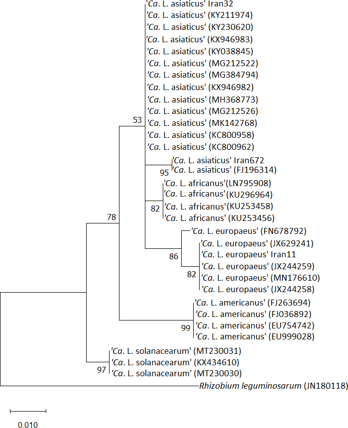
The evolutionary analyses of the identified ‘Ca. Liberibacter’ species were conducted in MEGA X (Kumar et al., 2018). The 420 bp partial sequences of 16S rDNA fragments belonging to the three identified ‘Ca. Liberibacter’ species were aligned with strains from GenBank, and phylogenetic analysis was performed using maximum likelihood methodology coupled with the Tamura-Nei model (Tamura and Nei, 1993) and 1000 replicates of the bootstrap for the statistical support of evolutionary branch lengths.
Results
PCR detection of ‘Ca. L. asiaticus’
DNA extracted from the leaf midribs of symptomatic and asymptomatic sweet orange trees were tested for the presence of ‘Ca. L. asiaticus’ by PCR using primer pair A2/J5. Amplicons of approximately 700 bp of the ribosomal protein rplKAJL rpoBC gene were obtained from all symptomatic but not from symptomless trees. Two PCR amplicons were directly sequenced and edited. The sequences were 100% identical to each other, and the BLAST analysis confirmed that the nucleotide sequences of the fragments exhibited 100% identity with the corresponding sequences from strains of ‘Ca. L. asiaticus’ available in the NCBI database (GenBank accession numbers AP014595, CP145497, KP210461). The infected trees showed symptoms of blotchy mottle, yellowing shoot, as well as small misshapen fruits, usually with color inversion (Figure 1).
OTU clustering and beta diversity
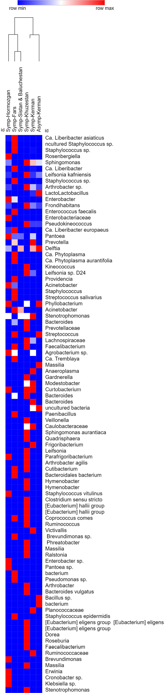
After removing the residual contaminant sequences from the quality-filtered libraries, 91 bacterial OTUs were obtained, including 1332 assigned reads (Suppl. Table 2, Figure 6). A number of 975, 137, 131, 55, 42, and 10 OTUs were identified from the surface-sterilized midrib and petiole tissue samples of Fars, Khuzestan, Hormozgan, Kerman (symptomatic), Kerman (asymptomatic), and Sistan and Baluchistan Provinces, respectively. These results indicate significantly reduced OTUs in average relative abundance in Khuzestan (7.11), Hormozgan (7.44) and Sistan and Baluchistan (17.72) provinces compared to the Far Province with the highest OTUs of 975.
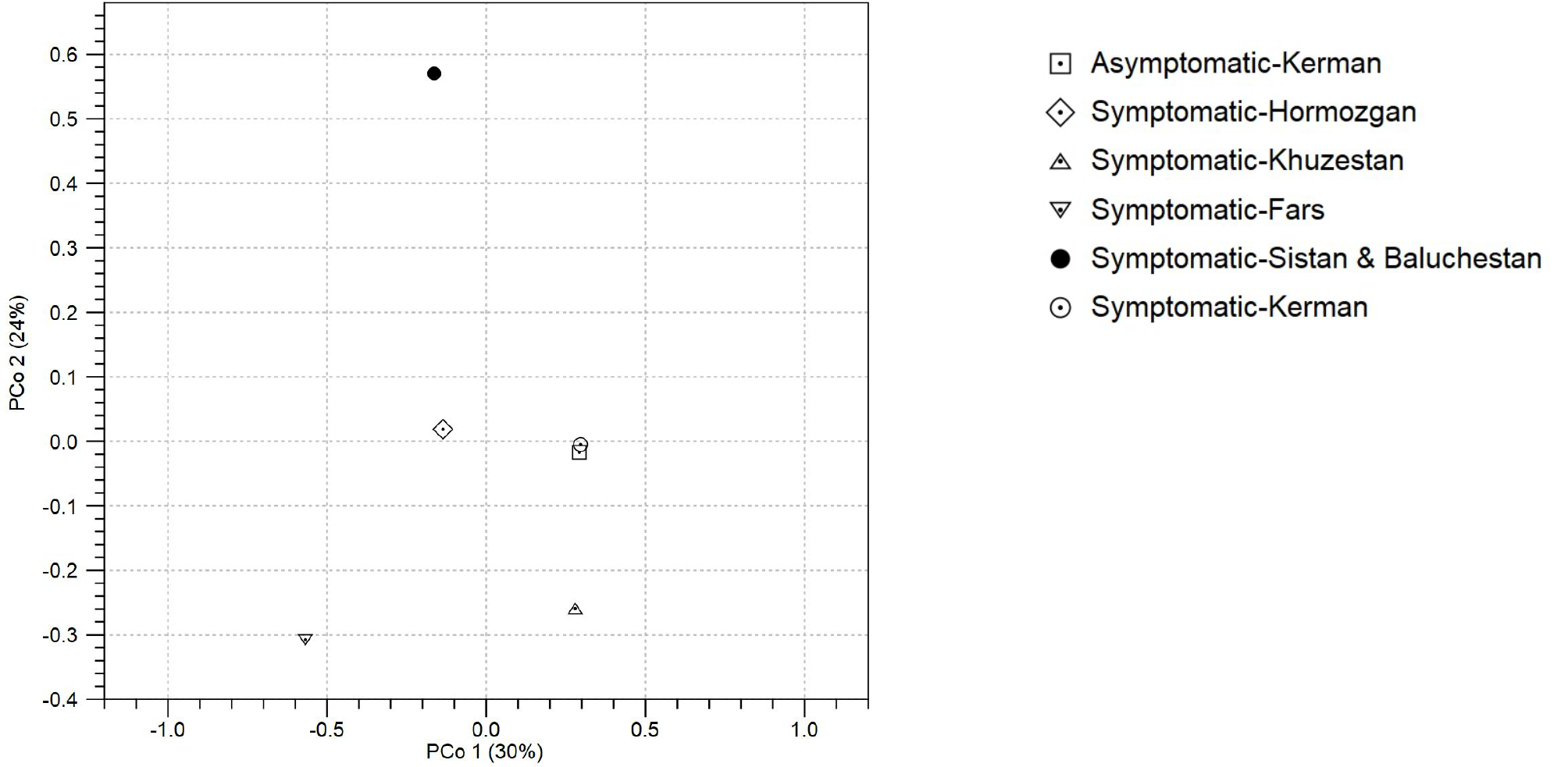
The microbial community distances (beta diversity) of the samples is depicted in Figure 3. Concerning the OTU-level PCoA (principal coordinates analysis) based on Bray-Curtis distances, the bacterial community structures of the five provinces differed from each other. Nonetheless, both samples including the infected (symptomatic) and non-infected (asymptomatic) samples from Kerman Province clustered together (Figure 3). PCo1 and PCo2 explained 30% and 24% of the variation in bacterial OTUs, respectively.
Microbial community structures of individual samples
The taxonomic distribution revealed that Alphaproteobacteria were the prevalent class in the Fars samples bacterial communities, accounting for 75.3% of all the detected taxa. Other well-represented phyla included 17.74% Bacilli, 2.36% Gamma-proteobacteria, 1.64% Mollicutes, 0.72% Bacteroidetes, 0.31% Clostridia, and 0.1% Actinobacteria (Suppl. Table 2, Figure 6). Similar trends are observed here, as seen in the OTU clustering and beta diversity analysis, indicating that Fars Province displays significantly different microbial community structure at the phylum and class level.
Overall, 75% of the microbial communities of the Fars samples were constituted mostly by ‘Ca. Liberibacter’ and 15.48% by Staphylococcus genera (Suppl. Table 2, all, 75% of the Figure 6). Furthermore, the most prevalent bacterial taxa in this region may not be the species with significant potential for inhibiting the colonization of ‘Ca. L. asiaticus’ particularly in relation to its impact on disease outcomes.
The presence of ‘Ca. L. asiaticus’ was confirmed not only in the Fars samples, but also in the other four symptomatic samples (Suppl. Table 2, Figure 6). Moreover, with the clustering of the OTUs at the 97% similarity threshold, two different OTUs were identified for ‘Ca. L. asiaticus’, presenting an evolutionary distance in the phylogenetic tree, as shown in Figure 4. With respect to the clustering of the OTUs at the 97% similarity threshold, ‘Ca. Liberibacter europaeus’ was detected in the symptomatic Fars samples. Moreover, the NCBI nucleotide blast of the 420 bp partial 16S sequenced rDNA fragment and the phylogenetic tree confirmed the similarity of the identified OTU to the reported ‘Ca. L. europaeus’ database (GenBank accession numbers JX629241, JX244259, MN176610) (Figure 4). Further, ‘Candidatus Phytoplasma aurantifolia = citri’ was detected in the microbial communities of the Fars samples. Multiple factors may play a role in simultaneous occurrence and development of ‘Ca. L. europaeus’ alongside other species, including ‘Ca. P. aurantifolia’ in this Province.
In general, the 16S amplicon metagenomic sequencing of DNA extracted from midrib and petiole tissues verified the presence of ‘Ca L. asiaticus’ in all five composite samples collected from the symptomatic sweet orange trees. No OTU of ‘Ca. Liberibacter’ and ‘Ca. Phytoplasma’ were found in the asymptomatic samples obtained from the Kerman groves.
The microbial communities of samples from Khuzestan Province consisted of 51.43% Actinobacteria, 17.14% Alphaproteobacteria, 14.29% Clostridia, 10% Bacteroidia, 5% Gamma-proteobacteria, and 2.14% Negativicutes. Among Actinobacteria, OTU belonging to Leifsonia kafniensis had the highest abundance in the microbial communities. In this region, the predominant bacterial taxa, especially L. kafniensis, along with other potentially relevant bacterial species, may play a significant role in influencing disease outcomes. This influence could be attributed to their ability to inhibit the colonization of ‘Ca. L. asiaticus’, which may explain the reduced incidence of this pathogen (Figure 5).
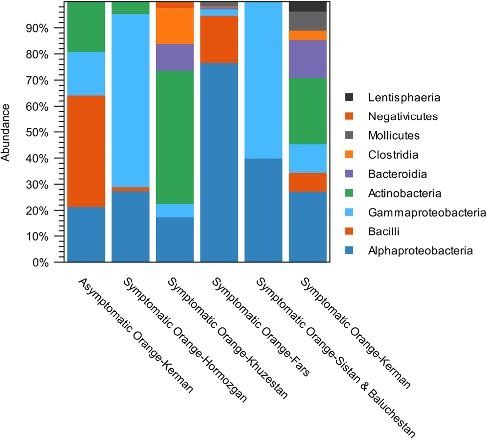
The bacterial communities of midrib and petiole tissues in the Sistan and Baluchistan samples contained 60% Gamma-proteobacteria and 40% Alphaproteobacteria. In this region, the most abundant bacterial taxa, Gamma-proteobacteria and Alphaproteobacteria may not be to that extent as the potentially relevant bacteria which could be influencing the disease outcomes by promoting or inhibiting ‘Ca. L. asiaticus’ colonization.
The midrib and petiole bacterial of the Kerman Province samples comprised 26.79% Actinobacteria, 26.79% Alphaproteobacteria, 14.3% Bacteroidia, 10.71% Gamma-proteobacteria, 7.14% Bacilli, 7.14% Mollicutes, 3.57% Clostridia, and 3.57% Lentisphaeria. However, the uninfected samples from Kerman Province contained 42.86% Bacilli, 21.43% Alphaproteobacteria, 19% Actinobacteria, and 16.67% Gamma-proteobacteria. The presence of the most abundant bacterial taxa, 42.86% Bacilli in the uninfected samples in this region in comparison with infected ones indicates the potential of the relevant bacterial species such as Bacillus spp. could be influencing the disease outcomes by inhibiting ‘Ca. L. asiaticus’ colonization.
An analysis of the bacterial community structure in symptomless samples from Kerman Province revealed a higher frequency of certain genera in comparison to symptomatic samples. The representatives of the class Bacilli, i.e. Lactobacillus spp. and Bacillus spp., in the symptomless sweet oranges in Kerman Province exhibited 12- and 4-fold changes compared to the infected samples from this province. These bacteria could be influencing the disease outcomes by inhibiting ‘Ca. L. asiaticus’ colonization. It may be in interaction with the prevailing climatic condition in this particular region
The bacterial communities of midrib and petiole tissues in the Hormozgan samples contained 66.4% Gamma-proteobacteria, 27.4% Alphaproteobacteria, 4.6% Actinobacteria, and 1.5% Bacilli. Approximately, 20% of the bacterial communities of the samples were constituted mostly by ‘Ca. Liberibacter’ species enclosing ‘Ca. L. europaeus’ in the sweet orange tree samples. The diverse bacterial taxa present in this region may not significantly impact disease outcomes by either promoting or inhibiting the colonization of ‘Ca. L. asiaticus.’
Discussion
In this study, microbial communities in the midrib and petiole tissues of HLB-infected sweet orange trees were investigated using 16S rRNA gene metagenomics sequencing. In previous report, the presence of ‘Ca. L. asiaticus’ in sweet orange and other local varieties of citrus trees was described in Sistan and Baluchistan and Hormozgan provinces, Iran (Faghihi et al., 2009). Both ‘Ca. L. asiaticus’ and phytoplasmas were detected in HLB-infected citrus trees. The HLB-associated phytoplasma from this study was a member of peanut witches’ broom (16SrII) phytoplasma group. The results presented here not only confirmed that ‘Ca. L. asiaticus’ is present in HLB disease under sampling groves in southern Iran, but also revealed that citrus midrib and petiole tissues could be colonized with a variety of bacteria. This variation could be defined as the influence of the geographical climatic parameters including temperature, precipitation and relative humidity percentage. This finding is in agreement with Camerota et al. (2012) and Daranas et al. (2019) in influence of the geographical climatic parameters and establishment of the biological control of bacterial plant diseases reported for Lactobacillus plantarum strains selected for their broad‐spectrum activity (Montero Castillo et al., 2015).
It is plausible that the progression and development of the disease in orange trees varied at the time of sampling, and it may be related to variations in the microbial populations associated with each region. Research has indicated that the composition of endophytic bacteria during the initial phases of ‘Ca. L. asiaticus’ infection differs from that observed in the later stages of the disease.
In this research, the highest identified microbial communities associated with the midrib and petiole tissues of HLB symptomatic sweet orange trees samples of Fars Province may stem from a variety of factors including disease development, vector activity, and climatic conditions. The various geographical regions of sample collection and the key climatic parameters such as average temperature, relative-humidity and the average annual precipitation in each region may be influenced the presence and distribution of the bacterial-communities associated with HLB-symptomatic orange-trees (Fitzpatrick et al., 2018; Grady et al., 2019). Moreover, the immune system, and interactions between microbes play a crucial role in shaping the structures of microbial communities (Naylor & Coleman-Derr, 2018).
The incidence and severity of the HLB disease may be affected by different geographical and climatic parameters in southern Iran. The disease is widespread in certain citrus-producing regions in the southern part of the country, notably in areas such as Jiroft (in orange) in Kerman Province and the Siyahoo region (in mandarin) in Hormozgan Province. In some orchards, the disease occurrence exceeds 50%, and even in these regions, orchards exposed to elevated levels of infection have been recorded.
The 16S amplicon metagenomics sequencing of DNA extracted from midrib and petiole tissues confirmed the presence of ‘Ca. L. asiaticus’ in all the five composite samples taken from the symptomatic sweet orange trees. No OTU of ‘Ca. Liberibacter’ and ‘Ca. Phytoplasma’ was detected in the asymptomatic samples belonging to the Kerman groves. There was an overall increase in the bacterial richness in the symptomatic Fars samples, mostly belonging to the class alpha-proteobacteria, especially ‘Ca. L. asiaticus’ species. The relative abundance of this bacterium in the Fars samples was 75% of the total identified OTUs, in agreement with the intensity of infection in the samples. The relative abundances of ‘Ca. L. asiaticus’ in Sistan and Baluchistan, Kerman, and Khuzestan provinces were 40, 3.5, and 0.7%, respectively.
With OTU clustering at the 97% similarity threshold, a ‘Ca. Liberibacter europaeus’ was identified in the symptomatic Fars samples (0.9% relative abundance). Moreover, the NCBI nucleotide blast of the 420 bp partial 16S sequenced rDNA fragment and the phylogenetic tree confirmed the similarity of the identified OTU with those previously reported. This is the first report of ‘Ca. L. europaeus’ in HLB-infected citrus (sweet orange) tree. This bacterium was first described in 2010 in asymptomatic pear trees in Italy (Raddadi et al., 2011). Nevertheless, it was also associated with mild symptoms when it was detected in Scotch broom shrub (Cytisus scoparius) in New Zealand (Thompson et al., 2013).
It is thought that ‘Ca. L. europaeus’ possibly co-exist with ‘Ca. Phytoplasma’ since both are found in the same vector, Cacopsylla pyri (Camerota et al., 2012). Interestingly, OTU of ‘Candidatus Phytoplasma’ was also identified in the Fars samples. The simultaneous incidence of multiple pathogens has been commonly observed in the vascular tissues of a single plant (Križanac et al., 2010; Nicolaisen et al., 2011; Arratia-Castro et al., 2016; Satta et al., 2016; Swisher et al., 2018). Likewise, different species/strains of phytopathogens may be hosted by the same individual insect vector, possibly being transferred together to the host plant. Occasionally, insect vectors are infected with various pathogens through feeding a variety of host plant species (Križanac et al., 2010; Raddadi et al., 2011; Swisher et al., 2018). The bacterial communities of insect vectors include a complex network of bacteria which have a significant influence on the biology of hosts (Ahmed et al., 2009; Camerota et al., 2012). Polyphagy as the ability of insects to feed from a variety of plants possibly influences the chance of plants to be infected by different pathogens. Microbial interactions inside insect vectors are highly complicated. However, the study of such multipartite interactions can help to develop microbial-based control strategies (Crotti et al., 2012; Saldaña et al., 2017). The simultaneous occurrence of ‘Ca. P. aurantifolia=citri’, ’Ca. L. asiaticus’, and ‘Ca. L. europaeus’ in the Fars citrus plants and their epidemiological role remain unknown, although it provides insights for further research.
Genome analysis indicates that ‘Ca. L. asiaticus’ lacks genes encoding some essential enzymes and other proteins, and relies on the presence of other microbiota within citrus phloem for its survival. Moreover, it is not cultured in axenic conditions and has only recently been successfully co-cultured within a microbial consortium, suggesting that its growth is likely highly reliant on associated microbial communities (Fujiwara et al., 2018).
Concerning other members of the orange bacterial communities, Staphylococcus spp., especially S. epidermidis, was detected as the second dominant microorganism in the Fars samples. Staphylococci, particularly S. epidermidis, are currently the most studied microorganisms due to their biofilm formation capacity. It has been suggested that phloem-limited phytopathogenic bacteria (e.g., ‘Ca. Phytoplasma’ and ‘Ca. Liberibacter’) depend on other phloem microbiota and phloem nutrients for the colonization of the plant host. It means that the biofilm formation of Staphylococci in host plants may facilitate the colonization of ‘Ca. Liberibacter’. However, mechanisms underlying such associations have not been well understood yet (Ahmed et al., 2009). Various reports have shown that the infection of citrus with HLB has a profound effect on the structure and composition of the citrus-associated bacterial communities (Fujiwara et al., 2018). The results of the current investigation open a new perspective for study on the function of plant bacterial communities on HLB synergistic and antagonistic agents. Co-infection with ‘Ca. Phytoplasma’ provides a solid hypothesis about its role in HLB-infected trees. No OTUs of ‘Ca. Liberibacter’ or ‘Ca. Phytoplasma’ were identified in the asymptomatic samples from the Kerman region. The analysis of the bacterial community’s composition in the asymptomatic (non-infected or healthy plant) samples from Kerman Province demonstrated that the frequency of certain genera, known as plant growth-promoting bacteria, was higher compared to the symptomatic samples. In this regard, the fold change of OTUs related to Bacillus spp. and Lactobacillus spp. in the non-infected samples from Kerman Province increased four and 12 times, respectively, compared to the symptomatic samples. Bacillus spp. and Lactobacillus spp. were previously identified from roots and leaves of ’Ca. L. asiaticus’-infected or uninfected citrus trees (Ginnan et al., 2018). Nevertheless, the higher frequency of both genera in the asymptomatic tree suggested a possible role of the mentioned bacteria in strengthening the citrus tree bacterial communities against pathogens. Today, it has been proved that plant bacterial communities are involved in plant health aspects such as growth, nutrition, immunity, infection, and protection against diseases under biotic and abiotic stresses (Vorholt, 2012; Mendes et al., 2013). Lactobacillus is a lactic acid bacterium with antagonistic activity. Generally, lactic acid bacteria produce antimicrobial compounds, inhibiting the growth of some bacterial species, and also, show antagonistic activities against some fungi (Daranas et al., 2019). It was mentioned earlier that biofilm-producing bacteria may have the ability to facilitate the colonization of phloem limited bacteria. It can be presumed that the antagonistic activity of Lactobacillus against certain groups like Staphylococcus spp. may indirectly control the phloem limited bacteria like ‘Ca. Liberibacter’. (Montero Castillo et al., 2015). It has been also demonstrated that treating citrus grafts and rootstocks with surfactin (SFC) of Bacillus subtilis indicated an effective antibacterial (eliminating or suppressing) activity against ’Ca. L. asiaticus’. SFC can also interact with plant cells by stimulating the induction of systemic immune resistance (Yang et al., 2018).
Bacterial cells situated in close proximity may have the capacity to alter their microenvironment. The composition of the microbial community plays a crucial role in the ability of ‘Ca. L. asiaticus’ to drive the progression of HLB. Other research groups have also documented the microbial diversity associated with HLBin planta. Certain plant growth-promoting bacteria, including Bacillus and Burkholderia, were identified in ‘Ca. L. asiaticus’ free leaf samples. In contrast, bacteria like Methylobacterium and Sphingobacterium were found in root samples taken from trees affected by HLB (Trivedi et al., 2011).
Some microorganisms extracted from plant tissues demonstrate potential as biocontrol agents against phytopathogens, as well as the ability to enhance plant growth and development.
A higher prevalence of Alcaligenaceae has been observed in asymptomatic samples compared to symptomatic samples from ‘Ca. L. asiaticus’ -infected citrus (Zhang et al., 2013). Some species may play a crucial role in mitigating the symptoms of HLB disease. Nevertheless, Methylobacterium was also identified in the root samples of citrus trees affected by HLB. The genus Methylobacterium was found to inhabit the xylem vessels of citrus plants, and the presence of abundant Methylobacterium species in citrus plants induces Citrus variegated chlorosis (CVC) disease through a synergistic interaction with Xylella fastidiosa (Araujo et al., 2002; Zhang et al., 2013). Consequently, the prevalence of the endophytic Methylobacterium may be linked to the HLB development.
Several additional factors may influence the population dynamics of plant endophytes. Even within a single plant species, the population density and concentration of endophytic bacteria may vary at various stages of disease progression.
It is plausible that the progression and development of the disease in orange trees varied at the time of sampling, and it may be related to variations in the microbial populations associated with each region. Research has indicated that the composition of endophytic bacteria during the initial phases of ‘Ca. L. asiaticus’ infection differs from that observed in the later stages of the disease. For instance, the bacterial populations of Methylobacterium spp. and Hymenobacter spp. were found to be more abundant in the leaf communities of trees exhibiting low disease levels. Conversely, their relative abundances were significantly diminished during the later stages of the disease (Ginnan et al., 2020).
The three predominant bacterial phyla including Proteobacteria, Bacteroidetes, and Actinobacteria, showed pronounced alterations in their relative abundance within the leaf and stem tissues as the severity of the disease increased (Ginnan et al., 2020).
The genus Massilia was identified in the non-infected samples from Kerman province, which were described recently in highly diverse environments, and certain species exhibited in vitro attributes related to plant growth promotion, including IAA production, siderophore production, and antagonistic activity toward Phytophthora infestans (Ofek et al., 2012). In addition, here also the most abundant bacterial taxa Lactobacillus spp. and Bacillus spp. in this region may influence the disease outcomes by inhibiting ‘Ca. L. asiaticus’ colonization (Ginnan et al., 2020).
A comprehensive comparison of the microbial communities among the different samples demonstrated that ‘Ca. Liberibacter’ was present in all the symptomatic samples. Due to focusing only on midrib and petiole tissues in the current study, a high number of OTUs was not really expected, especially since the surfaces of the leaf samples were well washed and sterilized before DNA extraction. As demonstrated by PCoA analysis based on Bray-Curtis dissimilarity distances, the bacterial community’s structures of the samples were different from each other. Nonetheless, the infected (symptomatic) and non-infected (asymptomatic) trees from Kerman Province clustered together, giving the impression that the geographical situation as significant abiotic factor has probably a substantial impact on the structure of the plant microbial communities.
The plant-associated microbiome is shaped by complex interactions between the plant host, microorganisms, and their surrounding environment.
A sustainable management strategy entails leveraging the citrus microbiome, as this microbial community is inherently compatible with the host plant. By occupying similar ecological niches as pathogens, the microbiome can effectively bolster the overall health of the plant and its defense mechanisms.
Future research aimed at elucidating the mechanistic interactions between antagonistic microbes and ‘Ca. L. asiaticus’ will facilitate the widespread implementation of sustainable management practices that utilize the microbiome to mitigate the progression of HLB disease.
Conclusions
This study aimed to investigate the bacterial communities of midribs and petiole tissues of HLB symptomatic orange (C. sinensis) trees from five major groves located in different geographical regions in southern Iran including Kerman, Sistan and Baluchistan, Fars, Hormozgan, and Khuzestan Provinces. Significant differences in the relative abundance of important bacterial species between HLB-infected sweet orange trees from different regions, highlight the impact of the geographic location on microbial community structure. The diverse geographical regions selected for sample collection, along with essential climatic factors including temperature, relative humidity, and average annual precipitation, significantly affected the presence and distribution of bacterial communities associated with HLB-symptomatic orange trees. This study marks the first detection of OTUs of ‘Ca. L. europaeus’ in sweet orange in Iran. The concurrent presence of ‘Ca. L. asiaticus’, ‘Ca. L. europaeus’ and ‘Ca. P. aurantifolia=citri’ in certain orange trees exhibiting symptoms of HLB offers valuable insights for future research endeavors. Due to the lack of control of healthy samples from other provinces in the country, making conclusions about similar microbial patterns under HLB diseases was impossible. In fact, understanding how the bacterial community’s influences and interacts with the citrus tree entails the implementation of multiple targeted experiments, aiming to understand plant-microbe and microbe-microbe interactions associated with HLB. Preparing a large microbial dataset in relation to variables associated with the plant health, defense, and disease could help to understand how these variables shape the citrus bacterial communities and identify individual microorganisms or consortia that play a role in HLB suppression or promotion by performing.
Contributions
This article was a part of PhD dissertation of Shiva Safarpour Kapourchali, PhD candidate of Islamic Azad University of Iran, Varamin-Pishva Branch. Mojdeh Malek organized the team members, submitted the proposal at the university and officially and scientifically supported the team. Ali Alizadeh Aliabadi designed the study, prepared samples, strongly and scientifically supported the team and revised the manuscript. Saeideh Rajaei conducted bioinformatic analysis, prepared laboratory space and revised the manuscript. Mohammad Mehdi Faghihi supported samples preparation, revised the manuscript and supported scenically the team. Mehdi Nasr Esfahani revised the manuscript and data curation. All authors read and approved the final manuscript.
Literature cited
Ahmed N.A., Petersen F.C., Scheie A., 2009. AI-2/LuxS is involved in increased biofilm formation by Streptococcus intermedius in the presence of antibiotics. Antimicrobial Agents and Chemotherapy 53(10): 4258–4263. https://doi.org/10.1128/AAC.00546-09
Alizadeh Aliabadi A., Foroutan A., Golmohamadi M., 2010. Occurrence of citrus greening caused by Candidatus Liberibacter asiaticus in Sistan-Baluchestan province. 19th Iranian Plant Protection Congress, 31 July-3 August 2010. Tehran. Iran. p. 525. (in Persian with English summary)
Alizadeh Aliabadi A., Ghasemi A., Salehi M., Faghihi M.M., Forootan A., 2013. Identification and distribution of citrus greening disease in Iran. Approved number: 14-16-16-8802-88001 and Farvast number: 44237 dated 16/04/2013.
Alizadeh A.A., Faghihi M.M., Salehi M., Ghasemi A., 2022. Dynamics of emergence and spread of citrus huanglongbing disease in Iran. Plant Pathology Science 11(2): 11–21. https://doi.org/10.2982/PPS.11.2.11
Araujo W., Marcon J., Maccheroni W Jr, Elsas JD, Vuurde JWL, et al., 2002. Diversity of endophytic bacterial populations and their interaction with Xylella fastidiosa in citrus plants. Applied Environmental Microbiology 68: 4906–4914. https://doi.org/10.1128/AEM.68.10.4906-4914.2002
Arratia-Castro A.A., Santos-Cervantes M.E., Arce-Leal Á.P., et al., 2016. Detection and quantification of ‘Candidatus Phytoplasma asterisʼ and ‘Candidatus Liberibacter asiaticusʼ at early and late stages of Huanglongbing disease development. Canadian Journal of Plant Pathology 38(4): 411–421. https://doi.org/10.1080/07060661.2016.1243586
Bazany K.E, Delgado‐Baquerzo M, Thompson A, Wang J‐T, Otto K, Adair R.C., et al. 2022. Management induce shifts in rhizosphere bacterial communities contribute to the control of pathogen causing citrus greenin disease. Journal of Sustainable Agriculture and Environment 1: 275–286. https://doi.org/10.1002/sae2.12029
Bendix C., Lewis J.D., 2018. The enemy within: phloem‐limited pathogens. Molecular Plant Pathology 19: 238–254. https://doi.org/10.1111/mpp.12526
Blaustein R.A., Lorca G.L., Meyer J.L., Gonzalez C.F., Teplitski M., 2017. Defining the core citrus leaf- and root-associated microbiota: Factors associated with community structure and implications for managing huanglongbing (citrus greening) disease. Applied Environmental Microbiology 83: e00210-17. https://doi.org/10.1128/AEM.00210-17
Bodenhausen N., Horton M.W., Bergelson J., 2013. Bacterial communities associated with the leaves and the roots of Arabidopsis thaliana. PloS One 8(2): e56329. https://doi.org/10.1371/journal.pone.0056329
Bolyen E., Rideout J.R., Dillon M.R., et al., 2019. QIIME 2: Reproducible, interactive, scalable, and extensible microbiome data science (2167-9843). Nature Biotechnology 37: 852–857. https://doi.org/10.1038/s41587-019-0209-9
Bosco D., D’Amelio R., Weintraub P., Jones P., 2009. Transmission specificity and competition of multiple phytoplasmas in the insect vector. Phytoplasmas: Genomes, Plant Hosts and Vectors, 293–308. https://doi.org/10.1079/9781845935306.02
Bové J.M., 2006. Huanglongbing: a destructive, newly-emerging, century-old disease of citrus. Journal of Plant Pathology 88: 7–37. https://doi.org/10.4454/jpp.v88i1.828
Camerota C., Raddadi N., Pizzinat A., … Alma A., 2012. Incidence of ‘Candidatus Liberibacter europaeus’ and phytoplasmas in Cacopsylla species and their host plants. Phytoparasitica 40(3): 213–221. https://doi.org/10.1007/s12600-012-0225-5
Cochran L., Samadi, M., 1976. Distribution of stubborn disease in Iran. International Organization of Citrus Virologists Conference Proceedings (1957–2010). https://doi.org/10.5070/C53tn9w703
Crotti E., Balloi A., Hamdi C., … Daffonchio D., 2012. Microbial symbionts: a resource for the management of insect‐related problems. Microbial Biotechnology 5(3): 307–317. https://doi.org/10.1111/j.1751-7915.2011.00312.x
Dala-Paula B.M., Plotto A., Bai J., Manthey J.A., Baldwin E.A., … Gloria M.B.A., 2019. Effect of huanglongbing or greening disease on orange juice quality, a review. Frontiers in Plant Science 9: 1976. https://doi.org/10.3389/fpls.2018.01976
da Graça J.V., 1991. Citrus greening disease. Annual Review of Phytopathology 29: 109–136. https://doi.org/10.1146/annurev.py.29.090191.000545
da Graça J.V., 2008. Biology, history and world status of Huanglongbing. Texas A & M University-Kingsville, Citrus Center, Weslaco TX 78596, USA.
Daranas N., Roselló G., Cabrefiga J., Donati I., 2019. Biological control of bacterial plant diseases with Lactobacillus plantarum strains selected for their broad‐spectrum activity. Annals of Applied Biology, 174(1): 92–105. https://doi.org/10.1111/aab.12476
Dominguez J.; Jayachandran K.; Stover E.; Krystel J.; Shetty K.G., 2023. Endophytes and plant extracts as potential antimicrobial agents against ‘Candidatus Liberibacter asiaticus’, causal agent of huanglongbing. Microorganisms 11: 1529. https://doi.org/10.3390/microorganisms11061529
Faghihi M.M., 2018. Investigation of association of phloem-limited prokaryotes with Siyahoo tangerine trees showing misshapen and inverted color change symptoms in fruit. 23rd Iranian Plant Protection Congress. 27–30 August 2018, Gorgan. IRAN.
Faghihi M., Salehi M., Bagheri A., Izadpanah K., 2009. First report of citrus huanglongbing disease on orange in Iran. Plant Pathology 58(4): 793–793. https://doi.org/10.1111/j.1365-3059.2009.02051.x
Faghihi M.M., Taghavi S.M., Salehi M., Golmohammadi M., 2016. First report of huanglongbing disease on Mexican lime in iran. In: Proceedings of the 20th International Organization of Citrus Virologist (IOCV) conference, April 10–15, 2016, Chongqing, China. p. 45.
Fitzpatrick C.R.; Copeland J.; Wang P.W.; Guttman D.S.; Kotanen P.M.; Johnson M.T.J., 2018. Assembly and ecological function of the root microbiome across angiosperm plant species. Proceedings of the National Academy of Sciences USA. 115, E1157–E1165. https://doi.org/10.1073/pnas.1717617115
Fujiwara K., Iwanami T., Fujikawa T., 2018. Alterations of ‘Candidatus Liberibacter asiaticus’-associated microbiota decrease survival of Ca. L. asiaticus in in vitro assays. Frontiers in Microbiology 9: 3089. https://doi.org/10.3389/fmicb.2018.03089
Ginnan N.A., Dang T., Bodaghi S., … Borneman J., 2018. Bacterial and fungal next generation sequencing datasets and metadata from citrus infected with ‘Candidatus Liberibacter asiaticus’. Phytobiomes 2(2): 64–70. https://doi.org/10.1094/PBIOMES-08-17-0032-A
Grady K.L., Sorensen J.W., Stopnisek N., Guittar J., Shade A., 2019. Assembly and seasonality of core phyllosphere microbiota on perennial biofuel crops. Nature Communications 10: 4135. https://doi.org/10.1038/s41467-019-11974-4
Hocquellet A., Toorawa P., Bové J.M., Garnier M., 1999. Detection and identification of the two Candidatus Liberobacter’ species associated with citrus huanglongbing by PCR amplification of ribosomal protein genes of the b operon. Molecular and Cellular Probes 13: 373–379. https://doi.org/10.1006/mcpr.1999.0263
Hu W., Wang X., Zhou Y., Li Z., Tang K., Zhou C., 2011. Diversity of the omp gene in Candidatus Liberibacter asiaticus in China. Journal of Plant Pathology 93(1): 211–214. https://doi.org/10.4454/jpp.v93i1.294
Klindworth A., Pruesse E., Schweer T., … Glöckner F.O., 2013. Evaluation of general 16S ribosomal RNA gene PCR primers for classical and next-generation sequencing-based diversity studies. Nucleic Acids Research 41(1): e1. https://doi.org/10.1093/nar/gks808
Križanac I., Mikec I., Budinščak Ž., Musić M.Š., Škorić D., 2010. Diversity of phytoplasmas infecting fruit trees and their vectors in Croatia. Journal of Plant Diseases and Protection 117(5): 206–213. https://doi.org/10.1007/BF03356362
Kumar Ghosh D., Bhose S., Motghare M., Gowa S., 2015. Genetic diversity of the Indian populations of ‘Candidatus Liberibacter asiaticus’ based on the tandem repeat variability in a genomic locus. Phytopathology. 105(8):1043–1049. https://doi.org/10.1094/PHYTO-09-14-0253-R
Kumar S., Stecher G., Li M., Knyaz C., Tamura K., 2018. MEGA X: molecular evolutionary genetics analysis across computing platforms. Molecular Biology and Evolution 35(6): 1547–1549. https://doi.org/10.1093/molbev/msy096
Limayem A., Martin E.M., Shankar S., 2024. Study on the citrus greening disease: Current challenges and novel therapies. Microbial Pathogenesis 192: 106688. https://doi.org/10.1016/j.micpath.2024.106688
Mendes R., Garbeva P., Raaijmakers J.M., 2013. The rhizosphere microbiome: significance of plant beneficial, plant pathogenic, and human pathogenic microorganisms. FEMS Microbiology Reviews 37(5): 634–663. https://doi.org/10.1111/1574-6976.12028
Mohkami A., Sattari R., Lori Z., Ehsani A., Nazemi A., 2011. First report of citrus huanglongbing in the Orzooiyeh region in Kerman province (Orzooiyeh). Iranian Journal of Plant Pathology 47: 105.
Montero Castillo P.M., Díaz A., 2015. Antagonistic action of Lactobacillus spp. against Staphylococcus aureus in cheese from Mompox-Colombia. Revista Facultad Nacional de Agronomía Medellín 68(2): 7721–7727. https://doi.org/10.15446/rfnam.v68n2.50991
Monazzah M., Nasr Esfahani M., Tahmasebi S., 2022. Genetic structure and proteomic analysis associated in potato to Rhizoctonia solani AG-3PT-stem canker and black scurf. Physiological and Molecular Plant Pathology 122: 101905. https://doi.org/10.1016/j.pmpp.2022.101905
Murray R., Schleifer K., 1994. Taxonomic notes: a proposal for recording the properties of putative taxa of procaryotes. International Journal of Systematic and Evolutionary Microbiology 44(1): 174–176. https://doi.org/10.1099/00207713-44-1-174
Naylor D., Coleman-Derr D., 2018. Drought stress and root-associated bacterial communities. Frontiers in Plant Science 8: 303756. https://doi.org/10.3389/fpls.2017.02223
Nicolaisen M., Contaldo N., Makarova O., Paltrinieri S., Bertaccini A., 2011. Deep amplicon sequencing reveals mixed phytoplasma infection within single grapevine plants. Bulletin of Insectology 64: S35–S36.
Ofek M., Hadar Y., Minz D., 2012. Ecology of root colonizing Massilia (Oxalobacteraceae). PloS One 7(7): e40117. https://doi.org/10.1371/journal.pone.0040117
Passera, A., Alizadeh, H., Azadvar, M., … Bianco P.A., 2018. Studies of microbiota dynamics reveals association of ‘Candidatus Liberibacter asiaticus’ infection with citrus decline. International Journal of Molecular Sciences 19(6): 1817. https://doi.org/10.3390/ijms19061817
Raddadi N., Gonella E., Camerota C., … Alma A., 2011. ‘Candidatus Liberibacter europaeus’ sp. nov. that is associated with and transmitted by the psyllid Cacopsylla pyri. Environmental Microbiology 13(2): 414–426. https://doi.org/10.1111/j.1462-2920.2010.02347.x
Rajaei S., Farshi R.S., Jazi M.M., Seyedi S., 2017. Efficient strategies for elimination of phenolic compounds during DNA extraction from Pistacia vera L. Agrivita Journal of Agricultural Science 39(3): 279–287. https://doi.org/10.17503/agrivita.v39i3.734
Saldaña M.A., Hegde S., Hughes G.L., 2017. Microbial control of arthropod-borne disease. Memórias do Instituto Oswaldo Cruz 112(2): 81–93. https://doi.org/10.1590/0074-02760160373
Salehi M., Rasoulpour R., 2016. First report of ‘Candidatus Liberibacter asiaticus’ associated with huanglongbing in Fars province. Iranian Journal of Plant Pathology 51(4): 563–566.
Salehi M., Faghihi M.M., Khanchezar A., Bagheri A., Izadpanah K., 2012. Distributioin of citrus huanglongbing disease and its vector in southern Iran. Iranian Journal of Plant Pathology 48(2): 195–208.
Satta E., Ramirez A.S., Paltrinieri S., Contaldo N., 2016. Simultaneous detection of mixed ‘Candidatus Phytoplasma asteris’ and ‘Ca. Liberibacter solanacearum’infection in carrot. Phytopathologia Mediterranea 55(3): 401–409. https://doi.org/10.14601/Phytopathol_Mediterr-18683
Safarpour K.S., Alizadeh Aliabadi A., Faghihi M.M., Rajaei S., Maleki M., 2022. Evaluation of ten citrus cultivars’ susceptibility to ‘Candidatus Liberibacter asiaticus’, citrus huanglongbing. Plant Protection 44: 1–18. https://doi.org/10.22055/ppr.2021.17131
Sagaram U.S., DeAngelis, K.M., Trivedi P., Andersen G.L., Lu S.E., Wang N., 2009. Bacterial diversity analysis of huanglongbing pathogen-infected citrus, using PhyloChip arrays and 16S rRNA gene clone library sequencing. Applied Environmental and Microbiology 75: 1566–1574. https://doi.org/10.1128/AEM.02404-08
Swisher K., Munyaneza J., Velásquez-Valle R., 2018. Detection of pathogens associated with psyllids and leafhoppers in Capsicum annuum L. in the Mexican states of Zacatecas, and Michoacan. Plant Disease 102: 146–153. https://doi.org/10.1094/PDIS-05-17-0758-RE
Tamura K., Nei M.,1993. Estimation of the number of nucleotide substitutions in the control region of mitochondrial DNA in humans and chimpanzees. Molecular Biology and Evolution 10(3): 512–526. Https://doi.org/10.1093/oxfordjournals.molbev.a040023
Tedersoo L., Drenkhan R., Anslan S., Morales-Rodriguez C., Cleary M., 2019. High‐throughput identification and diagnostics of pathogens and pests: overview and practical recommendations. Molecular Ecology Resources 19(1): 47–76. https://doi.org/10.1111/1755-0998.12959
Thompson S., Fletcher J., Ziebell H., … Pitman, A.R., 2013. First report of ‘Candidatus Liberibacter europaeus’ associated with psyllid infested Scotch broom. New Disease Reports 27(6): 2044-0588.2013. https://doi.org/10.5197/j.2044-0588.2013.027.006
Trivedi P., Duan Y., Wang N., 2010. Huanglongbing, a systemic disease, restructures the bacterial community associated with citrus roots. Applied Environmental and Microbiology 76: 3427–3436.https://doi.org/10.1128/AEM.02901-09
Villechanoux S., Garnier M., Renaudin J., Bovè J.M., 1993. The genome of the non-cultured bacterial-like organism associated with citrus greening disease contains the nusGrplKAJL-rpoBC gene cluster and the gene for a bacteriophage DNA polymerase. Current Microbiology 26:161–166. https://doi.org/10.1007/BF01577372
Vorholt J.A., 2012. Microbial life in the phyllosphere. Nature Reviews Microbiology 10(12): 828–840. https://doi.org/10.1038/nrmicro2910
Wickham H., 2016. Ggplot2: Elegant Graphics for Data Analysis. Springer; New York, NY, USA.
Warnes G.R., Bolker B., Bonebakker L., Gentleman R., Huber W., Liaw A., Lumley T., … Galili T., 2020. Gplots: Various R Programming Tools for Plotting Data. Package version 3.0.1.1. Available online: https://CRAN.R-project.org/package=gplots
Xu J., Zhang Y., Zhang P., Trivedi P., Riera N., Wang Y., … Wang N., 2018. The structure and function of the global citrus rhizosphere microbiome. Nature Communications 9: 4894. https://doi.org/10.1038/s41467-018-07343-2
Yang C., Zhong Y., Powell C.A., … Zhang M., 2018. Antimicrobial compounds effective against ‘Candidatus Liberibacter asiaticus’ discovered via graft-based assay in citrus. Scientific Reports 8(1): 1–11. https://doi.org/10.1038/s41598-018-35461-w
Zhang M., Powell C.A., Benyon L.S., Zhou H., Duan Y., 2013. Deciphering the bacterial microbiome of citrus plants in response to ‘Candidatus Liberibacter asiaticus’-infection and antibiotic treatments. PLoS One 8(11): e76331. https://doi.org/10.1371/journal.pone.0076331