https://doi.org/10.36253/phyto-15660
Accepted: January 10, 2025; Published: May 15, 2025
New or Unusual Disease Reports
First report of Calosphaeria pulchella causing canker and branch dieback of sour cherry trees (Prunus cerasus) in North Macedonia
1 Department for Plant Protection and Environment, Faculty of Agriculture, Goce Delcev University - Stip, UNILAB laboratory, Krste Misirkov, 10-А, P.O. 201, 2000, Stip, Republic of North Macedonia
2 Department of Microbiology and Microbial Biotechnology, Institute of Biology, Faculty of Natural Sciences and Mathematics-Skopje, Ss. Cyril and Methodius University in Skopje, Republic of North Macedonia
*Corresponding author. E-mail: sasa.mitrev@ugd.edu.mk
Summary. Symptoms of Calosphaeria canker were observed on sour cherry trees in the Stip region of North Macedonia. Fungal isolates were obtained from the infected twigs and branches showing external symptoms of twig dieback, occasionally followed by amber-coloured gummy exudates and brown to black vascular streaking in tissue cross-sections. The fungus was identified as Calosphaeria pulchella (Pers.: Fr.) J. Schröt (syn. Calosphaeriophora pulchella Réblová, L. Mostert, W. Gams & Crous), based on its morphology, and this identification was confirmed by sequence comparison in the GenBank database using the internal transcribed spacer region (RBK02_ITS1-ITS4) of the rDNA. The sequences had 100% identity and 100% query coverage with C. pulchella reference isolate SJC-6+ITS Internal transcribed spacer 1. Pathogenicity tests conducted on sour cherry and apricot trees showed that the obtained isolates were pathogenic to sour cherry, but not to apricot. This is the first report of Calosphaeria pulchella on sour cherry trees in North Macedonia, and in the Balkan region. This study increases understanding of the status of C. pulchella as a pathogen in this region.
Keywords. ITS, fungus, Calosphaeria canker.
Contents
Pathogen isolation and pathogenicity tests
Morphological characterization of isolated fungi
DNA extraction, PCR amplification and sequencing
Disease symptoms on naturally infected sour cherry trees
Morphological and cultural features of isolated fungi
Molecular identification of isolates
Introduction
Trunk and branch canker diseases have been reported in many countries, particularly indicating the prevalence of these diseases among Prunus spp., and the increasing concerns among related fruit production industries. Common symptoms of these diseases include wood necroses and cankers on the plant trunks or scaffold branches, necroses of woody tissues, and gummoses. As a result of host zylem and phloem disruption, movement of water and nutrients may cease, resulting in death of cambial and bark tissues and shoot and branch dieback. Over time, significant parts of affected trees or whole trees may die, while other trees in an orchard may remain unaffected.
Literature has reported that Cytospora canker, also known as perennial canker, Eutypa dieback, and Calosphaeria canker, are the most common diseases in cherry trees (Trouillas et al. 2012). Most studies referred to canker in sweet cherries, while information on the disease in sour cherries is scarce. Pruning wounds are the main entry points for the pathogens causing these diseases. Calosphaeria pulchella (Pers.: Fr.) J. Schröt (syn. Calosphaeriophora pulchella Réblová, L. Mostert, W. Gams & Crous) is associated with Calosphaeria canker in sweet cherry trees, and has been reported on sweet cherry in Spain (Berbegal et al., 2014; Berbegal and Armengol, 2018), Australia (Trouillas et al., 2012), California (Trouillas et al. 2010), France (Réblová et al., 2004), Italy (Cainelli et al., 2017), Germany (Bien and Damm, 2020), Chile (Auger et al. 2020), and in Turkey (Özben and Uzunok, 2023). The fungus also occurs on peach (Prunus persica) (Grinbergs et al., 2023), nectarine (P. persica) (Trouillas et al., 2012), and almond (P. dulcis) (Arzanlou and Dokhanchi, 2013). Relevance of the pathogen on other Prunus spp. is insufficiently explored. Perithecia of the pathogen are commonly produced beneath the periderms of dead and diseased branches. Asexual fruiting bodies of the pathogen have not been observed in nature, but an Acremonium-like anamorph was observed in culture by Réblová et al. (2004), for which Calosphaeriophora was proposed.
Rain and sprinkle irrigation favour the release of C. pulchella ascospores, which are the primary inocula (Trouillas et al., 2012). Canker has been noticed in established sour cherry orchards in the region of Stip during the last 10 years., in the Eastern part of North Macedonia. Although symptoms of canker are commonly observed on individual trees in sour cherry orchards in the Republic of North Macedonia, no study has identified the causal pathogens in this country. There is also no literature on occurrence of canker in sour cherries in the rest of the Balkan Peninsula, nor in Serbia, which are important areas for sour cherry production in the Balkans and Europe.
The aim of the present study was to characterize the causal agent of sour cherry canker in orchards of the Stip province, using phenotypic characteristics and provide description of morphological and molecular features of the pathogen.
Materials and methods
Experimental sites
The investigated commercial sour cherry tree orchards were located in the region of Stip, covering the area of 60 ha in the Eastern part of North Macedonia. During 2019, symptoms of canker were observed in approx. 10% of the cv. Oblachinska trees, which is a leading sour cherry variety grown in North Macedonia and the Balkan region. Harvested fruit is mainly used for industrial processing, although fresh consumption is also important because the fruit have high sugar and antioxidant contents (Karakashova et al., 2022). The trees were 5 to 10 years old at the time of the investigation. Spacings between trees in the orchards were 2.5 m within rows and 4 m between rows. Regular agrotechnical measures applied to the orchards include drip irrigation and annual pruning in early spring. Winter treatments included copper fungicides and insecticidal oil applications, while growing season regular protection was applied against Monilinia, using fungicides based on boscalid, pyraclostrobin, captan, or dithianon.
Pathogen isolation and pathogenicity tests
Symptomatic branches, suggesting canker disease, were collected from ten sour cherry trees. The bark was removed, and small pieces of wood were cut between diseased and healthy tissues. These wood samples were sterilized using 56% ethanol for approx. 30 s, double rinsed in sterile distilled water, dried, and then placed in Petri dishes containing potato dextrose agar (PDA; Biolife). The dishes were then maintained in the dark at 25°C until mycelium developed. Cultures were purified by transferring single hyphal tips onto new sterile PDA Petri plates. The fungal isolates obtained have been deposited in the culture collection of the UNILAB laboratory.
Pathogenicity of the isolates was assessed by inoculating healthy 5-year-old sour cherry plants (cv. Oblachinska) in May 2019. Circular injuries were made on the scaffold branches using a sterile knife. Inoculations were made using water suspension (108 CFU) from actively growing 7-d-old mycelium cultures, after which the inoculated wounds were covered with sterile cotton and plastic film (Figure 5 A). Water suspensions of sterile PDA were used as a negative inoculation control, following the same procedure for inoculation. Inoculated branches and trees were examined for symptoms every 2 weeks for the following 3 months. Pathogenicity of the isolates to apricots (cv. ‘Roxana’) was also assessed, using the procedures described above.
Morphological characterization of isolated fungi
Isolates were tentatively identified using available literature (Réblová et al., 2004; Trouillas et al., 2012). Colony morphology was assessed after 20 d incubation on PDA at 25°C, with cultures maintained in the dark to characterize the fungus anamorph. Colony colour was assessed, and size, colour and shape of conidiophores, phialides, and conidia were observed using an Olympus BX41 light microscope. Measurements fungus characteristics (n = 100) were conducted in water at ×1,000 magnification using an ocular micrometer. Images of the fungi captured using a digital camera (Olympus U-CMAD3, Mini Vid, LW Scientific).
DNA extraction, PCR amplification and sequencing
Identification of the isolates was achieved using polymerase chain reaction (PCR) by amplification of the internal transcribed spacer region (ITS) of the rDNA. Total genomic DNA was extracted according to the protocol of Möller et al. (1992). DNA amplification of the ITS region (Internal Transcribed Spacer region of the rDNA) was achieved using primers ITS1 (5ʹTCCGTAGGTGAACCTGCGG3ʹ) and ITS4 (5ʹTCCTCCGCTTATTGATATGC3ʹ) (Ferrer et al., 2001). PCR reactions were carried out in a thermal cycler (Eppendorf AG, No.5332) as follows: initial denaturation for 3 min at 95°C; amplification for 40 cycles of of 30 sec each; denaturation at 95°C, 1 min of primer annealing at 55–60°C and 45 sec for extension at 72°C, followed by a final step at 72°C for 5 min (Ferrer et al., 2001). PCR fragments were separated on 1.5% agarose gel (UltraPureTM Agarose, Invitrogen), stained in ethidium bromide and visualized under UV light. PCR-amplified fragments were used to construct libraries with rapid barcoding for Oxford Nanopore sequencing on the MinION device, using a Flongle flow cell. Total genomic DNA was extracted according to the protocol of Moller et al. (1992). The sequencing run produced 1,354 sample-specific reads in FASTQ format, which were then analyzed for taxonomic classification through the wf-metagenomics workflow on the EPI2ME platform. This workflow leveraged the NCBI Targeted Loci database, encompassing 16S rDNA, 18S rDNA, and ITS regions, to ensure accurate taxonomic assignment. Additionally, a consensus FASTA sequence of the ITS region was generated using the Medaka pipeline (https://github.com/nanoporetech/medaka), and BLASTn analysis was carried out to identify sequences with significant alignments.
Results and Discussion
Disease symptoms on naturally infected sour cherry trees
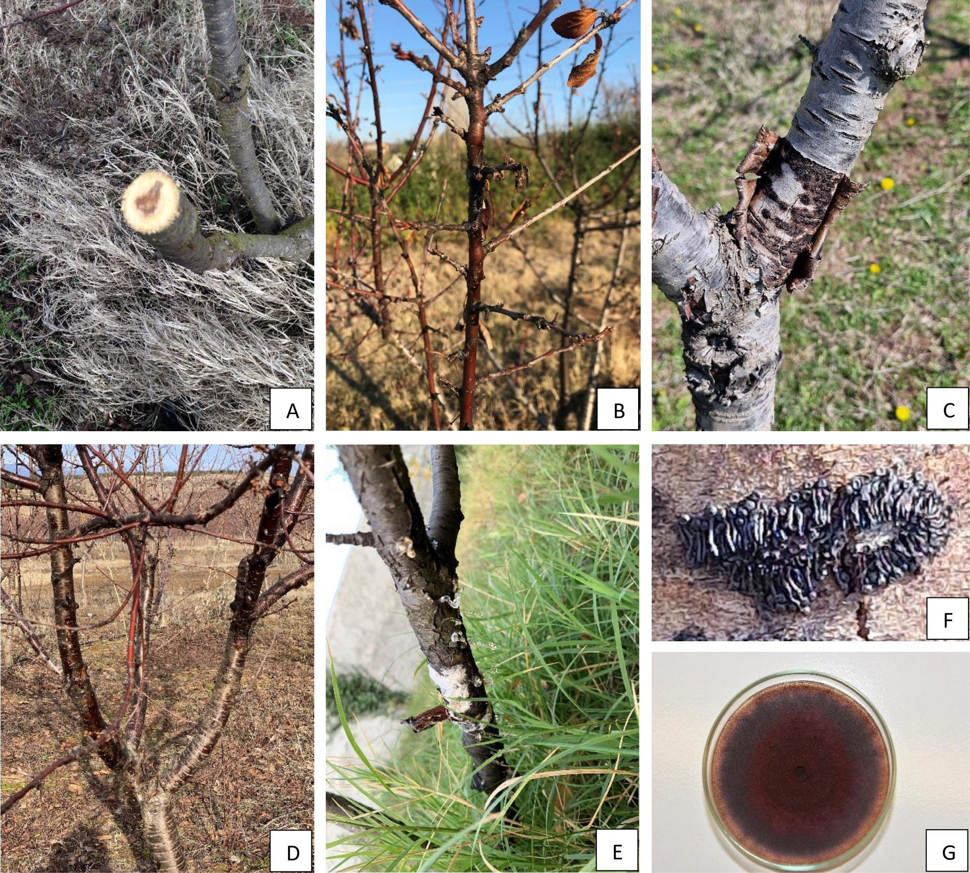
Canker symptoms were observed on cv. ‘Oblachinska’ sour cherry trees that had been growing under stress conditions (lack of water), with the symptoms appearing on wood and bark of twigs, branches and trunks. Trees in early stages of disease showed less visible symptoms, particularly in old scaffold branches and trunks, whereas symptoms on young twigs were more noticeable, including twig dieback and dead buds, occasionally with amber-coloured gummy exudate (Figure 1 B). Cankers were usually observed on host plant scaffold branches and trunks in latter stages of infections, and were often accompanied by gum exudation that leached from the cankers. Brown to black vascular streaking was visible in branch cross-sections, as circular discolouration within the tissue which advanced gradually from the heartwood into the sapwood (Figure 1 A). Symptoms of dead arm, twig, and branch dieback, and leaf desiccation were also observed (Figure 1 D). In severe cases and mostly in older trees, periderm was detached from the phloem, and perithecia bodies in circinate groups were present (Figure 1, C and F). Occasionally, large parts of affected trees were evident, and some trees were dead. These observed symptoms indicated the presence of Calosphaeria canker.
Morphological and cultural features of isolated fungi
Ten fungal isolates were obtained from sour cherry trees which had trunk or branch cankers and symptomatic young twigs. All the isolates had similar appearances, and their colonies were slow growing and distinct. After 15 d at 25°C, the isolate colonies had ceramide red moderate aerial mycelium with white margins.. After 30 d incubation, the colony colour became more intense dark purple (Figure 1 G). Young hyphae were hyaline with red pigmentation developing in cells, which had striped appearance when examined with light microscopy (Figure 2 A). Well-developed hyphae were completely red (Figure 2, A, B, and D). Individual hyphae grew in circular formations, giving a unique appearance (Figure 2 B). Conidiophores were phialidic, unbranched, and hyaline while conidiogenous cells (phialides) were hyaline, terminal or lateral, and monophialidic. Two types of phialides, which differed in size and shape, were observed on aerial mycelium. Type I phialides were the shortest, and were vase shaped adelophialides (Figure 2 F). Type II phialides were elongate ampulliform and slightly attenuated at the bases (Figure 2, D, and E). Conidia were hyaline, aseptate, allantoid to suballantoid in shape, with mean dimensions 2.7–9.3 × 1.3–2.5 μm (Figure 2 G). Perithecia were black, flask-shaped, with elongated necks, and were arranged in dense circinate groups beneath the periderm (Figure 1 F).
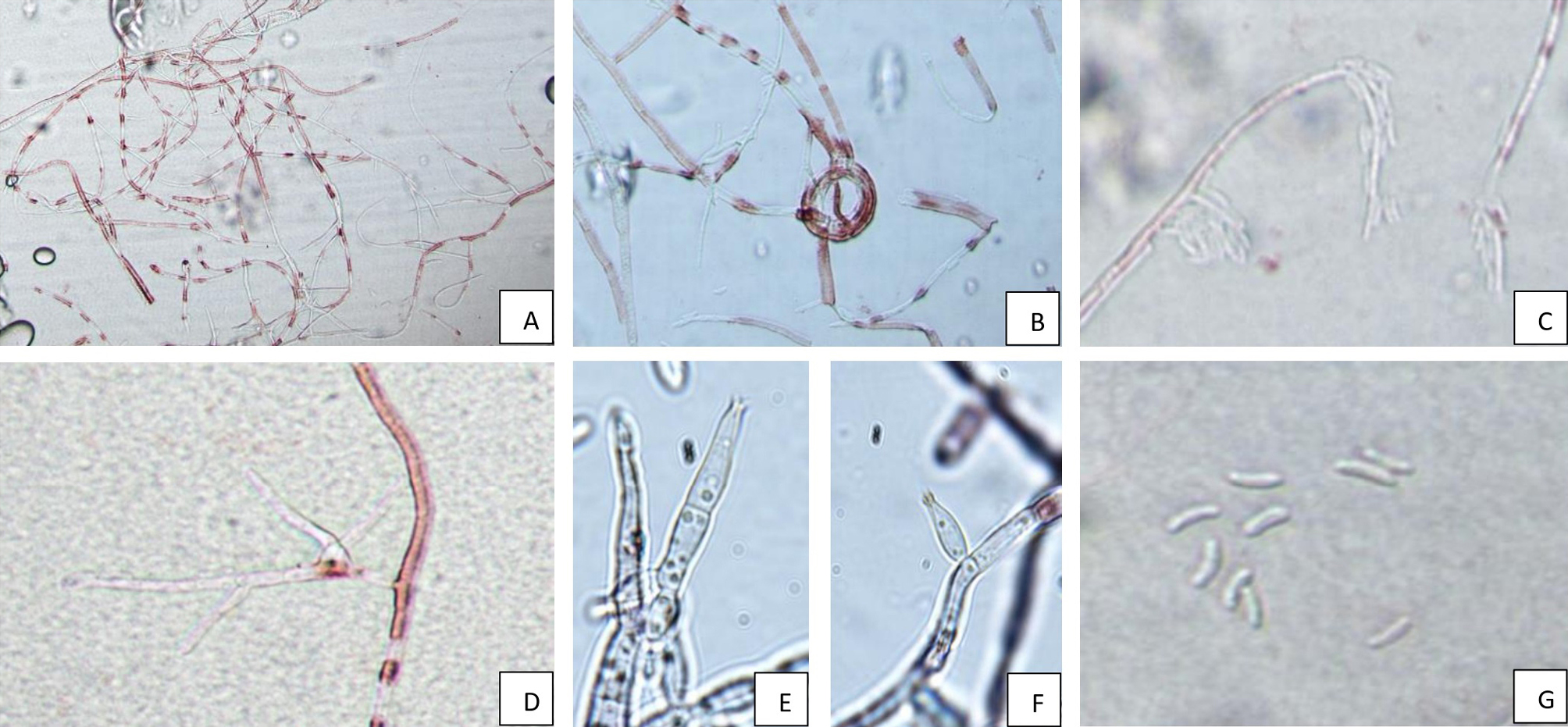
Morphological characteristics of the anamorph observed in culture, and teleomorph structures found in the field, accorded with those described as Calosphaeria pulchella by Réblová et al. (2004) and Trouillas et al. (2012).
Molecular identification of isolates
PCR fragments were separated on 1.5% agarose gel, stained in ethidium bromide, and visualized under UV light. The PCR products corresponded to size 330 bp for ITS. Three out of four samples indicated of C. pulchella. The identity of the fungus was further confirmed by sequencing, using one representative isolate. The taxonomic assignment conducted using the wf-metagenomics workflow identified a single hit corresponding to C. pulchella. Subsequent BLASTn analysis of the consensus sequence revealed a significant match to C. pulchella isolate SJC-61. Identification of C. pulchella isolates was confirmed by sequence comparison in the GenBank database using the internal transcribed spacer region (RBK02_ITS1-ITS4) of the rDNA. Sequences showed 100% identity and 100% query coverage with C. pulchella reference isolate SJC-6+ITS Internal transcribed spacer 1. The ITS sequence of one of the isolates obtained in this study was deposited into the GenBank (AN PP063991.1).
Pathogenicity tests
Pathogenicity tests showed that the C. pulchella isolates were pathogenic to sour cherry, but not to apricot. Observed symptoms on inoculated trees were very similar to the naturally occurring symptoms. Cankers appeared on inoculated trunks and scaffold branches, showing extensive hyaline gummy exudate (Figure 1 E), followed by dieback and gummoses on shooting twigs, as well as leaf decay and dieback of inoculated trunks. Development of the C. pulchella teleomorph was not observed on the inoculated branches. The pathogen was re-isolated from symptomatic twigs at 100% recovery rate, and its identity was confirmed, thus fulfilling Koch’s postulates. No recovery of C. pulchella was obtained from the non-inoculated branches.
Conclusions
Disease symptoms, and the morphological and molecular analyses in this study confirmed the presence of C. pulchella on sour cherry in North Macedonia, and this fungus and its anamorph are well-known as plant pathogenic Ascomycetes. The circinate groups of perithecia that occurred on host trunks and beneath the periderm are important signs for easy and quick diagnoses of Calosphaeria canker in the field. Pathogenicity of C. pulchella is well documented on sweet cherry, peach, nectarine and almond, but its relevance on sour cherry and on the other Prunus spp. is sinsufficiently explored. Only the study of Bien and Damm (2020) has reported isolation of C. pulchella from sour cherry, and that described a phylogenetic analysis of wood pathogens on Prunus trees in Germany.
Results from the present study confirm the pathogenicity of the C. pulchella isolates to sour cherry, and have shown that these isolates were not pathogenic to apricot. These results are the first identification of Calosphaeria pulchella (syn. Calosphaeriophora pulchella) causing canker on sour cherry in North Macedonia, in the Balkan region, and elsewhere in Central Europe.
Further research is required to determine the potential impacts of Calosphaeria canker on sour cherry and its potential to cause dieback diseases. It was observed that trees grown under stress conditions, particularly excess water, promote disease development. Winter treatments with copper fungicides and insecticidal oils, as well as chemical treatments based on the multi-site contact fungicides captan and dithianon, and the succinate-dehydrogenase inhibitor boscalid and the quinone outside inhibitor pyraclostrobin, which are used for regular protection against Monilinia in investigated general area of this study, have been shown to be ineffective against C. pulchella. Therefore, specific management strategies must be developed to reduce the impact of Calosphaeria canker, to provide sustainability in sour cherry production. If C. pulchella is not managed, it could result in substantial economic losses for sour cherry producers.
This research has increased knowledge of the aetiology of canker diseases affecting sour cherry trees and suggest that more attention should be given to Calosphaeria canker disease on sour cherries in the Balkans.
Literature cited
Auger J., Pozo L., Rubilar M., Briceño N., Osorio-Navarro C., Esterio M., 2020. First Report of canker and branch dieback of sweet cherry trees caused by Calosphaeria pulchella in Chile. Plant Disease 105(1): 2017. https://doi.org/10.1094/PDIS-05-20-1026-PDN
Arzanlou M., Dokhanchi H., 2013. Calosphaeria canker of almond caused by Calosphaeria pulchella in Iran. Archives of Phytopathology and Plant Protection 46(2): 215–226. https://doi.org/10.1080/03235408.2012.737256
Berbegal M., García-Jiménez J., Armengol J., 2014. First report of Calosphaeria pulchella causing canker and branch dieback of sweet cherry trees in Spain. Plant Disease 98: 1008–1009. https://doi.org/10.1094/PDIS-01-14-0027-PDN
Berbegal M., Armengol J., 2018. Pruning practices influence infection and dissemination of Calosphaeria pulchella, the cause of Calosphaeria canker of sweet cherry. Phytopathologia Mediterranea 57(1): 3−7. https://doi.org/10.14601/Phytopathol_Mediterr-21185
Bien S., Damm U., 2020. Prunus trees in Germany—a hideout of unknown fungi? Mycological Progress 19(7): 667–690. https://doi.org/10.1007/s11557-020-01586-4
Cainelli C., Longa C.M.O., Franchini S., Angeli G., Prodorutti D., 2017. Calosphaeria canker of sweet cherry in Trentino (north-eastern Italy). IOBC/WPRS BULLETIN 123: 195-197.
Grinbergs D., Chilian J., Isla M., Otarola J., 2023. First report of Calosphaeria pulchella causing cankers in peach (Prunus persica) in Chile. Plant Disease 107(10): 3314.
Ferrer C., Colom F., Frases S., Mulet E., Abad L.J., Alio J.L., 2001. Detection and identification of fungal pathogens by PCR and by ITS2 and 5.8S ribosomal DNA typing in ocular infections. Journal of Clinical Microbiology 39(8): 2873–2879. https://doi.org/10.1128/JCM.39.8.2873-2879.2001
Karakashova L., Bogdanovska V., Stojanov M., Milenkovska F.B., Velkoska-Markovska L., … Durmishi N., 2022. Total anthocyanins in fresh fruit and compote of “Oblacinska” sour cherry (Prunus cerasus L.). Journal of Agricultural, Food and Environmental Sciences 76(3): 25–30. https://doi.org/10.55302/jafes22763025k
Möller E.M., Bahnweg G., Sandermann H., Geiger H.H., 1992. A simple and efficient protocol for isolation of high molecular weight DNA from filamentous fungi, fruit bodies, and infected plant tissues. Nucleic Acids Research, 1992, Vol. 20, No. 22, 6115-6116.
Özben S., Uzunok S., 2023. First report of Calosphaeria pulchella causing canker and branch
dieback of cherry in Turkey. Journal of Plant Pathology 105:339 https://doi.org/10.1007/s42161-022-01255-3
Réblová M., Mostert L., Gams W., Crous, P.W., 2004. New genera in the Calosphaeriales: Togniniella and its anamorph Phaeocrella, and Calosphaeriophora as anamorph of Calosphaeria. Studies in Mycology 50(2): 533-550.
Trouillas F.P., Lorber J.D., Peduto F., Grant J., Coates W.W., … Gubler W.D., 2010. First report of Calosphaeria pulchella associated with branch dieback of sweet cherry trees in California. Plant Disease 94:1167. https://doi.org/10.1094/pdis-94-9-1167a
Trouillas F.P., Peduto F., Lorber J.D., Sosnowski M.R., Grant J., … Gubler W.D., 2012. Calosphaeria canker of sweet cherry caused by Calosphaeria pulchella in California and South Australia. Plant Disease 96(5):648-658. https://doi.org/10.1094/PDIS-03-11-0237