https://doi.org/10.36253/phyto-15597
Accepted: January 21, 2025; Published: May 15, 2025
Research Papers
Root and crown rot caused by Fusarium pseudograminearum in the euhalophyte Salicornia europaea: pathogenicity and mycotoxin production in plants grown in soilless culture
1 Department of Agriculture, Food and Environment, University of Pisa, Via del Borghetto 80, I-56124 Pisa, Italy
2 Interdepartmental Research Center Nutrafood “Nutraceuticals and Food for Health”, University of Pisa, Via del Borghetto 80, I-56124 Pisa, Italy
*Corresponding author. E-mail: emiliano.dellicompagni@phd.unipi.it
Summary. Salicornia europaea L. is a euhalophyte increasingly cultivated as a high-value green vegetable. In July 2021, root and crown rot occurred on 6-month-old S. europaea plants grown in peat-filled pots under a greenhouse, affecting 25% of plants. The causal agent was identified as Fusarium pseudograminearum O’Donnell & T. Aoki using morphological and molecular analyses. An experiment to assess the pathogenicity of this fungus to S. europaea was conducted with 96 seedlings in hydroponic culture. Half of these plants were inoculated with a conidial suspension of F. pseudograminearum. At 24 days post inoculation (dpi), half of the plants were transferred into a new hydroponic system, while the other plants were transplanted into pots. At 80 dpi, all inoculated plants grown in pots had shoot browning and desiccation symptoms, while these symptoms developed more slowly in 70% of the hydroponically grown inoculated plants. A qualitative symptom severity assessment scale showed that disease severity was greater (63%) in pot-grown plants than in hydroponically grown plants (46%). Fusarium pseudograminearum was consistently reisolated from diseased plants in both cultivation systems (62% from pots and 83% from hydroponics) fulfilling Koch’s postulates. Production of deoxynivalenol (DON) and zearalenone (ZEA) was investigated in vitro and in planta. Traces of DON (0.029 ± 0.012 mg kg-1) were found in severely damaged plants grown in hydroponics. In the in vitro test, F. pseudograminearum isolates from wheat crops in Spain (isolate ColPat-351) and Italy (isolate PVS Fu-7) were also assessed, and all tested isolates produced considerable amounts of ZEA. Fusarium pseudograminearum isolates obtained from S. europaea produced more DON (6.81 ± 0.24 mg kg-1, on average) than the Italian isolate PVS Fu-7 (0.37 ± 0.06 mg kg-1), while DON production by the Spanish isolate ColPat-351 was less than the limit of detection (< 0.25 mg kg-1). This is the first report of root and crown rot caused by F. pseudograminearum on S. europaea.
Keywords. Deoxynivalenol, fungi, glasswort, hydroponic system, plant disease, zearalenone.
Contents
Origin of infected samples and isolation of fungal isolates
Monoconidial cultures and storage conditions
DNA extraction and molecular identification of isolates
Artificial inoculations of Salicornia europaea seedlings grown in pots and in hydroponics
Mycotoxins production: fungal isolates and culture conditions
Amplification of mycotoxin-related genes
In vitro production of mycotoxins
In planta production of mycotoxins
Artificial inoculations and plant disease assessments
Production of mycotoxins in vitro and in planta
Introduction
Salicornia europaea L. (syn. Salicornia perennans Willd. subsp. perennans, Amaranthaceae), commonly known as glasswort or sea asparagus, is a euhalophyte that is widespread in many regions of the Northern Hemisphere, especially in the Mediterranean region (Lombardi et al., 2022). This plant thrives in saline ecosystems and can tolerate up to 1000 mM NaCl, with optimal growth at 200–400 mM NaCl (Cárdenas-Pérez et al., 2021). For this reason, it has received considerable interest as an alternative crop in saline agriculture, especially in desert areas and marginal lands (Araus et al., 2021). Salicornia europaea has a wide range of applications as a food and non-food crop. This plant is a good candidate for biodiesel production because of its high seed oil contents, for the remediation of saline soils, and for pharmaceutical purposes (Ventura and Sagi, 2013; Cárdenas-Pérez et al., 2021). It has edible shoots, raw or cooked, and is increasingly cultivated as a high-priced green vegetable (Araus et al., 2021; Cárdenas-Pérez et al., 2021). Despite the broad range of agronomic usages of S. europaea, little attention has been paid to phytopathological issues for this plant.
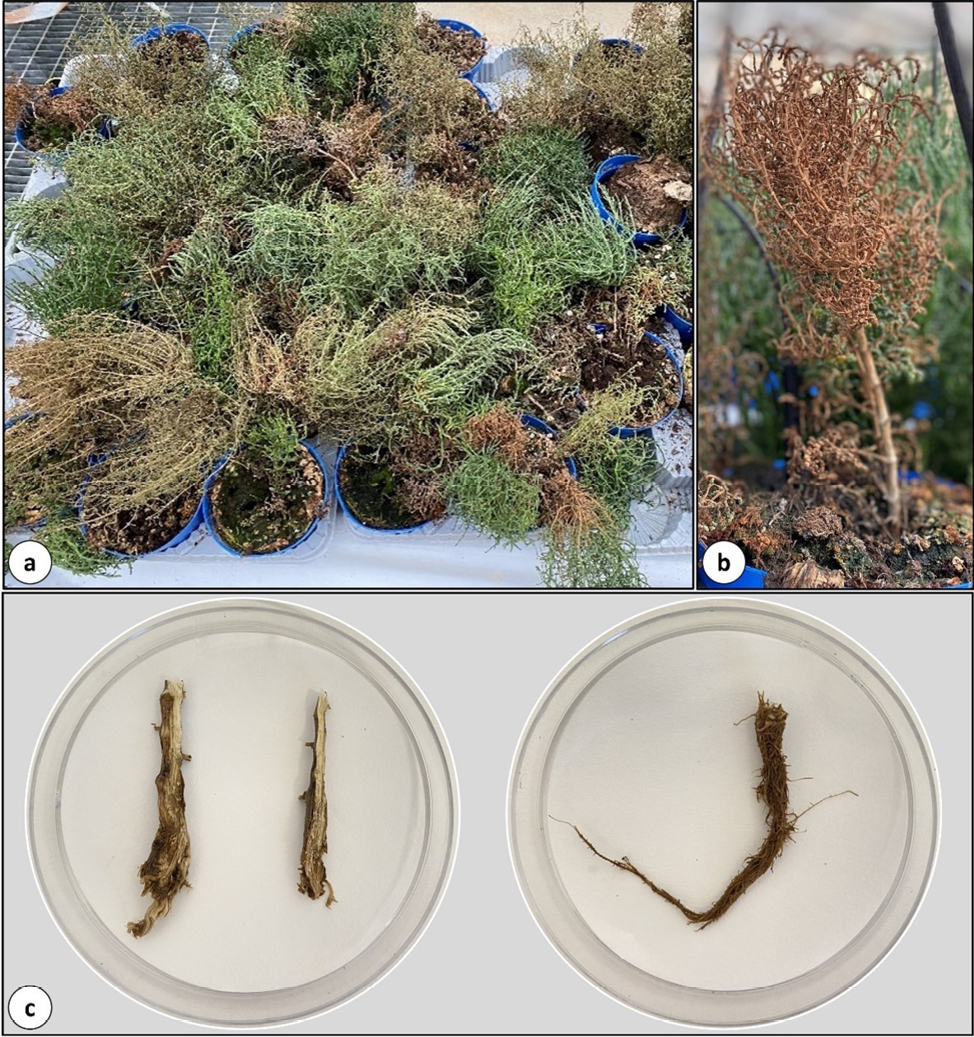
In July 2021, at the University of Pisa, severe root and crown rot symptoms were observed in pot-grown S. europaea plants purchased from a nursery in northern Italy (Figure 1, a, b, and c). Symptoms appeared on 25% of 6-month-old seed-propagated plants. The causal agent of the disease was identified as Fusarium pseudograminearum O’Donnell & T. Aoki. This fungus was previously described as F. graminearum Schwabe Group 1 and was identified for the first time in Australia in 1983 (Burgess et al., 1987). Subsequently, Aoki and O’Donnell (1999a) described the novel species F. pseudograminearum, based on morphological and molecular observations.
Fusarium pseudograminearum is a heterothallic Ascomycete, although the formation of perithecia by the teleomorph Gibberella coronicola T. Aoki & O’Donnell has been rarely observed (Aoki and O’Donnell, 1999b). This fungus is a soil-borne pathogen, primarily causing Fusarium crown rot (FCR) on small grain cereals, which is a common disease in warm, dry regions (Chakraborty et al., 2006; Poole et al., 2013; Sabburg et al., 2015). The fungus is widespread in Asia, especially China where it is becoming the predominant cause of FCR (Xu et al., 2017; Zhou et al., 2019; Deng et al., 2020), and in Australia (Obanor et al., 2013; Kazan and Gardiner, 2018). In Europe, the occurrence of F. pseudograminearum is unclear, as the pathogen has only been reported a few times. It was first identified as the cause of root rot on durum wheat in Foggia, southern Italy, by Balmas (1994), when it was still designated as F. graminearum Group 1. In 2016, F. pseudograminearum was found in Cordoba, southern Spain, causing FCR on Triticum aestivum (Agustí-Brisach et al., 2018). The pathogen has been described on wheat in the Mediterranean countries Algeria (Abdallah-Nekache et al., 2019), Syria (Alkadri et al., 2013), Tunisia (Kammoun et al., 2009), and Turkey (Tunali et al., 2008). However, Akinsanmi et al. (2007) noted that some alternative host plants (monocots and dicots) were infected by F. pseudograminearum, indicating that the pathogen is not host-specific. In Croatia, this fungus was first recorded on naturally infected rotted apples (Sever et al., 2012), and seeds of the wild legume Vicia cracca (Miličević et al., 2013). An endophytic F. pseudograminearum was also isolated from coastal dunegrass (Leymus mollis), improving plant growth and salinity resistance (Shan et al., 2021). Due to the uncertainty of the occurrence of the pathogen, the European Food Safety Authority (EFSA) has recently proposed to designate F. pseudograminearum as a potential quarantine pathogen for the European Union (EFSA Panel Health, 2022).
A key feature in several Fusarium species (including F. culmorum, F. graminearum, and F. pseudograminearum) is their ability to produce trichothecene B mycotoxins. This class of mycotoxins includes deoxynivalenol (DON), nivalenol (NIV), and acetylated derivatives (3-acetyldeoxynivalenol, 3ADON; 15-acetyldeoxynivalenol, 15ADON). Based on the type of trichothecene produced, each isolate can be assigned to a specific chemotype (3ADON, 15ADON, NIV). Each chemotype is determined by the trichothecene gene cluster (tri), composed of ten to 12 contiguous genes. The number of functional genes in this cluster varies depending on the species and chemotype (Pasquali and Migheli, 2014). Zearalenone (ZEA) is another mycotoxin of interest that exhibits strong estrogenic effects in mammals. ZEA is a polyketide mycotoxin synthesized from the acetate-polymalonate pathway, in which four genes are involved: pks4, pks13, zeb1, and zeb2 (Nahle et al. 2021). Determining the mycotoxins produced by an isolate is essential for studying the dynamics of a fungal population in a specific agricultural area, and to evaluate toxigenic risks of specific chemotypes in food and feed.
Root and crown rot caused by F. pseudograminearum on cultivated S. europaea plants has not been previously described. Therefore, the aims of the present study were: (i) to assess the pathogenicity and evaluate disease severity caused by a selected isolate of F. pseudograminearum towards S. europaea plants grown in two different growing systems (pots and hydroponics); and (ii) to determine the mycotoxins produced by three isolates of F. pseudograminearum from S. europaea compared to two other European isolates of F. pseudograminearum.
Materials and methods
Origin of infected samples and isolation of fungal isolates
Severe root and crown rot symptoms were observed on seed-propagated S. europaea plants (approx. 6 months old) that had been purchased from a commercial nursery in northern Italy. The plants were grown in pots filled with peat (Figure 1, a and b). Symptoms affected 25% of the plants.
To isolate the putative causal agent, the roots and crowns of affected plants were gently washed under running tap water. Symptomatic tissues (Figure 1 c) were then cut into small pieces and surface sterilized with sodium hypochlorite (NaOCl; 1% available chlorine) for 2 min, rinsed twice for 2 min in sterile distilled water, and then plated onto Potato Dextrose Agar plates (PDA, 42 g L-1, BioLife, Milan, Italy) amended with streptomycin (0.3 g L-1, Sigma-Aldrich, Saint Louis, MO, USA). The plates were then incubated at 25 ± 1°C under fluorescent light and checked daily for mycelium development. After 4 days of incubation, hyphae emerging from infected plant tissues were transferred onto new PDA plates.
Monoconidial cultures and storage conditions
Colony morphology, and macroconidia shapes observed under an optical microscope indicated that the putative causal agent of the disease belonged to the genus Fusarium. Further analyses, both morphological and molecular, were carried out by generating monoconidial cultures of three selected isolates (designated 3B, PD-A, and PD-B), each obtained from a different diseased plant. An isolate of F. oxysporum (DAFE SP21-23), a fungus often associated with S. europaea plant tissues but not pathogenic, was included in the study as a negative control. For preservation of the isolates, PDA plugs derived from the above-described monoconidial cultures were transferred onto Synthetic Nutrient-poor Agar (SNA), which contained: 1 g L-1 KH2PO4; 1 g L-1 KNO3; 0.5 g L-1 MgSO4·7H2O; 0.5 g L-1 KCl; 0.2 g L-1 glucose; 0.2 g L-1 sucrose; and 20 g L-1 agar (Nirenberg, 1981). The cultures were stored at 4°C. All the chemicals used in the culture medium were obtained from Sigma-Aldrich (Saint Louis, MO, USA).
Species identification
Morphological observations
The three monoconidial isolates, derived from three different plants, were identified at species level by combining a morphological and molecular approach. Colony morphology, pigmentation, and macroconidia sizes were compared to the published descriptions of Aoki and O’Donnell (1999a). Suspensions of macroconidia were obtained by rinsing 2-week-old PDA plates, which had been incubated at 25 ± 1°C in complete darkness, with 10 mL of sterile distilled water and gently scraping the mycelium with a sterile glass Drigalski spatula. The suspensions were then filtered through a layer of sterile Miracloth (Calbiochem, San Diego, CA, USA) and were observed using an optical microscope (model Dialux 22, Leitz, Wetzlar, Germany). Micrograph images were captured using a Leica DFC 450C digital camera, and fungal structures were measured using the software Leica Application Suite X Version 3.1.1.17751 (Leica Microsystems Ltd., Heerbrugg, Switzerland). For each isolate, at least 50 macroconidia with different numbers of septa (1- 3- and 5-septa) were measured.
DNA extraction and molecular identification of isolates
Five agar discs (diam. 6 mm) were cut from 1-week-old monoconidial PDA plates and were inoculated into 50 mL tubes each containing 25 mL of Yeast Malt Broth: 3 g L-1 yeast extract; 3 g L-1 malt extract; 5 g L-1 peptone; 10 g L-1 glucose. The tubes were then kept for 5 days at room temperature (22–25°C) under constant stirring at 60 rpm on the Multi-RS 60 programmable rotator (Biosan, Riga, Latvia), with a rotation angle of 75°. Mycelium was then collected by filtering each liquid culture through a layer of sterile Miracloth (Calbiochem, San Diego, CA, USA), washed twice with sterile distilled water, dried on sterile filter paper, and then stored at -20°C for DNA extraction.
Genomic DNA was extracted with the Genesig® Easy DNA/RNA extraction kit (Primer Design Ltd, UK), using the method described by Spada et al. (2023). DNA was quantified using the QubitTM DNA BR Assay Kit in a Qubit™ 4 Fluorometer (Invitrogen by Thermo Fisher Scientific Inc., Eugene, OR, USA). A fragment of the ef-1α gene was amplified according to O’Donnell et al. (1998), using the EF-1/EF-2 primer pair. PCR was carried out in a 25 μL reaction mix with GoTaq Green Master Mix 2X (Promega Corporation, Madison, WI, USA), 0.1 μM of each primer, 30-50 ng of DNA, and adding nuclease-free water to the volume. PCR conditions were: initial denaturation at 95°C for 8 min, followed by 35 cycles of 95°C for 30 sec, 53°C for 1 min, and 72°C for 1 min, and final extension at 72°C for 5 min. A negative control (no DNA) was included in the reaction. PCR products were analyzed by electrophoresis in 0.5× Tris-Borate-EDTA (TBE) buffer with 1% (w/v) agarose gels and detected by UV fluorescence after GelRed™ staining (Biotium Inc., CA, USA) according to the manufacturer’s instructions. The 100 bp DNA ladder (Promega, Madison, WI, USA) was used as a molecular size marker. Amplicons were purified with the QIAquick PCR purification kit (QIAGEN, Milan, Italy), following the manufacturer’s instructions. The purified amplicons were sent to BMR Genomic (Padua, Italy) for Sanger sequencing in both directions, with the same set of primers used for amplification. Consensus sequences were edited with BioEdit v 7.7 and used as queries in BLASTn searches of the GenBank database hosted by NCBI. To further confirm the species, the species-specific primers Fp1-1 and Fp1-2 were used, targeting a 523 bp fragment in the ef-1α gene (Aoki and O’Donnell, 1999a). As described above, each PCR was conducted in a 25 μL reaction mix using 0.24 μM of each primer and setting the thermal conditions described by the authors.
Artificial inoculations of Salicornia europaea seedlings grown in pots and in hydroponics
To assess the pathogenicity of F. pseudograminearum, inoculations of S. europaea plants were carried out. The plants were grown in pots and hydroponics (floating raft system). One isolate (F. pseudograminearum 3B) was used in this experiment. Seeds of S. europaea were purchased from Alsagarden (Niederhaslach, France), and were sown into polystyrene trays containing stone wool plugs (Grodan, Roermond, The Netherlands), which were kept under a greenhouse for germination (Figure 2 a). Plants were selected for phenotypic uniformity based on height and number of shoots. At 46 days after sowing (das), seedlings (n = 96) were removed from stone wool plugs and transferred to a small-scale floating raft system (Figure 2 b). This consisted of polystyrene rafts (7 x 10 cm, each containing eight plants) placed in a 200 mL dark-colored polypropylene tank filled with nutrient solution. The solution contained 14.0 mM NO3, 2.0 mM NH4, 2.0 mM H2PO4, 10.0 mM K, 4.5 mM Ca, 2.0 m M Mg, 5.0 mM SO4, 40.0 μM Fe, 40.0 μM B, 3.0 μM Cu, 10.0 μM Zn, 10.0 μM Mn, and 1.0 μM Mo. The nutrient solution in each hydroponic tank was continuously aerated to supply oxygen to the plants and maintain the inoculum in a homogeneous suspension. The plants were maintained in a growth chamber (25.0 ± 1.5°C, PAR 150 μmol m2 s-2 from LED tubes) for 11 days to promote root development. Subsequently, at 57 das, plant (n = 48) roots were inoculated with a suspension of F. pseudograminearum macroconidia (final concentration 105 macroconidia mL-1) (Figure 2 c1), and the other half with an equal volume of sterile distilled water (Figure 2 c2). Macroconidia suspensions were obtained as described above. At 24 days post inoculation (dpi; 81 das), half of the inoculated plants (n = 24) were transferred into 1 L pots filled with sterile peat, and the other half were transferred into 50 L tanks filled with 25 L of nutrient solution. The same method was used for control plants (those treated with sterile distilled water), and all plants were moved under a greenhouse (Figure 2 d). The plants had been randomly selected for transplanting in pots or hydroponics. The pots were sub-irrigated daily with nutrient solution, while in the hydroponic tanks, the nutrient solution was periodically added to maintain the initial volume.
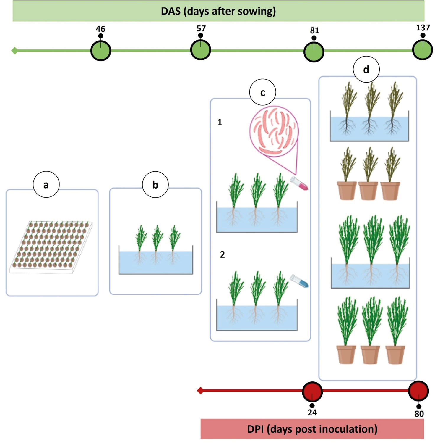
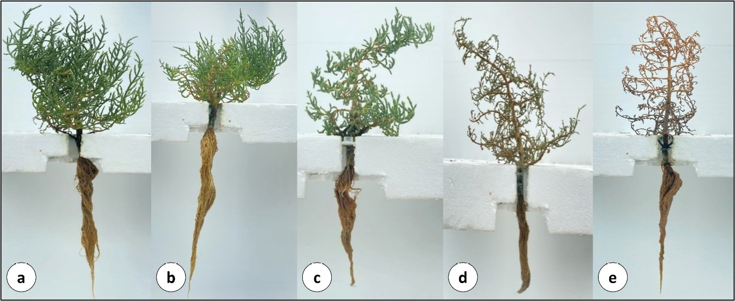
The trial lasted until 80 dpi (corresponding to 137 das), when disease symptoms (shoot browning) were assessed using a six-point empirical qualitative ordinal scale (0 to 5): 0 = no symptoms; 1 = browning on the main stem; 2 = browning on the basal lateral shoots; 3 = complete browning of the main stem and primary lateral shoots; 4 = almost complete browning, pale-green secondary shoots; and 5 = completely desiccated (dead) plant (Figure 3, a to e). A score was assigned to each plant and, for each treatment (i.e. potted-inoculated plants, hydroponics-inoculated plants, potted-non-inoculated plants, and hydroponic-non-inoculated plants), the plants (n = 24) were randomly divided into three groups of eight plants each. The McKinney Index (MKI; McKinney, 1923), which incorporates disease incidence and severity, was calculated for each group using the following formula:
where d = disease score, f = disease frequency (for a given group of plants), N = total number of observed plants, and D = the greatest level of disease infection on the empirical qualitative ordinal scale. Disease incidence was determined by counting the plants showing symptoms of at least 1 on the empirical scale.
At 80 dpi, re-isolations were carried out (as previously described) from plant tissues for the fulfillment of Koch’s postulates. In addition to plant roots and crowns, the first nine segments of the main stem and the two basal lateral shoots were plated, and the development of resulting organisms was assessed.
At 80 dpi, control and inoculated plants were also assessed for shoot fresh weight (SFW), shoot dry weight (SDW), and shoot succulence. For the inoculated plants, samples were collected from asymptomatic plants grown in hydroponics and from plants grown in pots with a disease severity score of 1. Shoot dry weights were measured after drying the fresh samples in a ventilated oven at 70°C until constant weight. Shoot succulence was calculated as the ratio of shoot water content to SDW.
Mycotoxins production: fungal isolates and culture conditions
The following isolates were used in the experiment on mycotoxin production: the three F. pseudograminearum isolates (3B, PD-A, and PD-B) from S. europaea, F. pseudograminearum ColPat-351 from wheat (Agustí-Brisach et al., 2018), F. pseudograminearum PVS Fu-7 from wheat (Balmas, 1994), and the non-pathogenic F. oxysporum DAFE SP21-23 isolate, obtained from S. europaea plant tissues in the present study, used as a negative control since this species does not produce DON or ZEA. The isolate DAFE SP21-23 was also used as further confirmation that only F. pseudograminearum isolates were DON and ZEA producers. All fungi were grown on PDA plates and maintained on SNA, as described before.
Amplification of mycotoxin-related genes
To determine whether F. pseudograminearum isolates were potentially mycotoxigenic, we first investigated if they had the gene pool required for mycotoxin biosynthesis. The tri5 gene and the pks4 gene were assessed in each isolate, as tri5 is involved in the biosynthesis of DON (Proctor et al., 1995; Desjardins et al., 1996), and pks4 is involved in the biosynthesis of ZEA (Lysøe et al., 2006).
A 650 bp fragment of the tri5 gene was amplified using the Tox5-1/Tox5-2 primer pair (Niessen and Vogel, 1998), and a 280 bp fragment of the pks4 gene using the PKS4F/PKS4R primers (Meng et al., 2010). PCR was carried out in a 25 μL reaction mix with GoTaq Green Master Mix 2X (Promega Corporation, Madison, WI, USA), 0.4 μM of each primer for tri5 and 0.5 μM for pks4, 30–50 ng of DNA, and adding nuclease-free water to the volume. Negative controls (no DNA) were included. The amplification conditions used were those described by the respective authors (cited above). Amplicons were purified and sequenced, and consensus sequences were generated as previously described. Amplification of fragments of the tri3 and tri12 genes (Starkey et al., 2007) was carried out in a multiplex PCR to find out whether the isolates belonged to a specific DON-chemotype (i.e. nivalenol, NIV; and acetylated forms of DON: 3-acetyldeoxynivalenol, 3ADON; and 15-acetyldeoxynivalenol, 15ADON). Primers 3CON, 3NA, 3D15A, and 3D3A were used to target the tri3 gene. Primers 12CON, 12NF, 12-15F, and 12-3F were used for the tri12 gene. Multiplex PCRs were carried out following the methods described by the authors.
In vitro production of mycotoxins
Production of DON and ZEA was evaluated in vitro in a model system that consisted of growing the F. pseudograminearum isolates on moistened barley grains. This system method was chosen so that the fungi were grown in optimal conditions for mycotoxin production (Blaney and Dodman, 2002; Clear et al., 2006; Kokkonen et al., 2010). Thus, 30 g of dried barley grains, obtained from a local organic farm, were rinsed overnight in 25 mL of sterile distilled water in 500 mL flasks and sterilized twice at 121°C for 20 min with 24 hours between the two heat treatments. Each flask was then inoculated with a PDA disk (diam. 10 mm) cut from an actively growing culture of each isolate. A mycelium-free PDA disk was used as a control. An experiment was established consisting of five biological replicates for each isolate. The fungi were grown for 3 weeks at room temperature, and the cultures were manually shaken to avoid grain clumps. After incubation, the grains were dried under a laminar flow cabinet for 48 hours and were ground with a coffee grinder.
Mycotoxin production was evaluated for DON and ZEA with the immunochromatographic RIDA® QUICK RQA ECO tests (R-Biopharm AG, Milan, Italy), according to the manufacturer’s instructions. The limits of detection (LOD) were 0.25 mg kg-1 for DON and 0.05 for ZEA, and the maximum measurable amounts were 50 mg kg-1 for DON and 1 mg kg-1 for ZEA. Sequential 10-fold dilutions were made, with the appropriate solvent, for samples outside these ranges until measurable amounts of mycotoxin were obtained. The DON test does not discriminate between acetylated forms of DON.
In planta production of mycotoxins
Mycotoxin production in S. europaea plants was assessed using LC-MS/MS, which is a commonly used multi-mycotoxin method to determine levels in feeds and foods to overcome specific matrix effects (De Santis et al., 2017; Iqbal, 2021). The method used (protocol: MP 213, rev. 3, 2022) detects 17 mycotoxins, including NIV, ZEA, DON, and the acetylated forms 3- and 15-acetyldeoxynivalenol.
According to the empirical scale of host plant symptoms (Figure 3), the plants were pooled and divided into three groups: no symptoms (score 0); mild symptoms (scores 1 to 2), and severe symptoms (scores 3 to 5). The plants were also divided based on the growing methods (i.e. in pots or hydroponics). For each group of plants, the analyses were carried out on three biological replicates. Therefore, at 80 dpi, the above-ground parts of each plant were cut and freeze-dried. Samples were sent to Biochemie Lab s.r.l. (Campi Bisenzio, Florence, Italy; Accredia No. 0195; https://www.accredia.it/) for LC-MS/MS analyses.
Statistical analysis
Data were subjected to 1-way ANOVA using CoStat 6.4 statistical software (Cohort Software, Monterey, CA, USA). Percentage data (disease incidence and MKI) were arcsin√% transformed before ANOVA. In the artificial inoculation experiment, for each cultivation method, plants (n = 24) were randomly divided into 3 groups (eight plants each), and disease incidence and MKI values were calculated for each group. The normality of the data was assessed using the Shapiro–Wilk test and homoscedasticity was tested using Bartlett’s test. Means were separated using Tukey’s honestly significant difference post-hoc (HSD) test (P ≤ 0.05).
Artificial inoculations of F. pseudograminearum on S. europaea plants were repeated twice in pots, using the same protocol, and both inoculations gave similar results. In addition, in the first experiment carried out in pots, the F. oxysporum isolate DAFE SP21-23 was also inoculated, but the inoculation did not cause any symptoms (data not shown). Only the results of the experiment conducted to compare the two growing methods (i.e. pots and hydroponics) are presented in this paper.
Results
Identification
Fusarium pseudograminearum colonies were isolated from 64% of the total specimens of diseased stems, roots, and crowns collected from S. europaea plants. Fusarium oxysporum colonies were also frequently observed, representing 45% of the total plated samples. On PDA, colonies appeared whitish to brownish-yellow on the upper surface, especially during the first few days of growth. After one week, reddish to dull-red pigmentation was visible. The reverse colony pigmentation ranged from pale brown to brownish yellow, becoming red, reddish-brown, and ruby after 2 weeks. Abundant and floccose aerial mycelium was typically observed during colony growth. The size of macroconidia was comparable to that described by Aoki and O’Donnell (1999a) (Table S1), although no 7-septate macroconidia were observed. Microconidia were absent. Three isolates (3B, PD-A, and PD-B) were selected for morphological and molecular studies.
The consensus sequences of the partial translation elongation factor 1 alpha (ef-1α) gene were 100% similar, in whole length, to those of F. pseudograminearum [GenBank accession numbers (AN) MG670539 to MG670541 and OM746828 to OM746837]. These sequences were deposited in GenBank under the AN PQ045864 to PQ045866. A species-specific PCR, with the Fp1-1/Fp1-2 primer pair, confirmed that the three isolates belonged to F. pseudograminearum (Figure S1).
Artificial inoculations and plant disease assessments
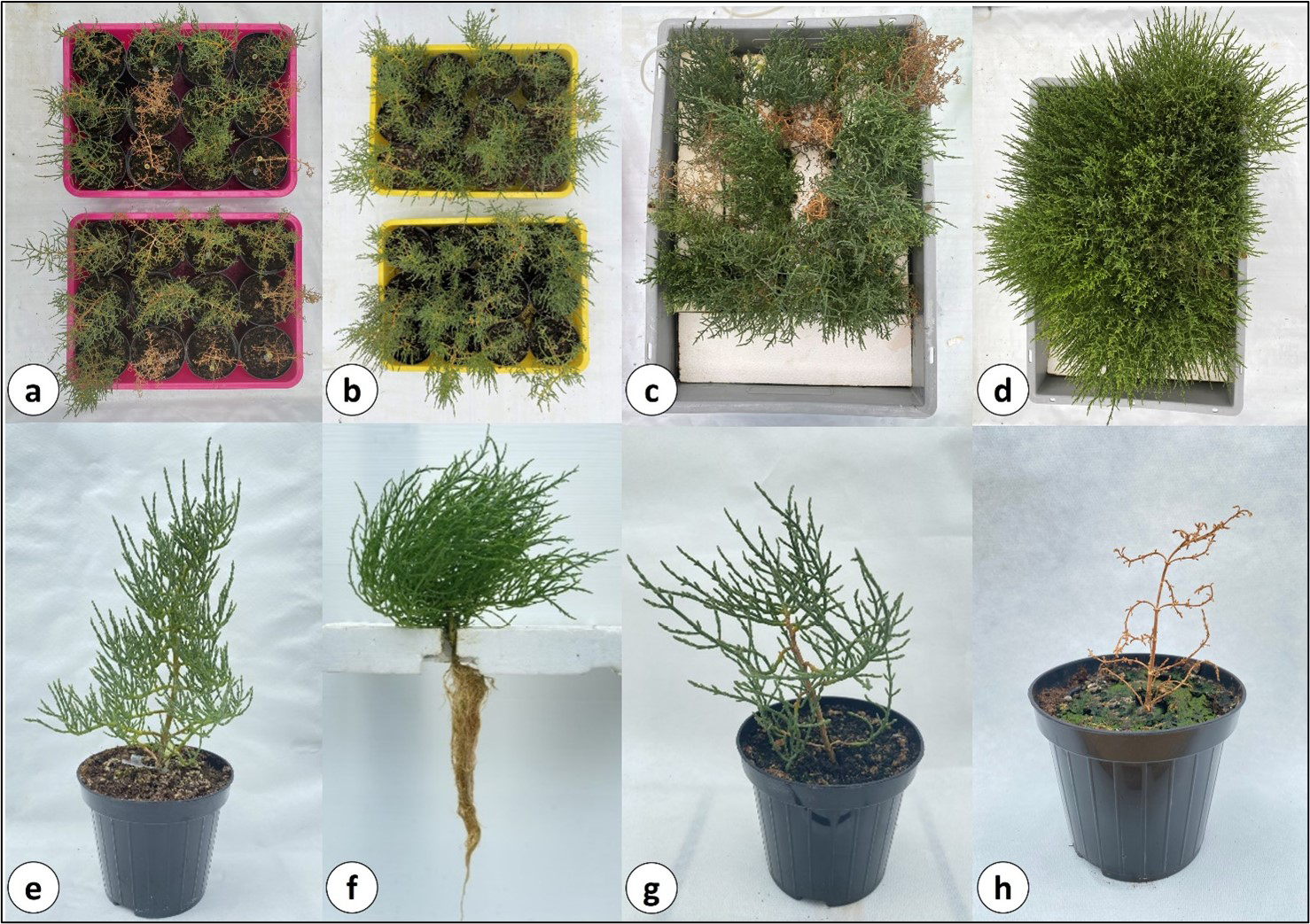
No symptoms were observed in control plants (Figure 4, b, d, e, and f). The plants cultivated in pots and hydroponics had different growth patterns (Table 1). The pot-cultivated plants tended to be woodier than those in hydroponics, especially at the base of the stem. In these cases, desiccation of at least 50% of the main stem of each affected plant was considered as score 1 on the empirical qualitative ordinal scale of disease symptoms (Figure 4 g). In addition to the control plants, asymptomatic inoculated plants (i.e. score 0), grown in hydroponics, and potted plants with a score of 1, were collected for the determination of shoot fresh weight (SFW), shoot dry weight (SDW), and succulence. Differences between inoculated and non-inoculated plants, within the same cultivation method, were statistically significant (P ≤ 0.05) for potted plants, but not for plants grown hydroponically (Table 1).
| Parameter | Hydroponics | Pots | ||
|---|---|---|---|---|
| Inoculated | Non-inoculated | Inoculated | Non-inoculated | |
| Shoot fresh weight (g/plant) | 56.3 ± 6.4 a | 62.9 ± 4.9 a | 7.3 ± 0.8 a | 16.7 ± 0.5 b |
| Shoot dry weight (g/plant) | 6.4 ± 0.6 a | 6.8 ± 1.1 a | 1.4 ± 0.2 a | 2.6 ± 0.2 b |
| Succulence (g/g) | 7.8 ± 0.4 a | 7.7 ± 1.2 a | 4.2 ± 0.3 a | 5.4 ± 0.3 b |
Symptoms of aerial parts of plants were characterized by acropetal patterns, initially affecting the stems and basal lateral shoots (Figure 4, a and c), following damage to the roots. The disease had a long asymptomatic phase. Initial wilting symptoms were observable at 24 dpi, when the plants were transplanted into pots or in hydroponics. There was no clear evidence of root or crown rot before transplanting. Clear signs of disease in the aboveground portions (i.e. desiccation of the main stems) were observed in pots 2 days after transplanting (26 dpi). The plants in hydroponics showed evidence of desiccation at 1 week after transplanting (31 dpi). These different rates of symptom appearance were very obvious at 80 dpi. First plant death in pots was recorded at 47 dpi (Figure 4 h) and at 61 dpi in hydroponics (Figure 5).
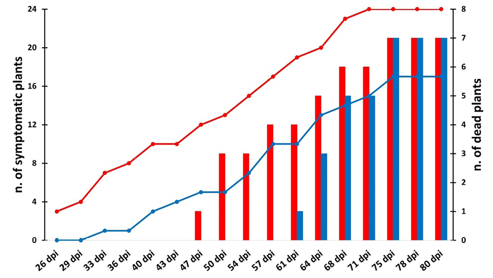
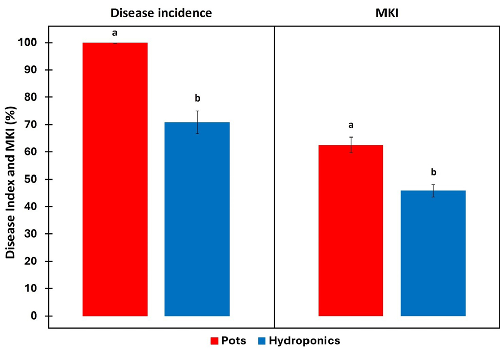
In pots, 100% (24/24) of the inoculated plants showed symptoms attributable to at least class 1 in the empirical qualitative ordinal scale of disease symptoms. In hydroponics, disease only affected 70.8% (17/24) of the plants, and seven out of 24 plants were asymptomatic and were assigned scores of zero. The number of dead plants (score 5) was the same at 80 dpi (seven) in pots and in hydroponics. Accordingly, disease incidence and McKinney Index were significantly greater in pot-grown plants than in those grown hydroponically (Figure 6).
Koch’s postulates were fulfilled by reisolating F. pseudograminearum from below- and above-ground tissues collected from both groups of plants, regardless of disease severity. The pathogen was mainly present in the roots and crowns. In pot-grown plants, F. pseudograminearum was re-isolated from 96% of sampled roots and 100% of sampled crown tissues, and 87% of root and 82% of crown samples of hydroponically grown plants. Pathogen re-isolation from above-ground tissues of potted plants was 41% from stems, 91% from first lateral branches, and 59% from second lateral branches. In hydroponics, re-isolations of F. pseudograminearum in above-ground plant tissues were higher, 85% from stems, 91% from first lateral branches, and 64% from second lateral branches (Table S2). Overall, re-isolations of F. pseudograminearum were greater from plants grown in the hydroponic system (83% of total plated tissues) than those grown in pots (62%). Fusarium pseudograminearum was also re-isolated from asymptomatic hydroponically grown plants.
Production of mycotoxins in vitro and in planta
In this experiment, in addition to the three isolates of F. pseudograminearum (3B, PD-A, and PD-B), two further isolates of F. pseudograminearum, PVS-Fu 7 (Balmas, 1994) and ColPat-351 (Agustí-Brisach et al., 2018) and one isolate of F. oxysporum (DAFE SP-21-23) isolated from S. europaea plants were included.
First, we tested whether the isolates had the gene pool required for the production of deoxynivalenol (DON) and zearalenone (ZEA) by amplifying a fragment of the tri5 (Niessen and Vogel, 1998) and pks4 (Meng et al., 2010) genes. All the isolates, except for F. oxysporum DAFE SP21-23, were positive for the 650 bp fragment of the tri5 gene (Figure S2), and the 280 bp fragment of the pks4 gene (Figure S3). Consensus sequences were used for BLASTn search to verify sequence identity and were deposited under the GenBank AN PQ045872 to PQ045876 for tri5 and PQ045867 to PQ045871 for pks4. For tri5, the sequences exhibited 98.65% (isolates 3B, PD-A, and PD-B) and 98.80% (isolates ColPat-351 and PVS-Fu 7) identity with the tri5 gene of F. pseudograminearum strains NRRL 28334, NRRL 28338, and 28062 (respectively, AN AY102583, AY102585, and AY102580). Lower sequence similarity was found for pks4. The sequences showed 95.02% (isolates 3B, PD-A, and PD-B), 95.37% (isolate ColPat-351), and 94.66% (isolate PVS-Fu 7) similarity with the pks4 gene of F. pseudograminearum CS3096 (AN: XM_009259983). The amplification of tri3 and tri12 genes (Starkey et al., 2007), with a multiplex PCR developed to determine the DON-chemotype, failed to produce any amplicons. Although several attempts were made, such as modifying the annealing temperature, concentration of DNA, primers combinations, and MgCl2 in the reaction mixture, the multiplex PCR yielded no results. Therefore, no specific DON-chemotype (i.e. NIV, 3ADON, 15ADON) could be assigned to the isolates.
The mycotoxigenic ability of the F. pseudograminearum isolates was assessed by growing them on barley grains. Table 2 shows the results for DON and ZEA production, assessed using the respective immunochromatographic test kits (Figures S4 and S5). Zearalenone was detected in all the samples with considerable yields. The PD-B isolate produced the least amount of toxin (40.01 ± 1.05 mg kg-1) while isolate ColPat-351 produced the greatest amount (69.1 ± 5.45 mg kg-1). In contrast, in the latter isolate the production of DON was below the LOD (0.25 mg kg-1), while isolate PVS-Fu 7 produced only a small amount of this mycotoxin (0.37 ± 0.06 mg kg-1). Interestingly, the F. pseudograminearum isolates obtained from S. europaea were higher DON producers, each with comparable yields. As expected, the F. oxysporum isolate DAFE SP21-23 did not produce the tested mycotoxins.
| Isolate | DON (mg kg-1) | ZEA (mg kg-1) |
|---|---|---|
| F. pseudograminearum 3B | 6.93 ± 0.43 a | 61.10 ± 2.17 ab |
| F. pseudograminearum PD-A | 6.72 ± 0.11 a | 52.94 ± 2.93 abc |
| F. pseudograminearum PD-B | 6.79 ± 0.19 a | 40.01 ± 1.05 c |
| F. pseudograminearum ColPat-351 | < LOD c | 69.10 ± 5.45 a |
| F. pseudograminearum PVS Fu-7 | 0.37 ± 0.06 b | 47.48 ± 4.49 bc |
| F. oxysporum DAFE SP21-23 | < LOD c | < LOD d |
| Control | < LOD c | < LOD d |
Mycotoxin production in planta was low. Deoxynivalenol was detected by LC-MS/MS only at 0.029 ± 0.012 mg kg-1 in severely damaged S. europaea plants (disease scores 3 to 5) grown in hydroponics, while all samples were negative for ZEA, NIV, 3ADON, and 15ADON. Since S. europaea plants were inoculated with the F. pseudograminearum 3B isolate, the results were consistent with those obtained from the molecular analyses, which did not identify any specific DON chemotype.
Discussion
This is the first report of root and crown rot caused by F. pseudograminearum on cultivated S. europaea plants. There are very few records of diseases affecting Salicornia species (Delli Compagni et al., 2024). The only report regarding cultivated species is for S. bigelovii, which was found to be susceptible to Botrytis cinerea in Mexico (Rueda Puente et al., 2014). Pathogenicity of this fungus was confirmed by spraying a conidial suspension on detached branches. There are no documented artificial inoculation experiments on S. europaea, since the pathogenic species found on this halophyte have only been described in wild plants (Delli Compagni et al., 2024). In the present study, artificial inoculations were carried out on seed-propagated plants of S. europaea.
The pathogenicity test showed that root and crown rot caused by F. pseudograminearum was less severe when test plants were grown hydroponically (MKI = 46%) than when grown in potting soil (MKI = 63%) and all the pot-grown plants developed browning symptoms. Disease incidence was less in the hydroponic system, with seven out of 24 plants remaining asymptomatic (Figure 6). These results indicate that disease severity depended on the cultivation method. At 80 dpi, SFW, SDW, and shoot succulence of inoculated, asymptomatic plants (i.e. score 0) grown in the hydroponic system resembled parameters measured for the control plants, whereas the same parameters in score 1 potted plants were significantly different from the respective controls (Table 1). Moreover, the potted plants also had disease symptoms soon after transplanting, while in hydroponics symptom development was delayed (Figure 5). However, mortality rates were the same between the two groups of plants (each with seven out of 24 plants), although in hydroponics the first plant death occurred 2 weeks later than in pots (Figure 5).
Hydroponic cultivation can reduce plant susceptibility to infectious diseases compared to soil cultivation. In a similar experiment, Maurer et al., (2023) found that sweet basil plants grown in a static hydroponic solution were much less sensitive to downy mildew caused by Peronospora belbahrii than those grown in soil. They stated that the higher antioxidant content in hydroponically grown basil plants could alleviate symptom development. Moulin et al. (1994) showed that several Pythium species (P. aphanidermatum, P. intermedium, P. irregulare, P. sylvaticum, and P. ultimum var. ultimum) caused damping-off of cucumber when grown in sand-peat soil, but only P. aphanidermatum caused disease in substrate (stone wool) and in hydroponic culture. These authors reported that symptoms were milder in hydroponics than in substrate. Moreover, inoculations with Streptomyces scabies resulted in less infection of potato plants grown hydroponically compared to pot-grown plants (Khatri et al., 2011). Similarly, rice artificially inoculated with Xanthomonas oryzae pv. oryzae, the causative agent of bacterial leaf blight, had lower disease severity in hydroponic culture than in soil (Song et al., 2016).
In the present study, the greater growth rate of hydroponically grown plants than those in potting soil, was likely due to the better supply of water and minerals, and oxygenation of roots compared to soil cultivation, which may have resulted in the reduced rate of development of F. pseudograminearum root and crown rot on S. europaea. In addition, F. pseudograminearum is a soil-borne pathogen and causes disease in warm and dry climates (Poole et al., 2013), with increased virulence in drought conditions (Kazan and Gardiner, 2018). In agricultural areas characterized by low moisture and high temperatures, F. pseudograminearum was found to be predominant compared to other Fusarium species (e.g. F. culmorum), which prefer cool and humid regions (Poole et al., 2013).
Little information is available on mycotoxins on Salicornia species. Lopes et al. (2020) reported that several S. ramosissima and Salicornia sp. samples were contaminated by aflatoxins in Portugal. These compounds were detected in plants collected in the wild, and also in those cultivated with conventional and organic methods, but no mycotoxins were detected from greenhouse and hydroponically cultivated plants.
In the present study, DON was detected in low amounts from inoculated plants. Traces of DON (0.029 ± 0.012 mg kg-1) were detected only in severely damaged (score 3–5) plants grown hydroponically. This result could be due to the greater water content of these plants compared to those grown in potting soil since high levels of water activity (aw) have been shown to be necessary for optimal production of trichothecenes by Fusarium spp. (Hope et al., 2005; Han et al., 2018; Rybecky et al., 2018; Belizán et al., 2019). Therefore, one hypothesis is that the inoculated F. pseudograminearum isolate produced the mycotoxin only in suitable conditions (such as high plant water content) and during an advanced stage of disease development. In wheat kernels, the most favorable conditions for DON and ZEA production by F. pseudograminearum were an aw of 0.97 and a temperature of 25°C (Cui et al., 2022).
When inoculated on barley grains, all the tested F. pseudograminearum isolates produced ZEA with considerable yields, similar to those documented by other authors (Blaney and Dodman, 2002; Clear et al., 2006; Alkadri et al., 2013). Furthermore, only the three F. pseudograminearum isolates obtained from S. europaea were high DON producers, each with similar production (Table 2). The other two European isolates had different mycotoxigenic activities. Low production of DON was detected in the Italian isolate F. pseudograminearum (PVS Fu-7), while production of DON by the Spanish isolate (ColPat-351), was below the LOD (Table 2). In addition, PCR targeting the tri3 and tri12 genes to discriminate between NIV and acetylated derivatives of DON producers (i.e. 3ADON and 15ADON) did not identify any specific DON chemotype. Deng et al. (2020), in a molecular analysis to determine the DON chemotypes of 372 isolates of F. pseudograminearum, showed that 196 isolates did not produce any fragments in tri3 and tri12 PCR assays, with the same primers used in this study. After LC-MS/MS analysis, isolates without a chemotype marker were assigned to the putative 15-ADON chemotype (Deng et al., 2020).
The present study is the first to record root and crown rot of S. europaea caused by F. pseudograminearum. This study also provides an important contribution to the current distribution and host range of this pathogenic fungus in Europe. In addition, the mycotoxin deoxynivalenol was detected in diseased plants, although only in small quantities and under specific conditions. Further studies are ongoing to assess the environmental conditions (e.g. salinity and temperature) that may promote mycotoxigenic activity in F. pseudograminearum and to determine potential mycotoxin exposure risk in S. europaea.
Acknowledgements
This research was funded by the project “HALOphytes grown in saline Water for the production of INnovative ready-to-eat salad – HALOWIN”, funded by the University of Pisa (project code PRA_2020_43). The authors are grateful to Grazia Puntoni (University of Pisa) for the technical support, and Prof. Virgilio Balmas (University of Sassari) and Prof. Carlos Agustí-Brisach (University of Cordoba) for providing the F. pseudograminearum isolates used in this study.
Literature cited
Abdallah-Nekache N., Laraba I., Ducos C., Barreau C., Bouznad Z., Boureghda H., 2019. Occurrence of Fusarium head blight and Fusarium crown rot in Algerian wheat: identification of associated species and assessment of aggressiveness. European Journal of Plant Pathology 154(3): 499–512. https://doi.org/10.1007/s10658-019-01673-7
Agustí-Brisach C., Raya-Ortega M.C., Trapero C., Roca L.F., Luque F., … Trapero A., 2018. First report of Fusarium pseudograminearum causing crown rot of wheat in Europe. Plant Disease 102(8): 1670. https://doi.org/10.1094/PDIS-11-17-1840-PDN
Akinsanmi O.A., Chakraborty S., Backhouse D., Simpfendorfer S., 2007. Passage through alternative hosts changes the fitness of Fusarium graminearum and Fusarium pseudograminearum. Environmental Microbiology 9(2): 512–520. https://doi.org/10.1111/j.1462-2920.2006.01168.x
Alkadri D., Nipoti P., Döll K., Karlovsky P., Prodi A., Pisi A., 2013. Study of fungal colonization of wheat kernels in Syria with a focus on Fusarium species. International Journal of Molecular Science 14(3): 5938–5951. https://doi.org/10.3390/ijms14035938
Aoki T., O’Donnell K., 1999a. Morphological and molecular characterization of Fusarium pseudograminearum sp. nov., formerly recognized as the Group 1 population of F. graminearum. Mycologia 91(4): 597–609. https://doi.org/10.1080/00275514.1999.12061058
Aoki T., O’Donnell K., 1999b Morphological characterization of Gibberella coronicola sp. nov., obtained through mating experiments of Fusarium pseudograminearum. Mycoscience 40(6): 443–453. https://doi.org/10.1007/BF02461021
Araus J.L., Rezzouk F.Z., Thushar S., Shahid M., Elouafi I.A., … Serret M.D., 2021. Effect of irrigation salinity and ecotype on the growth, physiological indicators and seed yield and quality of Salicornia europaea. Plant Science 304: 110819. https://doi.org/10.1016/j.plantsci.2021.110819
Balmas V., 1994. Root rot of wheat in Italy caused by Fusarium graminearum Group I. Plant Disease 78: 317A. https://doi.org/10.1094/PD-78-0317A
Belizán M.M.E., Gomez A. de los, Terán Baptista Z.P., Jimenez C.M., Sánchez Matías M. del H., … Sampietro D.A., 2019. Influence of water activity and temperature on growth and production of trichothecenes by Fusarium graminearum sensu stricto and related species in maize grains. International Journal of Food Microbiology 305: 108242. https://doi.org/10.1016/j.ijfoodmicro.2019.108242
Blaney B.J., Dodman R.L., 2002. Production of zearalenone, deoxynivalenol, nivalenol, and acetylated derivatives by Australian isolates of Fusarium graminearum and F. pseudograminearum in relation to source and culturing conditions. Australian Journal of Agricultural Research 53(12): 1317–1326. https://doi.org/10.1071/AR02041
Burgess L.W., Klein T.A., Bryden W.L., Tobin N.F., 1987. Head blight of wheat caused by Fusarium graminearum Group 1 in New South Wales in 1983. Australasian Plant Pathology 16: 72–78. https://doi.org/10.1071/APP9870072
Cárdenas-Pérez S., Piernik A., Chanona-Pérez J.J., Grigore M.N., Perea-Flores M.J., 2021. An overview of the emerging trends of the Salicornia L. genus as a sustainable crop. Environmental and Experimental Botany 191: 104606. https://doi.org/10.1016/j.envexpbot.2021.104606
Chakraborty S., Liu C.J., Mitter V., Scott J.B., Akinsanmi O.A., … Simpfendorfer S., 2006. Pathogen population structure and epidemiology are keys to wheat crown rot and Fusarium head blight management. Australasian Plant Pathology 35: 643–655. https://doi.org/10.1071/AP06068
Clear R.M., Patrick S.K., Gaba D., Roscoe M., Demeke T., … Turkington T.K., 2006. Trichothecene and zearalenone production, in culture, by isolates of Fusarium pseudograminearum from western Canada. Canadian Journal of Plant Pathology 28(1): 131–136. https://doi.org/10.1080/07060660609507279
Cui H., Wang S., Yang X., Zhang W., Chen M., … Wang S., 2022. Predictive models for assessing the risk of Fusarium pseudograminearum mycotoxin contamination in post-harvest wheat with multi-parameter integrated sensors. Food Chemistry: X 16:100472. https://doi.org/10.1016/j.fochx.2022.100472
De Santis B., Debegnach F., Gregori E., Russo S., Marchegiani F., … Brera C., 2017. Development of a LC-MS/MS method for the multi-mycotoxin determination in composite cereal-based samples. Toxins 9(5): 169. https://doi.org/10.3390/toxins9050169
Delli Compagni E., Pardossi A., Pecchia S., 2024. Fungal and fungal-like diseases of halophytes in the Mediterranean basin: a state-of-the-art review. Horticulturae 10(4): 313. https://doi.org/10.3390/horticulturae10040313
Deng Y.Y., Li W., Zhang P., Sun H.Y., Zhang X.X., … Chen H.G., 2020 Fusarium pseudograminearum as an emerging pathogen of crown rot of wheat in eastern China. Plant Pathology 69(2):240–248. https://doi.org/10.1111/ppa.13122
Desjardins A.E., Proctor R., Bai G., McCormick S., Shaner G., … Hohn T.M., 1996 Reduced virulence of trichothecene-nonproducing mutants of Gibberella zeae in wheat field tests. Molecular Plant-Microbe Interactions. 9(9): 775. http://doi.org/10.1094/MPMI-9-0775
EFSA Panel on Plant Health (PLH), Bragard C, Baptista P, Chatzivassiliou E, Di Serio F, Gonthier P, … Reignault P.L., 2022 Pest categorisation of Fusarium pseudograminearum. EFSA Journal 20:e07399. https://doi.org/10.2903/j.efsa.2022.7399
Han Z., Shen Y., Di Mavungu J.D., Zhang D., Nie D., Jiang K., … Zhao Z., 2018. Relationship between environmental conditions, TRI5 gene expression and deoxynivalenol production in stored Lentinula edodes infected with Fusarium graminearum. World Mycotoxin Journal 11(2): 177–186. https://doi.org/10.3920/WMJ2017.2245
Hope R., Aldred D., Magan N., 2005. Comparison of environmental profiles for growth and deoxynivalenol production by Fusarium culmorum and F. graminearum on wheat grain. Letters in Applied Microbiology 40(4): 295–300. https://doi.org/10.1111/j.1472-765X.2005.01674.x
Iqbal S.Z., 2021. Mycotoxins in food, recent development in food analysis and future challenges; a review. Current Opinion in Food Science 42: 237–247. https://doi.org/10.1016/j.cofs.2021.07.003
Kammoun L.G., Gargouri S., Hajlaoui M.R., Marrakchi M., 2009. Occurrence and distribution of Microdochium and Fusarium species isolated from durum wheat in northern Tunisia and detection of mycotoxins in naturally infested grain. Journal of Phytopathology 157(9): 546–551. https://doi.org/10.1111/j.1439-0434.2008.01522.x
Kazan K., Gardiner D.M., 2018. Fusarium crown rot caused by Fusarium pseudograminearum in cereal crops: recent progress and future prospects. Molecular Plant Pathology 19(7): 1547–1562. https://doi.org/10.1111/mpp.12639
Khatri B.B., Tegg R.S., Brown P.H., Wilson C.R., 2011. Temporal association of potato tuber development with susceptibility to common scab and Streptomyces scabiei-induced responses in the potato periderm. Plant Pathology 60(4): 776–786. https://doi.org/10.1111/j.1365-3059.2011.02435.x
Kokkonen M., Ojala L., Parikka P., Jestoi M., 2010. Mycotoxin production of selected Fusarium species at different culture conditions. International Journal of Food Microbiology 143(2): 17‒25. https://doi.org/10.1016/j.ijfoodmicro.2010.07.015
Lombardi T., Bertacchi A., Pistelli L., Pardossi A., Pecchia S., … Sanmartin C., 2022. Biological and agronomic traits of the main halophytes widespread in the Mediterranean region as potential new vegetable crops. Horticulturae 8(3): 195. https://doi.org/10.3390/horticulturae8030195
Lopes M., Castilho M., Sanches-Silva A., Freitas A., Barbosa J., … Ramos F., 2020. Evaluation of the mycotoxins content of Salicornia spp.: a gourmet plant alternative to salt. Food Additives & Contaminants: Part B 13(3): 162–170. https://doi.org/10.1080/19393210.2020.1741692
Lysøe E., Klemsdal S.S., Bone K.R., Frandsen R.J.N., Johansen T., … Giese H., 2006. The PKS4 gene of Fusarium graminearum is essential for zearalenone production. Applied Environmental Microbiology 72(6): 3924–3932. https://doi.org/10.1128/AEM.00963-05
Maurer D., Sadeh A., Chalupowicz D., Barel S., Shimshoni J.A., Kenigsbuch D., 2023. Hydroponic versus soil-based cultivation of sweet basil: impact on plants’ susceptibility to downy mildew and heat stress, storability and total antioxidant capacity. Journal of the Science of Food and Agriculture 103(15): 7809–7815. https://doi.org/10.1002/jsfa.12860
McKinney H.H., 1923. Influence of soil temperature and moisture on infection of wheat seedlings by Helminthosporium sativum. Journal of Agricultural Research 26: 195–218.
Meng K., Wang Y., Yang P., Luo H., Bai Y., Yao B., 2010. Rapid detection and quantification of zearalenone-producing Fusarium species by targeting the zearalenone synthase gene PKS4. Food Control 21(2): 207–11. https://doi.org/10.1016/j.foodcont.2009.05.014
Miličević T., Kaliterna J., Ivić D., Stričak A., 2013. Identification and occurrence of Fusarium species on seeds of common vetch, white lupin and some wild legumes. Poljoprivreda 19(1): 25–32.
Moulin F., Lemanceau P., Alabouvette C., 1994. Pathogenicity of Pythium species on cucumber in peat-sand, rockwool and hydroponics. European Journal of Plant Pathology 100: 3–17. https://doi.org/10.1007/BF01871963
Nahle S., El Khoury A., Atoui A., 2021. Current status on the molecular biology of zearalenone: its biosynthesis and molecular detection of zearalenone producing Fusarium species. European Journal of Plant Pathology 159: 247‒258. https://doi.org/10.1007/s10658-020-02173-9
Niessen M.L., Vogel R.F., 1998. Group specific PCR-detection of potential trichothecene-producing Fusarium-species in pure cultures and cereal samples. Systematic and Applied Microbiology 21(4): 618–631. https://doi.org/10.1016/S0723-2020(98)80075-1
Nirenberg H.I., 1981. A simplified method for identifying Fusarium spp. occurring on wheat. Canadian Journal of Botany 59(9): 1599–1609. https://doi.org/10.1139/b81-217
O’Donnell K., Kistler H.C., Cigelnik E., Ploetz R.C., 1998. Multiple evolutionary origins of the fungus causing Panama disease of banana: concordant evidence from nuclear and mitochondrial gene genealogies. Proceedings of the National Academy of Science 95(5): 2044–2049. https://doi.org/10.1073/pnas.95.5.2044
Obanor F., Neate S., Simpfendorfer S., Sabburg R., Wilson P., Chakraborty S., 2013. Fusarium graminearum and Fusarium pseudograminearum caused the 2010 head blight epidemics in Australia. Plant Pathology 62(1): 79–91. https://doi.org/10.1111/j.1365-3059.2012.02615.x
Pasquali M., Migheli Q., 2014. Genetic approaches to chemotype determination in type B-trichothecene producing Fusaria. International Journal of Food Microbiology. 189: 164‒182. https://doi.org/10.1016/j.ijfoodmicro.2014.08.011
Poole G.J., Smiley R.W., Walker C., Huggins D., Rupp R., … Paulitz T.C., 2013. Effect of climate on the distribution of Fusarium spp. causing crown rot of wheat in the Pacific Northwest of the United States. Phytopathology 103(11): 1130–1140. https://doi.org/10.1094/PHYTO-07-12-0181-R
Proctor R.H., Hohn T.M., McCormick S.P., 1995. Reduced virulence of Gibberella zeae caused by disruption of a trichothecene toxin biosynthetic gene. Molecular Plant-Microbe Interactions 8(4): 593–601. https://doi.org/10.1094/mpmi-8-0593
Rueda Puente E.O., Hernandez Montiel L.G., Holguin Peña J., Murillo Amador B., Rivas Santoyo F.J., 2014. First report of Botrytis cinerea Pers. on Salicornia bigelovii Torr. in North-West México. Journal of Phytopathology 162(7-8): 513–515. https://doi.org/10.1111/jph.12205
Rybecky A.I., Chulze S.N., Chiotta M.L., 2018. Effect of water activity and temperature on growth and trichothecene production by Fusarium meridionale. International Journal of Food Microbiology 285: 69–73. https://doi.org/10.1016/j.ijfoodmicro.2018.07.028
Sabburg R., Obanor F., Aitken E., Chakraborty S., 2015. Changing fitness of a necrotrophic plant pathogen under increasing temperature. Global Change Biology 21(8): 3126–3137. https://doi.org/10.1111/gcb.12927
Sever Z., Ivić D., Kos T., Miličević T., 2012. Identification of Fusarium species isolated from stored apple fruit in Croatia. Archives of Industrial Hygiene and Toxicology 63(4): 463–470. https://doi.org/10.2478/10004-1254-63-2012-2227
Shan X., Zhu Y., Redman R., Rodriguez R.J., Yuan Z., 2021. The chromosome-scale genome resource for two endophytic Fusarium species, F. culmorum and F. pseudograminearum. Molecular Plant-Microbe Interactions 34(6): 703–706. https://doi.org/10.1094/MPMI-07-20-0205-A
Song A., Xue G., Cui P., Fan F., Liu H., … Liang Y., 2016.The role of silicon in enhancing resistance to bacterial blight of hydroponic- and soil-cultured rice. Scientific Reports 6: 24640. https://doi.org/10.1038/srep24640
Spada M., Pugliesi C., Fambrini M., Palpacelli D., Pecchia S., 2023. Knockdown of BMP1 and PLS1 virulence genes by exogenous application of RNAi-Inducing dsRNA in Botrytis cinerea. International Journal of Molecular Sciences 24(5): 4869. https://doi.org/10.3390/ijms24054869
Starkey D.E., Ward T.J., Aoki T., Gale L.R., Kistler H.C., O’Donnell K., 2007. Global molecular surveillance reveals novel Fusarium head blight species and trichothecene toxin diversity. Fungal Genetics and Biology 44(11): 1191–1204. https://doi.org/10.1016/j.fgb.2007.03.001
Tunali B., Nicol J.M., Hodson D., Uçkun Z., Büyük O., … Bağci H., 2008. Root and crown rot fungi associated with spring, facultative, and winter wheat in Turkey. Plant Disease 92(9): 1299–1306. https://doi.org/10.1094/PDIS-92-9-1299
Ventura Y., Sagi M., 2013. Halophyte crop cultivation: the case for Salicornia and Sarcocornia. Environmental and Experimental Botany 92: 144–153. https://doi.org/10.1016/j.envexpbot.2012.07.010
Xu F., Song Y.L., Wang J.M., Liu L.L., Zhao K., 2017. Occurrence of Fusarium crown rot caused by Fusarium pseudograminearum on barley in China. Plant Disease 101(5): 837. https://doi.org/10.1094/PDIS-10-16-1436-PDN
Zhou H., He X., Wang S., Ma Q., Sun B., Ding S., … Li H., 2019. Diversity of the Fusarium pathogens associated with crown rot in the Huanghuai wheat-growing region of China. Environmental Microbiology 21(8): 2740–2754. https://doi.org/10.1111/1462-2920.14602