https://doi.org/10.36253/phyto-15575
Accepted: April 28, 2025; Published: May 15, 2025
Research Papers
First report of Xanthomonas arboricola on oleander
1 Hungarian University of Agriculture and Life Sciences, Buda Campus, Institute of Plant Protection, H-1118, Budapest Ménesi Road 44, Hungary
2 Széchenyi István University, Department of Plant Sciences, Albert Kázmér Faculty of Mosonmagyaróvár, 2. Vár square, Mosonmagyaróvár; Hungary; ELKH-SZE PhatoPlant-Lab, Széchenyi István University, 2. H-9200, Mosonmagyaróvár Vár square, Hungary
*Corresponding author E-mail: karacs.vegh.anita@uni-mate.hu
Summary. Nerium oleander L is a long-lasting flowering vegetatively propagated ornamental plant of the Mediterranean region, where it is a major imported flowering pot plant. Only a few bacteria can infect it such as Pseudomonas savastanoi pv. nerii, Agrobacterium tumefaciens and Xylella fastidiosa. Between 2018 and 2022 we collected several infected plant parts in our country. In 2020 we observed atypical symptoms on the leaves and stems, which were not clearly similar to the known bacterial infection of oleander. In our work, we aimed to identify the pathogen. The isolates formed yellow-coloured bacterial colonies on King-B and on YDC agar, were Gram-negative, oxidase negative and induced hypersensitive reaction on tobacco leaves. The biochemical properties were determined by API 20E and API 50CH tests. Brown necrosis was observed on oleander leaves in a pathogenicity test. Multilocus sequence analysis was used for molecular identification of the pathogen. Three housekeeping genes (gyrB, fyuA and rpoD) were amplified. According to symptoms, colony morphology, biochemical features, pathogenicity and molecular methods, the pathogen was identified as Xanthomonas arboricola. This is the first report of the plant pathogenic Xanthomonas spp. on oleander.
Keywords. Nerium oleander, Xanthomonas, bacterial disease, Hungary.
Contents
Introduction
Nerium oleander L. is an evergreen tropical and subtropical plant of the Apocynaceae family. It is a vegetatively propagated ornamental plant. The plant is appreciated for its striking flower of different colours. Only a few bacteria can infect oleander during cultivation, but only Pseudomonas savastanoi pv. nerii (Janse) Young, Dye & Wilkie causes significant damage. The typical symptoms are knots or galls on stems, twigs, leaves and seedcases. The knots are few millimetres, soft and green in the early stages of infection. Then it grows to a few centimetres, turns brown (Smith, 1906) and decays as the periderm blocks the source of water and nutrients. The severity of symptoms is influenced by phytohormone production (Themsah et al., 2010). Xanthomonas (Dowson 1939) is a large genus of plant pathogenic Gram-negative bacteria, which are obligate aerobes and generally rod shaped with a single polar flagellum (Bradbury, 1984). Many members of this genus are yellow-pigmented because of xanthan production (Kennedy and Bradshaw, 1984). The Xanthomonas genus comprises 45 species (Parte et al., 2020) which show a high degree of host plant specificity. The range of host plants is mostly a single plant or a few plants from the same botanical family (Ryan et al., 2011). However, it is difficult to define the exact range of host plants, as pathogenicity tests are only carried out on a few cultivated plants (Bull and Koike, 2015). The symptoms are ranging from water-soaked spots, wilting to cankers (Rudolph, 1993). Xanthomonas spp. cause serious diseases on important crops such as rice, citrus, banana, cabbage, tomato and walnut (Kennedy and Bradshaw, 1984; Ryan et al., 2011). Additionally, Xanthomonas produces a polysaccharide called xanthan gum, which is used in industry as a thickener and emulsifier (Backer et al., 1998).
Materials and methods
We have been investigating oleander knot in Hungary since 2018. Only a few cases of atypical symptoms were observed during the nationwide sampling of approximately 300 samples. In 2020 two infected plant samples, one from Southern Great Plain (Kecskemét) and one from Northern Great Plain (Nyírbátor) were collected from hobby gardeners. Symptoms of brown, necrosis on the leaf, and untypical canker on the stem were observed. The symptomatic plant parts were delivered to the laboratory of the Department of Plant Pathology, Hungarian University of Agriculture and Life Sciences. The samples were surface-sterilized, homogenized and streaked onto King’s B agar (King et al., 1954). The agar plates were incubated at room temperature (RT) for 48 to 72 h, and pure cultures of bacterial isolates were obtained by colony subculturing. Hypersensitive reaction (HR) was tested on tobacco leaves (Nicotiana tabacum L. Xanthi) using pure bacterial suspension of 5×10⁷ cells mL-1. The suspension was determined with a spectrophotometer set at λ = 560 nm. Leaves were assessed at 24 and 48 h post-inoculation. Gram feature was determined by the KOH test (Powers, 1995). Biochemical analysis was performed using the API 20E and API 50CH kit (BioMérieux). Colony morphology was observed on Yeast Extract-Dextrose-Calcium Carbonate agar (YDC). For pathogenicity test 1 year old oleander plants were used. Stem internodes, between the first and the second leaves were injected using sterile syringes containing bacterial suspension of 5×10⁷ cells mL1 and 2 leaves of each plant were also lacerated with the inoculant syringe needle. Sterilized distilled water was used for injection as a negative control. 3 plants were used for each replicate. The inoculated plants were maintained at RT with a relative humidity greater than 90% for 1 week. Symptoms were observed daily for 3-week post-inoculation. To determine host plants specificity of isolates detached leaves of apricot (Prunus armeniaca L.), walnut (Juglans regia L.), pepper (Capsicum annum L.), kohlrabi (Brassica oleraceae L convar. acephala var. gongylodes), cabbage (B. oleracea convar. capitata), geranium (Pelargonium zonale L’Hér ex Aiton), poplars (Populus alba L., P. nigra) and willow (Salix alba L.) were used. The surface-sterilized leaves were infected in two ways: inoculated with bacterial colonies using sterilised toothpicks and transferred with bacterial suspension of 5×10⁷ cells mL1 using a sterilized paintbrush. Positive control leaves were infected with bacteria from Gene Bank of Institute of Plant Protection. Negative control leaves were inoculated with sterilized distilled water. The leaves were incubated at hight relative humidity. 5 leaves were used for replicates. The cabbage and the kohlrabi were bought in a supermarket and the leaves of apricot, walnut, pepper and geranium were collected in a hobby garden. Symptoms were observed daily for 2 weeks after inoculation.
Multilocus sequence analysis (MLSA) was conducted using three housekeeping genes (fyuA, rpoD, gyrB) and forty strains (Table 1). The polymerase chain reaction (PCR) cycling conditions for fyuA and rpoD genes were 3 min at 94°C, then 30 s at 94°C, 30 s at 54°C, 1 min at 72°C for 30 cycles, then 10 min at 72°C (Young et al., 2008). The PCR cycling conditions for gyrB were 2.5 min at 94°C, then 30 s at 94°C, 45 s at 50°C, 1 min at 68°C for 34 cycles, then 7 min at 68°C (Parkinson et al., 2007). The re-isolated bacteria were confirmed by 16S rDNA PCR using 63F and 1389R primers. The PCR cycling conditions were 5 min at 94°C, then 15 s at 95°C, 30 s 55°C, 90 s 72°C for 30 cycles, then 10 min at 72°C (Osborn et al., 2000). Primers for partial sequences of fyuA, rpoD gyrB and 16S rDNA are listed in Table 2. The amplification was visualised on a 1% (w/v) agarose gel in 1 × TBE buffer with ECOSafe (Biocenter). The PCR products were cleaned with the High Pure PCR Product Purification Kit (Roche Diagnostics GmbH). The nucleotide sequence of the PCR amplified DNA fragment was determined and compared with sequences from the National Center for Biotechnology Information (NCBI) database, using the Basic Local Alignment Search Tool (BLAST) program. Homologous sequences from other members of the Xanthomonas spp. were included in the phylogenetic analysis for comparisons. For phylogenetic analysis Mega 11.1 was used (Tamura and Nei, 1993; Tamura et al., 2021).
| Strain | Xanthomonas spp. | NCBI Accession Numbers | ||
|---|---|---|---|---|
| gyrB | fyuA | rpoD | ||
| X1 | arboricola | PQ094467 | PQ094469 | PQ094471 |
| X2 | arboricola | PQ094468 | PQ094470 | PQ094472 |
| CFBP 1159 | arboricola pv. corylina | EU499002 | EU498895 | EU499121 |
| CFBP 2528 | arboricola pv. juglandis | EU498951 | EU498852 | EU499070 |
| CFBP 2535 | arboricola pv. pruni | EU498853 | EU498854 | EU499072 |
| Xp10 | arboricola pv. pruni | MN520634 | MN520626 | MN520630 |
| CFBP 4924 | axonopodis pv. axonopodis | EU498952 | EU498853 | EU499071 |
| LMG 982 | axonopodis pv. axonopodis | EU498981 | EU498914 | EU499100 |
| CFBP 3836 | axonopodis subsp. alfalfae | EU499001 | EU498894 | EU499120 |
| CFBP 2524 | axonopodis pv. begoniae | EU498962 | EU498909 | EU499081 |
| ICMP 7493 | axonopodis pv. citri | EU499024 | EU498910 | EU499143 |
| ICMP 10022 | axonopodis pv. citri | EU499045 | EU498930 | EU499165 |
| CFBP 2526 | axonopodis pv. glycines | EU499003 | EU498896 | EU499122 |
| CFBP 7153 | axonopodis pv. manihotis | EU499006 | EU498898 | EU499125 |
| CFBP 6546 | axonopodis pv. phaseoli | EU499015 | EU498904 | EU499134 |
| CFBP 7663 | axonopodis pv. phaseoli | EU498968 | EU498864 | EU499087 |
| ICMP 7462 | axonopodis pv. ricini | EU499023 | EU498771 | EU499142 |
| LMG 8122 | campestris pv. campestris | EU499018 | EU498907 | EU499137 |
| CFBP 2350 | campestris pv. campestris | EU498948 | EU498849 | EU4999067 |
| CFBP 5828 | campestris pv. raphani | EU498982 | EU498877 | EU499101 |
| CFBP 7270 | dyei | GQ183103 | GQ183117 | GQ183090 |
| CFBP 7261 | dyei pv. eucalypti | GQ183102 | GQ183115 | GQ183088 |
| CFBP 2157 | fragariae | EU499000 | EU498893 | EU499119 |
| NCPPB 2949 | fragariae | EU499012 | EU498901 | EU499131 |
| ICMP 6646 | fragariae | EU499019 | EU498908 | EU499138 |
| ICMP 1661 | hortorum pv. hederae | EU498987 | EU498880 | EU499106 |
| CFBP 4925 | hortorum pv. hederae | KY984200 | KYP984167 | KYP984233 |
| KBNS163 | hortorum pv. pelargoni | KP900004 | KP899987 | KP899953 |
| KBNS165 | hortorum pv. pelargoni | KP900006 | KP899889 | KP899955 |
| ICMP 12013 | oryzae pv. oryzicola | EU499050 | EU498935 | EU499170 |
| CFBP 2286 | oryzae pv. oryzicola | EU499007 | EU498899 | EU499126 |
| ICMP 16690 | euvesicatoria pv. perforans | EU499059 | EU498944 | EU499179 |
| NCPPB 762 | pisi | EU498976 | EU498872 | EU499095 |
| CFBP 3123 | populi | EU499035 | EU498919 | EU499155 |
| NCPPB 989 | vasicola | EU498974 | EU498870 | EU499093 |
| CFBP 2543 | vasicola | EU498992 | EU498885 | EU499111 |
| ICMP 3490 | vasicola | EU498994 | EU498887 | EU499113 |
| NCPPB 422 | vesicatoria | EU498954 | EU498855 | EU499073 |
| ICMP 696 | vesicatoria | EU498980 | EU498876 | EU499099 |
| ICMP 115 | vesicatoria | EU498956 | EU498857 | EU499075 |
| Sequence | Forward | Reverse |
|---|---|---|
| fyuA | XfyuA1F | XfyuA1R |
| AGCTACGAYGTGCGYTACGA | GTTCACGCCRAACTGGTAG | |
| rpoD | XrpoD1F | XrpoD1R |
| TGGAACAGGGCTATCTGACC | CATTCYAGGTTGGTCTGRTT | |
| gyrB | XgyrPCR2F | Xgyrrsp1 |
| AAGCAGGGCAAGAGCGAGCTGTA | CAAGGTGCTGAAGATCTGGTC | |
| 16S rDNA | 63F CAGGCCTAACACATGCAAGTC |
1389R ACGGGCGGTGTGTACAAG |
Results and Discussion
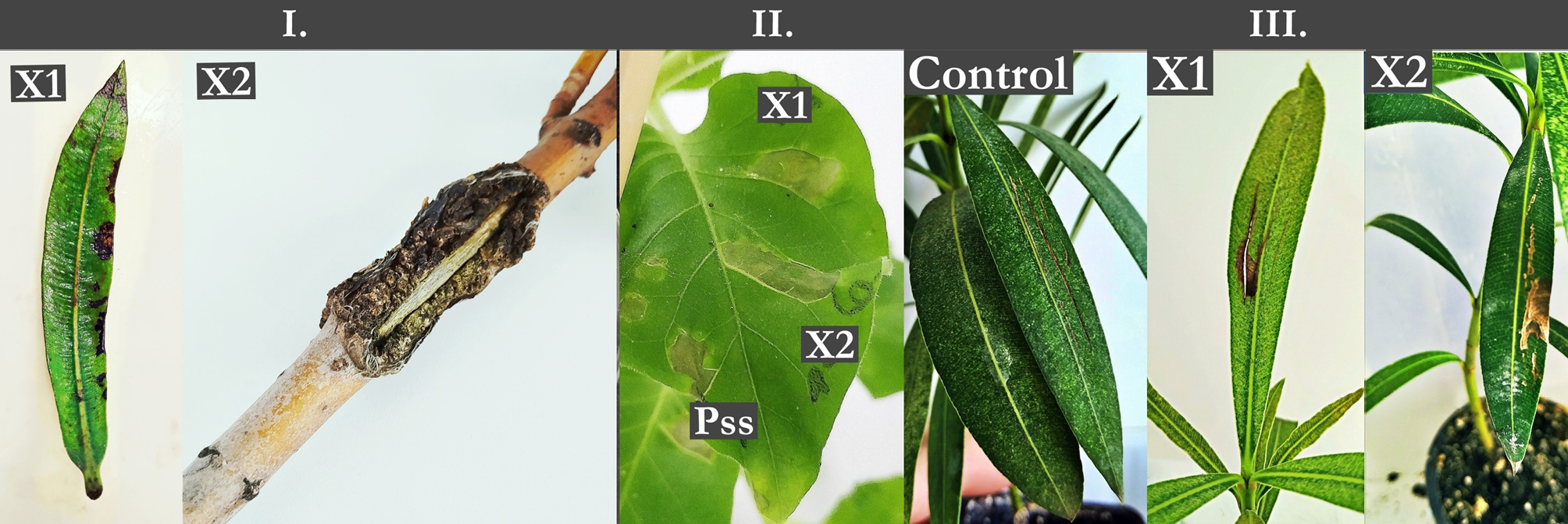
Between 2018 and 2022 we collected several infected oleander plant parts in Hungary. First of all, we brought into focus Pseudomonas savastanoi pv. nerii, however other pathogens (among others Botrytis cinerea, Cercospora spp.) were also investigated. In 2020 we observed two atypical symptoms, one from Southern Great Plain (Kecs-kemét) and one from Northern Great Plain (Nyírbátor). On the stem splitting of the canker bark were observed, which was not typical symptoms of oleander canker. In addition, there was no isolation of Pseudomonas spp. from the sample. On the leaf margins and main stem of the leaf scattered brown spots and watery necrosis were observed (Figure 1). It was assumed that the symptoms were caused by bacteria, which are unknown pathogens of oleander. Our previous research confirmed this hypothesis, as we isolated for the first time the pathogen Serratia marcescens bacteria on oleander leaf and seedcase (Fodor et al., 2022). The colonies of both isolates (X1, X2) were yellow-colored, smooth-edged, slightly convex on King-B and on YDC agar. The KOH tests were positive, so both isolates were Gram-negative. The isolates induced HR on tobacco leaves 24 h post-inoculation (Kennedy and Bradshaw, 1984; Trébaol, et al., 2000; Vicente and Holub, 2013) (Figure 1).
There was little variation in the biochemical properties of the isolates. The API 20NE test gave a positive reaction during sixteen reactions (NO3, GLU, ESC, GEL, PNPG, GLU, ARA, MNE, MAN, NAG, MAL, GNT, CAP, ADI, MLT, CIT). Only the production of arginine dihydrolase (ADH) showed differences (X1-negative, X2-positive) (Table 3). On the API 50CH kit the isolates differed in eight tests (DXYL, ARB, SAL, CEL, MEL, TRE, GLGY, XLT). The X2 isolate showed positive results on D- Xylose, arbutin, cellobiose melibiose, trehalose, glycogen, xylitol, while the X1 isolate showed negative results. On salicin and xylitol tests of the isolate X2 showed a positive reaction, while X1 showed a negative reaction (Table 4). Vauterin and co-authors (1995) studied the biochemical properties of two X. populi strains. Our isolates show difference from their isolates in arabinose (ARA) utilization (positive), while in 11 reaction (NAG, CEL, GAL, MAN, TRE, SOR, TUR, XLT, GLY) our isolates were differed their isolates by half and half. The result X. populi of López and co-authors (2018) showed differences only in mannitol (MAN) assimilation.
| NO3 | TRP | GLU | ADH | URE | ESC | GEL | PNPG | GLU | ARA | |
|---|---|---|---|---|---|---|---|---|---|---|
| X1 | + | - | + | + | - | + | + | + | + | + |
| X2 | + | - | + | - | - | + | + | + | + | + |
| MNE | MAN | NAG | MAL | GNT | CAP | ADI | MLT | CIT | PAC | |
| X1 | + | + | + | + | + | + | + | + | + | - |
| X2 | + | + | + | + | + | + | + | + | + | - |
| 0 | GLY | ERY | DARA | LARA | RIB | DXYL | LXYL | ADO | MDX | GAL | |
|---|---|---|---|---|---|---|---|---|---|---|---|
| X1 | - | - | - | - | - | - | - | - | - | - | + |
| X2 | - | - | - | - | - | - | + | - | - | - | + |
| GLU | FRU | MNE | SBE | RHA | DUL | INO | MAN | SOR | MDM | MDG | |
| X1 | + | + | + | - | - | - | - | - | - | - | - |
| X2 | + | + | + | - | - | - | - | - | - | - | - |
| NAG | AMY | ARB | ESC | SAL | CEL | MAL | LAC | MEL | SAC | TRE | |
| X1 | + | + | - | - | + | - | + | - | - | + | - |
| X2 | + | + | + | - | - | + | + | - | + | + | + |
| INU | MLZ | RAF | AMD | GLGY | XLT | GEN | TUR | LYX | TAG | DFUC | |
| X1 | - | - | - | + | - | + | - | - | - | - | + |
| X2 | - | - | - | + | + | - | - | - | - | - | + |
| LFUC | DARL | LARL | GNT | 2KG | 5KG | ||||||
| X1 | + | - | - | - | - | - | |||||
| X2 | + | - | - | - | - | - | |||||
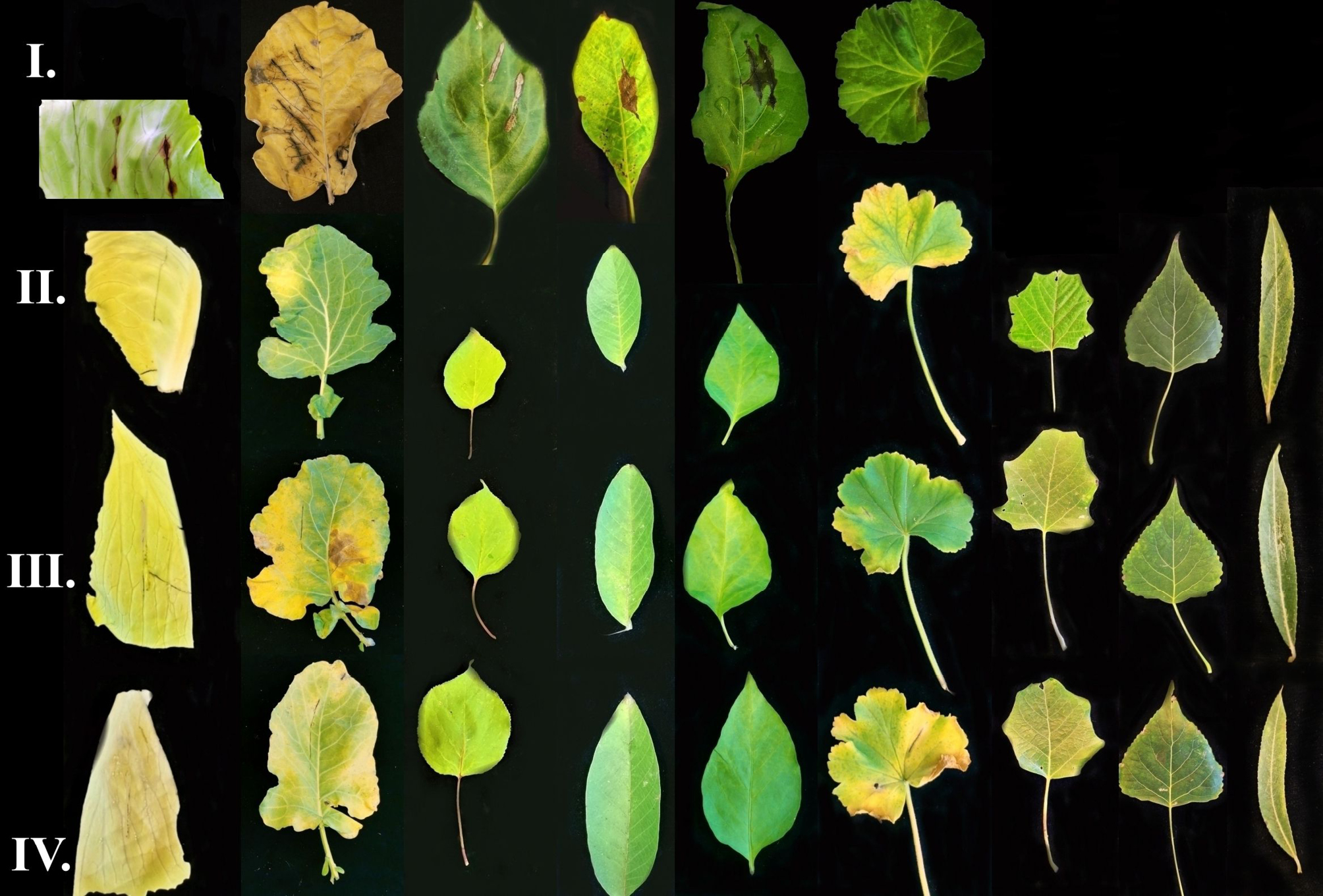
Two weeks after inoculation, necrosis was observed on the oleander leaves around the injection points. Control showed no symptoms. Although the isolated bacteria did not cause knots. It is assumed that the X2 isolate did not produce tumour. The pathogen of interest was the only microorganism re-isolated from lesions on the different inoculated plants, which was confirmed by colony morphology and PCR. The Koch’s postulates were fulfilled only in the case of X1 isolate. The X2 isolate were also pathogen of oleander, however the symptoms experienced during isolation were not observed during pathogenicity test (Figure 1). Leaves of apricot, walnut, pepper, kohlrabi, cabbage, geranium, poplars and willow did not show symptoms. There was no difference between infected and negative control leaves. However, the positive control isolates caused necrosis around the inoculation points (Figure 2).
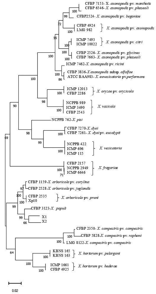
The phylogenetic tree was constructed using fyuA, rpoD, gyrB housekeeping genes, which was separated two branches (Figure 3). On one branch X. oryzae, X. pisi, X. dey, X. vesicatoria, X. fragariae and pathotypes of X. axonopodis were separated. On the other branch, X. arboricola and the pathotypes of X. campestris, X. hortorum and X. populi were found. The X1 and X2 isolate were on a separate branch with pathotype of X. arboricola (X arboricola pv. corylina, X. arboricola pv. juglandis, X. arboricola pv. pruni) and X. populi. Both isolates were similar and related to a Xanthomonas arboricola pv. pruni isolate from Montenegro (Xp10). They were on the same branch with X. populi (CFBP 3123). However, our isolates were different. They did not cause symptoms on Populus spp. and Salix spp. leaves, which plants are the only host of X. populi (De Kam, 1984). This indicates that our isolates may belong to a different pathotype, which needs to be confirmed by further studies- e.g. molecular testing, DNS-DNS hybridization, fatty acid analysis. To the best of our knowledge, this is the first report of the Xanthomonas as a pathogen of oleander. Additionally, a Xanthomonas spp. have been identified from olive (Olea europaea L), which caused brown necrosis and canker on the stream (Taylor et al., 2001; Young et al., 2010). Our isolates caused only on the oleander leaves necrosis in the pathogenicity test. They did not induce canker, although they may play a role in the evolve of on oleander knot symptoms.
Acknowledgement
This work was supported by the ELKH TKI (project number: 3200107). The research was funded by the “VP4-10.2.2.-20.Ex situ conservation of genetic resources of rare and endangered plant species and microorganisms”.
References
Backer A., Katzen F., Puhler L., Ielpi L., 1998. Xanthan gum biosynthesis and application: a biochemical/genetic perspective. Applied Microbiology and Biotechnology 50: 145–152. https://doi.org/10.1007/s002530051269
Bradbury J.F., 1984. Genus II Xanthomonas (Dowson 1939). In: Bergey’s Manual of Systematic Bacteriology (Krieg, N.R., Holt, J.G. ed) vol. 1. The Williams & Wilkins Co., Baltimore, 199-210.
Bull C.T., Koike S.T., 2015. Practical benefits of knowing the enemy: modern molecular tools for diagnosing the etiology of bacterial diseases and understanding the taxonomy and diversity of plant-pathogenic bacteria. Annual Review of Phytopathology 53: 157–180. https://doi.org/10.1146/annurev-phyto-080614-120122
De Kam M., 1984. Xanthomonas campestris pv. populi, the causal agent of bark necrosis in poplar. Netherlands Journal of Plant Pathology 90: 13–22. https://doi.org/10.1007/BF02014178
Fodor A., Palkovics L., Végh A., 2022. First report of Serratia marcescens from oleander in Hungary. Phytopathologia Mediterranea 61: 311–317. https://doi.org/10.36253/phyto-13354
Kennedy J.F., Bradshaw I.J. 1984. Production, properties and applications of xanthan. Progress in Industrial Microbiology 19, 319–371.
King E.O., Ward M.K., Raney D.E., 1954. Two simple media for the demonstration of pyocyanin and fluorescin. Journal of Laboratory and Clinical Medicine 44: 301–307.
López M.M., Lopez-Soriano P., Garita-Cambronero J., Beltrán C., Taghouti G., … Marco-Noales E., 2018. Xanthomonas prunicola sp. nov., a novel pathogen that affects nectarine (Prunus persica var. nectarina) trees. International Journal of Systematic and Evolutionary Microbiology 68(6): 1857–1866. https://doi.org/10.1099/ijsem.0.002743
Osborn A.M., Moore E.R., Timmis K.N., 2000. An evaluation of terminal-restriction fragment length polymorphism (T-RFLP) analysis for the study of microbial community structure and dynamics. Environmental Microbiology 2: 39–50. https://doi.org/10.1046/j.14622920.2000.00081.x
Parkinson N., Aritua V., Heeney J., Cowie C., Bew J., Stead D., 2007. Phylogenetic analysis of Xanthomonas species by comparison of partial gyrase B gene sequences. International Journal of Systematic and Evolutionary Microbiology 57(12): 2881–2887. https://doi.org/10.1099/ijs.0.65220-0
Parte A.C., Carbasse S.J., Meier-Kolthoff J.P., Reimer L.C., Göker M., 2020. List of Prokaryotic names with Standing in Nomenclature (LPSN) moves to the DSMZ. International Journal of Systematic and Evolutionary Microbiology 70(11): 5607–5612. https://doi.org/10.1099/ijsem.0.004332
Powers E.M., 1995. Efficacy of the Ryu nonstaining KOH technique for rapidly determining gram reactions of food-borne and waterborne bacteria and yeasts. Applied and Environmental Microbiology 61: 3756–3758. https://doi.org/10.1128/aem.61.10.3756-3758.1995
Rudolph K., 1993. Infection of the plant by Xanthomonas. In: Xanthomonas (Swings J. G., Civerolo E. L. ed) Chapman and Hall, London. 193–264.
Ryan R.P., Vorhölter F.J., Potnis N., Jones J.B., Van Sluys M.A., … Dow, J.M. 2011. Pathogenomics of Xanthomonas: understanding bacterium-plant interactions. Nature Reviews Microbiology 9(5): 344–355. https://doi.org/10.1038/nrmicro2558
Smith C.O. ,1906. A Bacterial disease of oleander. Bacillus oleae (Arcang.) Trev. Botanical Gazette, 42(4): 301–310.
Tamura K., Nei M., 1993. Estimation of the number of nucleotide substitutions in the control region of mitochondrial DNA in humans and chimpanzees. Molecular Biology and Evolution 10(3): 512–526. https://doi.org/10.1093/oxfordjournals.molbev.a040023
Tamura K., Stecher G., Kumar S., 2021. MEGA 11: Molecular Evolutionary Genetics Analysis Version 11. Molecular Biology and Evolution. 38(7): 3022–3027. https://doi.org/10.1093/molbev/msab120
Taylor R.K., Hale C.N., Hartill, W.F.T., 2001. A stem canker disease of olive (Olea europaea) in New Zealand. New Zealand Journal of Crop and Horticultural Science 29(3): 219–228. https://doi.org/10.1080/01140671.2001.9514181
Temsah M., Hanna L., Saad A.T., 2010. Histological pathogenesis of Pseudomonas savastanoi on Nerium oleander. Journal of Plant Pathology 92(2): 407–413.
Trébaol G., Gardan L., Manceau C., Tanguy J. L., Tirilly Y., Boury S., 2000. Genomic and phenotypic characterization of Xanthomonas cynarae sp. nov., a new species that causes bacterial bract spot of artichoke (Cynara scolymus L.). International Journal of Systematic and Evolutionary Microbiology 50(4): 1471–1478.
Vauterin L., Hoste B., Kersters K., Swings J., 1995. Reclassification of Xanthomonas. International Journal of Systematic and Evolutionary Microbiology 45(3): 472–489. https://doi.org/10.1099/00207713-45-3-472
Vicente J.G., Holub E.B., 2013. Xanthomonas campestris pv. campestris (cause of black rot of crucifers) in the genomic era is still a worldwide threat to brassica crops. Molecular Plant Pathology 14(1): 2–18. https://doi.org/10.1111/j.1364-3703.2012.00833.x
Young J.M., Park, D.C., Shearman H.M., Fargier E., 2008. A multilocus sequence analysis of the genus Xanthomonas. Systematic and Applied Microbiology 31(5): 366–377. https://doi.org/10.1016/j.syapm.2008.06.004
Young J.M., Wilkie J.P., Park D.C., Watson D.R.W., 2010. New Zealand strains of plant pathogenic bacteria classified by multi‐locus sequence analysis; proposal of Xanthomonas dyei sp. nov. Plant Pathology 59(2): 270–281. https://doi.org/10.1111/j.1365-3059.2009.02210.x