https://doi.org/10.36253/phyto-15328
Accepted: April 24, 2025; Published: May 15, 2025
Research Papers
Isolation and identification of Fusarium spp. associated with Fusarium wilt of chickpea (Cicer arietinum L.) in Algeria
1 Plant Protection Laboratory, Abdelhamid Ibn Badis University of Mostaganem, 27000 – Algeria
2 Laboratory protection, Valorization of Coastal Marine Resources and Molecular Systematics, Abdelhamid Ibn Badis University of Mostaganem, 27000 – Algeria
3 Animal and Applied Physiology Laboratory, Abdelhamid Ibn Badis University of Mostaganem, 27000 – Algeria
4 Research Center of Biodiversity and Ecology, UMR5300, CNRS- Paul Sabatier University- National Polytechnic Institute INP-ENSAT, Avenue de l’Agrobiopole 31326 Castanet-Tolosan, France
*Corresponding author. E-mail: djamel.mahiout@univ-mosta.dz
Summary. Chickpea (Cicer arietinum L.) is an important vegetable crop in many Mediterranean countries, and Fusarium is known to cause wilt in these crops. Fusarium oxysporum f. sp. ciceris and Fusarium redolens are the only species which have been reported as the causes of Fusarium wilt in Algeria. Fusarium isolates (74) were obtained from roots of chickpea plants showing symptoms of wilting and necroses in xylem tissues. The plants were from 11 eleven principal provinces for chickpea crops in Algeria. Laboratory identifications for 31 isolates were achieved by sequencing their transcription elongation Factor 1- α (Ef-1 α) gene regions. Four principal species were identified including F. oxysporum (20 isolates), F. redolens (ten isolates), F. solani (one isolate) based on PCR using species-specific primers, and three formae speciales were identified within Fusarium oxysporum. Fusarium redolens and F. oxysporum f. sp. ciceris were dominant in the sampling sites. Fusarium redolens was identified in the western region of Algeria, and F. oxysporum f. sp. ciceris was not isolated from this region. In contrast, from the eastern region, F. redolens was not isolated but F. oxysporum f. sp. ciceris was detected. Pathogenicity tests showed that chickpea was susceptible to various Fusarium species. Inoculated plants exhibited gradual wilting that eventually affected entire plants. This study provides new knowledge of the distribution of Fusarium species causing chickpea wilt in Algeria. Precise identification and the localization of pathogen species is important for developing Fusarium wilt management strategies, including development of wilt resistant chickpea cultivars.
Keyswords. Koch’s postulates, transcription elongation factor 1-α gene.
Abbreviations: FR: Fusarium redolens; FS: Fusarium solani; FO: Fusarium oxysporum; FOC: Fusarium oxysporum f. sp. ciceris; FOL: Fusarium oxysporum f. sp. lactuceae. Transcription elongation Factor 1-α gene (EF-1α).
Contents
Plant sampling, and isolation and maintenance of fungi
Molecular characterization of Fusarium isolates
Assessment of Koch’s postulates for representative fungi
Introduction
Chickpea (Cicer arietinum L.) is an important pulse crop for human nutrition, and is a major component of food production systems that are resilient to climate change. Chickpea is a preferred food legumes (Siddique et al., 2000) in some regions because of its multiple uses. The high protein content of the seeds (approx. 40% of seed weight), has potential human health benefits, including reduced risk of diabetes, cardiovascular disease, and cancer (Merga and Haji, 2019). Chickpea also contributes to soil fertility by fixing atmospheric nitrogen in the soil through symbiosis with rhizobia, which is especially important in dry climate areas. However, chickpea crops are often affected by telluric diseases such as Fusarium wilt, Verticillium wilt and those caused by Sclerotinia sp. and Rhizoctonia sp. (Nene et al., 2012).
Fusarium wilt is now widespread in most chickpea production areas of Africa, southern Europe, the Americas, and Asia. This disease is caused by several Fusarium species, and accurate and rapid identification of the responsible pathogens is important for development of appropriate and efficient management of Fusarium wilt of chickpea (Tekeoğlu et al., 2017). In Algeria, Fusarium wilt of chickpea has been attributed to Fusarium oxysporum f. sp. ciceris (FOC), but in January 2022 Zaim and Bekkar (2022) reported a second pathogen, F. redolens (FR), causing this disease in the western region of the country.
Formal identification of the pathogen responsible for chickpea-wilt in Algeria was made using classical methods, including morphological characteristics and fulfilment of Koch’s postulates. Fusarium oxysporum (FO) was defined by morphological criteria, including the shape of the microconidia, macroconidia and conidiophore structure (false head on short phialides formed on hyphae). This morphological identification is problematic, however, because of the diversity of non-pathogenic and saprophytic isolates in soil. Fusarium isolates are usually tested on the chickpea ILC-482 genotype, which is susceptible to all races of F. oxysporum f. sp. ciceris (Jimenez-Diaz et al., 1989), and is supposed to confirm the identity of the pathogen.
Identification of Fusarium spp. based only on morphology is difficult, because informative morphological characteristics are limited, and microscopic traits can be influenced by environmental conditions, so that their plasticity and intergradation make them subject to misinterpretation (Leslie et al., 2001). For example, distinguishing F. redolens from F. oxysporum can be particularly challenging, because this relies on differences in size of their macroconidia (Gordon, 1952), and intermediate conidium forms may occur (Baayen and Gams, 1988). For these reasons, the taxonomic position of F. redolens has been problematic. Whereas Booth (1971) considered this fungus to be a variety of F. oxysporum, Nelson et al., (1983) considered F. redolens and F. oxysporum to be synonymous. The use of DNA-based methodologies now makes it possible to differentiate between different species of Fusarium (Jimenez-Gasco and Jimenez-Diaz., 2003; Jiménez-Fernandez et al., 2011).
During spring of 2020 and 2021, the present study investigated occurrence of Fusarium wilt of chickpea in the north of Algeria. Samples were collected from eleven provinces. Wilting and yellowing symptoms were observed on aerial parts of diseased chickpea plants, and 74 fungal isolates were obtained from roots. To identify these isolates, their translation elongation factor 1-alpha (EF-1α) gene regions were sequenced.
Materials and methods
Plant sampling, and isolation and maintenance of fungi
During the chickpea growing seasons of spring 2020 and 2021, chickpea plants with yellowing and wilting symptoms on their areal parts were collected from 23 farmer fields in different areas in north western and eastern Algeria. These areas were in to 11 provinces, five in the western region and six in the eastern region. The major chickpea sowing method in the west is strip sowing, whereas in the east it is broadcast sowing.
Tissue pieces (5 × 5 mm) were taken from diseased tap root tissues of wilted chickpea plants, and were surface-sterilized in 2% sodium hypochlorite for 30 sec, then rinsed three times in sterile distilled water, and dried on sterile paper. Samples were then plated on potato dextrose agar (PDA), and incubated at 27°C in the dark for 7 d. Growing tips of hyphae developing from in cultures were then transferred onto fresh PDA. Monoconidial isolates were prepared from cultures of fungi obtained from each region, and cultures were stored at 4°C.
A total of 74 Fusarium sp. isolates were obtained, of which 31 were sequenced using the EF-1α gene. The 31 isolates were chosen based on macroscopic characteristics (colony colour and appearance on PDA). Two to four representative isolates of the morphotypes from each province were considered.
DNA extractions
The selected fungal cultures were grown on PDA 25 ± 2°C in the dark for 7 d. For each extraction, 100 mg of mycelium was collected by scraping the colonies from three plates per isolate. DNA of each isolate was extracted using a commercial NucleoSpin Plant II kit (Macherey-Nagel), following the supplier’s instructions. The quality and concentration of extracted DNA were verified after 1% agarose gel electrophoresis, using a NanoDrop spectrophotometer (at 260/280 nm).
Molecular characterization of Fusarium isolates
The translation elongation factor-1alpha (EF-1α) gene region was amplified by PCR, using the universal primers EF-728F (CATYGAGAAGTTCGAGAAGG) and EF-2 (GGARGTACCAGTSATCATGTT) (Carbone and Kohn, 1999), in a 25 μL reaction volume containing 1.5 mM MgCl2 (Promega), 0.2mM dNTPs (Invitrogen), 2 μg mL-1 fungal DNA, 0.5µM primers each, 1× Taq Buffer (Promega), and 1-unit Taq polymerase (Promega).
Amplifications were carried out in a thermal cycler (Icycler Bio-Rad), with an initial denaturation step for 5 min at 95°C, followed by 35 cycles each of 30 sec of denaturation at 95°C, 30 sec at annealing temperature (52°C), and 45 sec at 72°C, and an extension step of 7 min at 72°C. Amplification products were separated by electrophoresis on a 1.5% agarose gels. The gels were stained with MidoriGreen (Nippon), and were visualized under UV light by using the Gel doc system (Biorad). The size marker was a 100 bp DNA ladder (Invitrogen). The PCR products were purified with a NucleoSpin® Gel and a PCR Clean-up kit (Macherey-Nagel), following the manufacturer’s instructions. PCR products of the EF-1α regions were sequenced by Sanger sequencing (Sanger et al., 1977) directly from the PCR products, without a cloning step, using Applied Biosystems BigDye Kit v3.1 and the primers used for PCR amplification of the fragments. Sequencing was performed in the direction (forward/reverse) for each amplicon. Sequencing was carried out by Gene Life Sciences.
The sequences obtained were analyzed and cleaned using the CHROMAS PRO software, and were then compared with those from the GenBank database using the BLAST Program (https://blast.ncbi.nlm.nih.gov/Blast.cgi Blast) to identify the isolates based on percentage homology with reference strains. A dendrogram was established using the UPGMA method algorithm to show the genetic diversity of the isolated fungi.
Assessment of Koch’s postulates for representative fungi
To verify the pathogenicity of Fusarium spp. isolates, five species [F. redolens (FR), F. solani (FS), F. oxysporum (FO), Fusarium oxysporum f. sp. ciceris (FOC), and F. oxysporum f. sp. lactuceae (FOL)] were inoculated individually onto plants of cultivar ILC-482 from ICARDA. The seeds were surface sterilized, and then pre-germinated on moist cotton wool, which provided favorable conditions for seedling germination (ISTA, 2015; Korter et al., 2023).
The pre-germinated seeds were then sown in plastic pots (30 × 20 cm) containing a sterile mixture of potting soil and sand (1:1 v:v). For each Fusarium sp., three pots each containing four plants were used. Seven d after sowing (3–4 leaf stage), the plants were removed from soil and their root systems were soaked for 30 min in a conidial suspension (106 conidia mL-1 (Gao et al., 1995; Ficcadenti et al., 2002). Five pots were used as controls, where the plants were soaked in sterile water without inoculum.
Results and Discussion
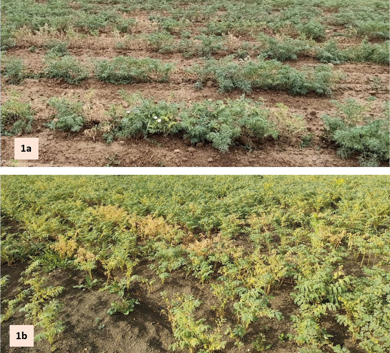
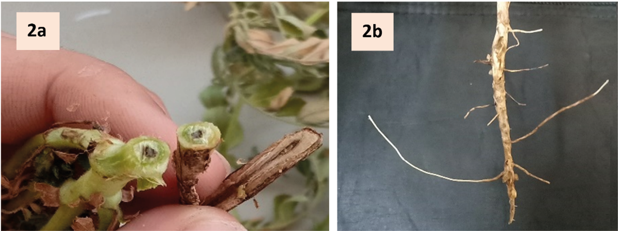
In the assayed chickpea fields, diseased plants were spread throughout each field, with incidence up to 30% (Figure 1, a and b). In addition to wilting and yellowing symptoms, the stems and root xylem tissues of the diseased plants had black discolourations (Figure 2 a). Brown necrotic lesions in the tap roots and necroses of lateral roots (Figure 2 b) also occurred on most of the sampled plants from the west of Algeria. The same symptoms were observed by Jimenez-Fernandez et al. (2011).
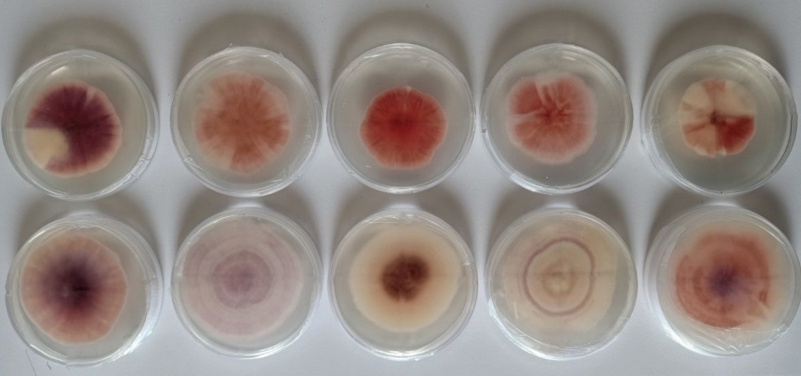
On PDA, colonies of the isolates had different morphotypes (Figure 3), with differences in colour, growth rates, and aspect of mycelium. All colonies initially had white to cream-coloured mycelium, which turned (after 3 to 7 d incubation) to purple and pink for isolates of F. redolens, purple tinged or slightly orange for F. solani, or cream or purple for F. oxysporum f. sp. ciceris and F. oxysporum. The mycelium texture was fluffy or appressed.
Microscopic observations of most of the isolates revealed septate mycelium which carried phialides bearing false-headed conidia (Figure 4).
The morphological and cultural characteristics on PDA identified the isolates as Fusarium sp., based on the descriptions of Booth (1977).
BLASTn sequence analyses with (https://blast.ncbi.nlm.nih.gov/Blast.cgi) identified the isolates as follows: ten isolates were F. redolens, six were F. oxysporum f. sp. ciceris, nine were F. oxysporum, five were F. oxysporum f. sp. Lactuceae, and one isolate was F. solani (Ascomycota, Pezizomycotina, Sordariomycetes, Hypocreomycetidae, Hypocreales, Nectriaceae) with 94.85 to 100% sequence similarity to the sequences of type isolates from Genbank.
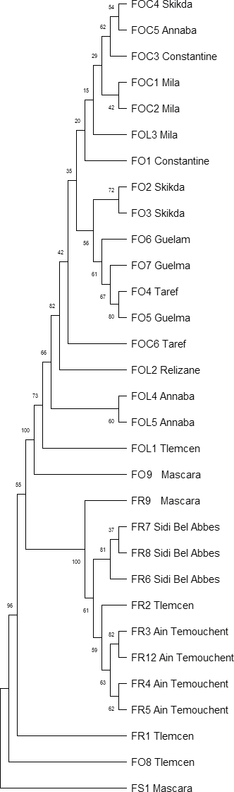
A dendrogram was constructed based on the EF-1α sequences, using the UPGMA method (Sneath and Sokal, 1973) to show phylogenetic relationships among the Fusarium isolates obtained from different regions of Algeria (Figure 5). The branches of the dendrogram were supported by bootstrap values, demonstrating the robustness of the phylogenetic relationships among these isolates. For 15 isolates from the western region, ten isolates (67%) were FR, four (27%) were FO of one forma specialis (F. oxysporum f. sp. lactuceae), and one isolate (6.7%) was FS. In contrast, among the 16 isolates from the eastern region of Algeria, all were FO, in which two formae speciales were identified, six of FOC, and three of FOL (Table 1).
| Region | Province | Code | Identification | Field coordinates | Percentage similarity | Blast Best Hit ID |
Accession number NCBI |
|---|---|---|---|---|---|---|---|
| Western region | Tlemcen | FOL1 | Fusarium oxysporum f. sp. lactuceae | 35°09’54.3”N+1°26’50.7”W | 98.64 | MK801786.1 | PQ899234 |
| FR1 | Fusarium redolens | 34°56’42.5”N+1°14’59.6”W | 94.85 | HQ731060.1 | PV388859 | ||
| FR2 | Fusarium redolens | 99.75 | HQ731060.1 | PV388860 | |||
| FO8 | Fusarium oxysporum | 35°09’54.3”N+1°26’50.7”W | 100.00 | ON703236.1 | PQ899228 | ||
| Ain Temouchent | FR12 | Fusarium redolens | 35°13’56.7”N+1°20’50.5”W | 99.16 | HQ731063.1 | PV388869 | |
| FR3 | Fusarium redolens | 35°13’56.7”N+1°20’50.5”W | 99.58 | MT305223.1 | PV388861 | ||
| FR4 | Fusarium redolens | 35°16’36.2”N+1°13’43.1”W | 99.58 | OR270920.1 | PV388862 | ||
| FR5 | Fusarium redolens | 35°17’43.2”N+1°05’10.6”W | 99.57 | OR105855.1 | PV388863 | ||
| Sidi Bel Abbes | FR6 | Fusarium redolens | 35°14’18.9”N+0°43’51.5”W | 99.79 | OR270920.1 | PV388864 | |
| FR7 | Fusarium redolens | 35°16’15.8”N+0°35’04.5”W | 99.79 | OR270920.1 | PV388865 | ||
| FR8 | Fusarium redolens | 35°23’25.0”N+0°29’03.4”W | 99.85 | MK172061.1 | PV388866 | ||
| Mascara | FR9 | Fusarium redolens | 35°26’56.5”N+0°08’48.9”E | 99.57 | HQ731060.1 | PV388867 | |
| FS1 | Fusarium solani | 35°24’23.9”N+0°18’27.0”E | 99.57 | HE647956.1 | OR234389 | ||
| FO9 | Fusarium oxysporum | 97.53 | MW361989.1 | - | |||
| Relizane | FOL2 | Fusarium oxysporum f. sp. lactuceae | 35°59’58.4”N+0°52’33.9”E | 99.35 | MH412703.1 | PQ899235 | |
| Eastern region | Mila | FOC1 | Fusarium oxysporum f. sp. ciceris | 36°30’22.9”N+6°16’35.1”E | 99.55 | FJ538240.1 | PQ899229 |
| FOC2 | Fusarium oxysporum f. sp. ciceris | 36°27’54.3”N+6°13’21.8”E | 100.00 | FJ538240.1 | PQ899230 | ||
| FOL3 | Fusarium oxysporum f. sp. lactuceae | 36°27’21.7”N+6°12’51.0”E | 99.85 | OP918954.1 | PQ899236 | ||
| Constantine | FOC3 | Fusarium oxysporum f. sp. ciceris | 36°17’58.0”N+6°38’41.8”E | 100.00 | FJ538241.1 | PQ899231 | |
| FO1 | Fusarium oxysporum | 36.294952, 6.641571 | 98.46 | PP795998.1 | PQ613583 | ||
| Skikda | FOC4 | Fusarium oxysporum f. sp. ciceris | 36°43’22.0”N+6°51’29.3”E | 100.00 | FJ538241.1 | PQ899232 | |
| FO2 | Fusarium oxysporum | 36°42’43.2”N+6°47’47.8”E | 100.00 | OQ511027.1 | PQ613583 | ||
| FO3 | Fusarium oxysporum | 99.85 | OQ511027.1 | PQ899231 | |||
| Annaba | FOC5 | Fusarium oxysporum f. sp. ciceris | 36°46’38.1”N+7°44’18.4”E | 98.85 | FJ538241.1 | PQ899233 | |
| FOL4 | Fusarium oxysporum f. sp. lactuceae | 99.57 | MW316853.1 | PQ899237 | |||
| FOL5 | Fusarium oxysporum f. sp. lactuceae | 99.78 | MW316854.1 | - | |||
| El Taref | FOC6 | Fusarium oxysporum f. sp. ciceris | 36°42’28.4”N+7°44’26.5”E | 98.81 | FJ538240.1 | - | |
| FO4 | Fusarium oxysporum | 99.85 | OQ511027.1 | PQ613586 | |||
| Guelma | FO5 | Fusarium oxysporum | 36°39’03.1”N+7°42’35.2”E | 99.85 | KF537337.1 | PQ613587 | |
| FO6 | Fusarium oxysporum | 100.00 | OQ511027.1 | PQ613588 | |||
| FO7 | Fusarium oxysporum | 99.85 | OQ511027.1 | PQ899227 |
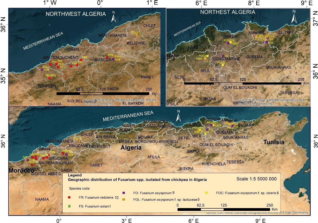
These results indicate that F. redolens is dominant in the western region of Algeria, while F. oxysporum prevails in the eastern region, where F. oxysporum f. sp. ciceris was the most frequently identified forma specialis among the isolates collected from the east. Fusarium oxysporum f. sp. ciceris was not isolated from the western region, and Fusarium redolens was not isolated from the eastern region of the country (Figure 6).
Koch’s postulates were confirmed by inoculating chickpea cultivar ILC-482 and re-isolating the fungi from the inoculated chickpea roots, which indicates that all the Fusarium isolates were infectious agents of diseased plants sampled from the chickpea fields. These results show that chickpea culture can be susceptible to different Fusarium species, and even to FOL that has been previously shown to be a pathogen of lettuce. All the inoculated plants developed progressive wilting. Although formae speciales of F. oxysporum are mainly described as highly specific, their host ranges have been found to increase. For example, only 53 of the 106 formae speciales listed by Edel-Hermann and Lecomte (2019) remain associated with a unique plant species. Similarly, Ramirez-Suero et al. (2010) showed susceptibility of Medicago truncatula to several F. oxysporum formae speciales, even from non-legume hosts.
In the present study, wilting symptoms appeared on inoculated plants at 21 d after inoculation for all of the Fusarium isolates. Leaves at the base of plants initially showed marginal yellowing before wilting (Figure 7). The symptoms then developed progressively towards the upper leaves, and most rapidly in plants inoculated with F. oxysporum f. sp. ciceris and F. redolens. Roots of inoculated plants had reduced root hairs and brown/black discolouration, which extended to the plant collars and sometimes caused collar narrowing (Figure 7b).
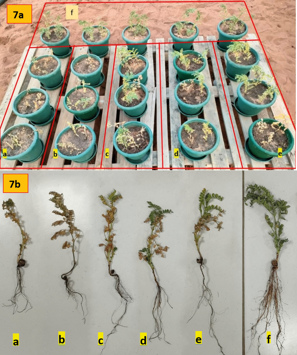
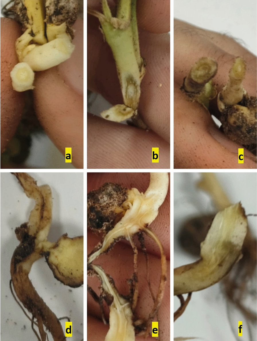
Longitudinal sections of primary roots of the diseased plants had dark vascular necroses, which were more or less dark according to the inoculated Fusarium isolates. Root xylem necroses were sometimes accompanied by wet rots, where plants were inoculated with F. solani or F. redolens (Figure 8). All non-inoculated (control) plants remained healthy. Symptoms on roots and leaves were more severe on plants inoculated with isolates of FOC or FR than on those inoculated with the other isolates (Figure 7, a and b).
To date, chickpea wilt in the western region of Algeria has been attributed to Fusarium oxysporum f. sp. ciceris, using morphological characteristics of the pathogenic fungi (Benfreha et al., 2014) or Koch’s postulates for the chickpea ILC-482 genotype (Tlemsani et al., 2015; Zaim, 2016). The present study indicates that FOC does not exist in the western region of Algeria. Using ITS and EF-1α, Zaim and Bekkar (2022) identified only Fusarium redolens of 20 Fusarium isolates from chickpea in the western region of this country. The present study further confirms the absence of FOC, since among the 15 Fusarium isolates that were obtained from the western region, ten were F. redolens, and the remaining five were FS (one isolate), FO (two isolate), and FOL (two isolates). Although only 31 of 74 isolates were sequenced in the present study, these results and those of Zaim and Bekkar (2022) confirm that FOC is absent from western Algeria.
Fusarium redolens has been reported to cause wilt of tomato (Edel-Hermann et al., 2012), and damping-off of Aleppo pine (Pinus halepensis) (Lazreg et al., 2013) in the North west of Algeria. Association of F. redolens with wilt symptoms of chickpea has been reported in Morocco, Spain, Lebanon, and Pakistan (Jimenez-Fernandez et al., 2011), Saskatchewan (Taheri, 2011), Tunisia (Bouhadida et al., 2017), Turkey (Tekeoğlu et al., 2017), and Iran (Chehriand Sattar, 2018).
The present study isolated F. solani from the western region of Algeria. This fungus was previously reported to infect chickpea in China, India, Spain, Pakistan, Iran, and other countries (Zhuang et al., 2005; Jiménez-Fernández et al.,2011). In Algeria, F. solani has been reported to cause wilt of potato (Solanum tuberosum) (Azil et al., 2021) and tomato (Solanum lycopersicum) (Abdesselem et al., 2016), as well as damping-off of Aleppo pine (Pinus halepensis) (Lazreg et al., 2014). Therefore, the present study is the first to show that F. solani is a chickpea pathogen in Algeria.
While F. redolens and F. oxysporum f. sp. ciceris are known to cause yellowing and wilt of chickpea, the reasons for their distinct geographic distributions in Algeria remains unclear. Several factors could explain this separation of the pathogens, including variations in climate, soil structure, and cropping systems. Further research is required to confirm these potential influences.
It is well-documented that plant pathogen prevalence can be linked to climatic conditions, such as temperature and rainfall, which differ across geographic regions. For example, F. graminearum tends to dominate in warm areas such as parts of the United States of America, while F. culmorum is common in cool, maritime regions such as the United Kingdom (Parry et al., 1995; Doohan et al., 2003). Similarly, optimal temperatures and moisture levels are critical for Fusarium sporulation and inoculum dispersal (Rossi et al., 2001), suggesting that local environmental factors may play a role in the distributions of FR and FOC.
Soil characteristics, including moisture and pH, also affect Fusarium spp. growth and resulting host disease development. Previous studies have shown that soil pH can influence Fusarium growth rates and expression of wilt symptoms (Chen et al., 2013; Gatch and du Toit, 2017). Additionally, resistance to Fusarium wilt has been observed in particular soils, with microbiological interactions such as competition for iron and/or carbon playing important roles (Elad and Baker, 1985b; Couteaudier and Alabouvette, 1990).
To fully understand the geographic distributions of FOC and FR, further research is required on effects on the fungi of soil physicochemical properties, microbial communities, and agricultural practices. It remains that FS and FOC could be serious future threats to chickpea cultivation in Algeria. It would also be worthwhile to test the aggressiveness of Fusarium spp. on different chickpea cultivars to understand whether those isolates have cultivar specificities.
The use of morphological traits to differentiate morphologically similar Fusarium spp., including F. oxysporum and F. redolens, may lead to incorrect pathogen identifications. However, accurate identification of pathogenic Fusarium spp. and F. oxysporum formae speciales is important for efficient management of diseases caused by these fungi, particularly when resistant cultivars are one of the few and most effective control measures for these diseases, as is the case for Fusarium wilt of chickpea (Nene and Reddy, 1987).
Identification of new pathogen species and races would help plant breeders to select appropriate germplasm for development of resistant crop cultivars. Identification of disease-causing Fusarium spp. may also help development of cultural practices for management of Fusarium wilt, which can be achieved with use of resistant cultivars and adjustment of sowing dates (Jiménez et al., 1991; Jalali and Chand, 1992; Jiménez et al., 1998; Landa et al., 2004; Navas-Cortés et al., 1998; Navas-Cortés et al., 2000), and appropriate crop rotations (Landa et al., 2006).
Literature cited
Abdesselem S.M., Nisserine H.K., Mebrouk K., Jamal E.H., Jos Eacute S., Eduardo G., 2016. Characterization of Fusarium oxysporum isolates from tomato plants in Algeria. African Journal of Microbiology Research 10, 1156–1163. https://doi.org/10.5897/ajmr2016.8161
Azil N., Stefanczyk E., Sobkowiak S., Chihat S., Boureghda H., Sliwka, J., 2021. Identification and pathogenicity of Fusarium spp. Associated with tuber dry rot and wilt of potato in Algeria. European Journal Plant Pathology 159, 495–509. https://doi.org/10.1007/s10658-020-02177-5
Baayen R.P., Gams W., 1988. The Elegans fusaria causing wilt disease of carnation. I. Taxonomy. Netherlands Journal of Plant Pathology 94: 273–288.
Benfreha F.Z., Henni D.E., Merzoug A., 2014. Fusarium wilt of Chickpea (Cicer arietinum L.) in Northwest Algeria. African Journal of Agricultural Research Vol. 9(1), 168-175, https://doi.org/10.5897/AJAR2013.6694
Booth C., 1971. The Genus Fusarium. Commonwealth Mycological Institute, Kew, Surrey, UK. Reprinted 1977, 237 pp.
Booth C., 1977. Fusarium Laboratory Guide to the Identification of the Major Species. Commonwealth Mycological Institute, Kew, Surrey, England, 58 pp.
Bouhadida M., Jendoubi W., Gargouri S., Beji M., Kharrat M., Chen W., 2017. First report of Fusarium redolens causing Fusarium yellowing and wilt of chickpea in Tunisia. Plant Disease 101. https://doi.org/10.1094/PDIS-08-16-1114-PDN
Carbone I., Kohn L.M., 1999. A method for designing primer sets for speciation studies in filamentous ascomycetes. Mycologia 91: 553–556. https://doi.org/10.2307/3761358
Chehri K.h., Sattar H.A., 2018. Detection of fumonisin chemotype produced by Fusarium proliferatum isolated from nuts in Iraq using specific PCR assays. Biological Journal of Microorganism 24: 21–27.
Chen L.H., Huang X.Q., Yang X.M., Shen Q.R., 2013. Modeling the effects of environmental factors on the population of Fusarium oxysporum in cucumber continuously cropped soil. Communications in Soil Science and Plant analysis 44: 2219–2232.
Couteaudier Y., Alabouvette C., 1990. Quantitative comparison of Fusarium oxysporum competitiveness in relation with carbon utilization. FEMS Microbiology Ecology 74: 261–268.
Doohan F.M., Brennan J., Cooke B.M., 2003. Influence of climatic factors on Fusarium species pathogenic to cereals. European Journal of Plant Pathology 109: 755–768. https://doi.org/10.1023/A:1026090626994
Edel-Hermann V., Gautheron N., Steinberg C., 2012. Genetic diversity of Fusarium oxysporum and related species pathogenic on tomato in Algeria and other Mediterranean countries. Plant Pathology 61: 787–800. https://doi.org/10.1111/j.1365-3059.2011.02551.x
Edel-Hermann V., Lecomte C., 2019. Current status of Fusarium oxysporum formae speciales and races. Phytopathology 109: 512–530. https://doi.org/10.1094/PHYTO-08-18-0320-RVW
Elad Y., Baker R., 1985b. The role of competition for iron and carbon in suppression of chlamydospores germination of Fusarium spp. by Pseudomonas spp. Phytopathology 75: 1053–1059.
Felsenstein J., 1985. Confidence limits on phylogenies: An approach using the bootstrap. Evolution 39: 783–791.
Ficcadenti N., Sestili S., Annibali S., Campanelli G., Belisario A., … Corazza L., 2002. Resistance to Fusarium oxysporum f. sp. melonis Race 1, 2 in Muskmelon Lines. Plant Disease, 86: 897–900.
Gao H., Beckman C.H., Mueller W.C., 1995. The rate of vascular colonisation as a measure of the genotypic interaction between various cultivars of tomato various formae or races of Fusarium oxysporum. Physiological Molecular Plant Pathology 46: 29–43.
Gatch E.W., du Toit L.J., 2017. Limestone-mediated suppression of Fusarium wilt in spinach seed crops. Plant Disease 101: 81–94. https://doi.org/10.1094/PDIS-04-16-0423-RE
Gordon W.L., 1952. The occurrence of Fusarium species in Canada, II. Prevalence and taxonomy of Fusarium species in cereal seeds. Canadian Journal of Botany 30: 236–238.
ISTA, 2015. International Rules for Seed Testing. International Seed Testing Association, Bassersdorf, Switzerland.
Jalali B.L., Chand H., 1992. Chickpea wilt. In: Plant Diseases of International Importance.. Diseases of Cereals and Pulses. Vol. I (Singh U.S., Mukhopadhayay A.N., Kumar J., Chaube H.S., ed). Prentice Hall, Englewood Cliffs, NJ, USA. 429–444.
Jimenez-Diaz R.M., Singh K.B., Trapero-Casas A., 1989. Races of Fusarium oxysporum f. sp. ciceri infecting chickpeas in southern Spain. In: Vascular Wilt Diseases of Plants (Tjamos E.C., Beckman C.H. ed.), NATO ASI Ser., Springer-Berlin, 1989, vol. H28, 515–520.
Jiménez-Díaz, R.M., Porta-Puglia A., Tivoli B., 1998. New approaches in the integrated management of legume diseases: Toward sustainable crop health. Pages 89–93. In: European Conference Grain Legumes, 3rd. Opportunities for High Quality, Healthy and Added-value Crops to Meet European Demands. European Association for Grain Legumes, ed. Valladolid, Spain.
Jiménez-Díaz R.M., Singh K.B., Trapero-Casas A., Trapero-Casas, JL, 1991. Resistance in kabuli chickpeas to Fusarium wilt. Plant Disease 75: 914–918.
Jimenez-Fernandez D., Navas-Cortes J.A., Montes-Borrego M., Jiménez-Díaz RM, Landa B.B., 2011. Molecular and pathogenic characterization of Fusarium redolens, a new causal agent of Fusarium yellows in chickpea. Plant Disease 95: 860–870. https://doi.org/10.1094/PDIS-12-10-0946
Jiménez-Gasco M.M., Jiménez-Díaz R.M., 2003. Development of a specific polymerase chain reaction based-assay for the identification of Fusarium oxysporum f. sp. ciceris and its pathogenic races 0, 1A, 5 and 6. Phytopathology 93: 200–209. https://doi.org/10.1094/PHYTO.2003.93.2.200
Korter G.O., Sunday A.A., Ehiagwina F.O., Lawal P.O., Akinola T.E., Kolawole S.K., 2023. Comparative studies of rockwool and cotton wool for tomatoes production: Nutrient balance, plant growth and fruit quality. International Research Journal of Modernization in Engineering Technology and Science 5: 19–25.
Landa B.B., Navas-Cortés J.A., Jiménez-Díaz R.M., 2004. Integrated management of Fusarium wilt of chickpea with sowing date, host resistance, and biological control. Phytopathology 94: 946–960. https://doi.org/10.1094/PHYTO.2004.94.9.946
Landa B.B., Navas-Cortés J.A., Jiménez-Gasco M.M., Katan J., Retig B., Jiménez-Díaz R.M., 2006. Temperature response of chickpea cultivars to races of Fusarium oxysporum f. sp. ciceris, causal agent of Fusarium wilt. Plant Disease 90: 365–374. https://doi.org/10.1094/PD-90-0365
Lazreg F., Belabid L., Sanchez J., Gallego E., Bayaa B., 2014. Pathogenicity of Fusarium spp. associated with diseases of Aleppo-pine seedlings in Algerian forest nurseries. Journal of Forest Science 60:1 15–120.
Lazreg F., Belabid L., Sanchez J., Gallego E., Garrido-Cardenas J.A., Elhaitoum A., 2013. First Report of Fusarium redolens as a Causal Agent of Aleppo Pine Damping-Off in Algeria. Plant Disease 97: 997. https://doi.org/10.1094/PDIS-12-12-1169-PDN
Leslie J.F., Zeller K.A., Summerell B.A., 2001. Icebergs and species in populations of Fusarium. Physiological and Molecular Plant Pathology. 59: 107–117. https://doi.org/10.1006/pmpp.2001.0351
Merga B., Haji J., 2019. Economic importance of chickpea: production, value, and world trade. Cogent Food & Agriculture, vol. 5 (1). https://doi.org/10.1080/23311932.2019.1615718
Nelson P.E., Toussoun T.A., Marasas W.F.O., 1983. Fusarium species: An illustrated Manual for Identification. Pennsylvania State University Press, University Park, USA.
Nene Y.L., Reddy M.V., 1987. Chickpea diseases and their control. In: The Chickpea. (Saxena M.C., Singh K.B., ed.). Oxon, UK. CAB-International, 233–270.
Nene Y.L., Reddy M.V., Haware M.P., Ghanekar A.M., Amin K.S., … Sharma M., 2012. Field Diagnosis of Chickpea Diseases and their Control. Information Bulletin No. 28 (revised). International Crops Research Institute for the Semi-Arid Tropics. 60 pp.
Parry D.W., Jenkinson P., McLeod L., 1995. Fusarium ear blight (scab) in small grain cereals – a review. Plant Pathology. https://doi.org/10.1111/j.1365-3059.1995.tb02773.x
Ramirez-Suero M., Khanshour A., Martinez Y., Rickauer M., 2010. A study on the susceptibility of the model legume plant Medicago truncatula to the soil-borne pathogen Fusarium oxysporum. European Journal Plant Pathology 126: 517–530. https://doi.org/10.1007/s10658-009-9560-x
Rossi V., Ravanetti A., Pattori E., Giosuè S., 2001. Influence of temperature and humidity on the infection of wheat spikes by some fungi causing Fusarium head blight. Journal of Plant Pathology 83: 189–198. https://doi.org/10.4454/JPP.V83I3.1128
Sanger F., Nicklen S., Coulson AR, 1977. DNA sequencing with chain-terminating inhibitors. Proceedings National Academy Sciences USA 74 (12): 5463–7. Bib code: 1977PNAS...74.5463S. https://doi.org/10.1073/pnas.74.12.5463. PMC 431765. PMID 271968
Siddique K.H.M., Brinsmead R.B., Knight R., Knights E.J., Paull J.G., Rose I.A., 2000. Adaptation of chickpea (Cicer arietinum L.) and faba bean (Vicia faba L.) to Australia. In R. Knight (Ed.), Linking Research and Marketing Opportunities for Pulses in the 21st Century (289–303). Dordrecht: Kluwe Academic Publishers. https://doi.org/10.1007/978-94-011-4385-1_26
Sneath P.H.A. and Sokal R.R. 1973. Numerical Taxonomy. Freeman, San Francisco. 600 pp.
Taheri A.E., 2011. First report of Fusarium redolens from Saskatchewan and its comparative pathogenicity. Canadian Journal Plant Pathology 33: 559. https://doi.org/10.1080/07060661.2011.620631
Tamura K., Nei M., Kumar S., 2004. Prospects for inferring very large phylogenies by using the neighbor-joining method. Proceedings of the National Academy of Sciences (USA) 101: 11030–11035.
Tamura K., Stecher G., Kumar S. 2021. MEGA 11: Molecular Evolutionary Genetics Analysis Version 11. Molecular Biology and Evolution. https://doi.org/10.1093/molbev/msab120.
Tekeoğlu M., Özkılınç H., Tunalı B., Kusmenoğlu İ., Chen W., 2017. Molecular identification of Fusarium spp. causing wilt of chickpea and the first report of Fusarium redolens in Turkey Mediterranean Agricultural Sciences 30 (1): 27–33. https://doi.org/10.22268/AJPP-39.4.231240
Tlemsani M., Bellahcene M., Fortas Z., 2015. Behavior study of chickpeas varieties (Cicer arietinum L.) against Fusarium wilt. Asian Journal of Biochemical and Pharmaceutical Research.
Zaim S., 2016. Essai de lutte biologique contre le Fusarium oxysporum f. sp. ciceris à l’aide des microorganismes de la rhizosphère de la culture du pois chiche. PhD Thesis, University of Mascara, Mascara, Algeria, 220 pp.
Zaim S., Bekkar A.A., 2022. First report of Fusarium redolens causing Fusarium yellows on chickpea in Algeria. Journal of Plant Pathology 104(2). https://doi.org/10.1007/s42161-022-01044-y
Zhuang W.Y., Guo L., Guo S.Y., Guo Y.L., Mao X.L., … Zhang X.Q., 2005. Fungi of Northwestern China. Ithaca (NY): Mycotaxon, 430 pp.