https://doi.org/10.36253/ijae-17559
Dynamic tunnelling nanotube formation characterizes AML–Mesenchymal Stromal cell crosstalk in the Bone Marrow Microenvironment
Department of Anatomical, Histological, Forensic & Orthopaedic Sciences, Section of Histology and Medical Embryology, Sapienza University of Rome, Rome, Italy
*Corresponding author. E-mail address: silvia.masciarelli@uniroma1.it
Abstract. Acute myeloid leukaemia (AML) is a rapidly progressing cancer that leads to the accumulation of immature blasts in the bone marrow (BM). The BM niche, in particular mesenchymal stromal cells (MSCs), supports and protects AML cells, leading to resistance to treatment and relapses. AML blasts actively interact with mesenchymal MSCs, reshaping the niche and engaging in direct cytoplasmic exchange through tunnelling nanotubes (TNTs). Using cocultures of MOLM-13 AML cells and MS-5 stromal cells labelled with distinct vital dyes, we observed the rapid formation of TNTs-like protrusions within the first hours of contact. Phalloidin staining confirmed their continuous F-actin structure, consistent with canonical TNT morphology. Notably, TNT frequency increased following treatment, indicating that therapeutic stress enhances AML–MSC crosstalk. These findings show that AML establishes fast, dynamic physical interactions with MSCs and that TNT formation is further stimulated by treatment, highlighting TNT-mediated crosstalk as a potential target to weaken stromal protection and improve therapeutic response.
Keywords: AML, BM microenvironment, tunnelling nanotubes, MSC.
Contents
Introduction
AML is a very aggressive and rapidly growing subtype of blood cancer. It is a genetically heterogeneous disease, in which haematopoietic stem cells undergo events of mutations, resulting in the deregulation of self-renewal, proliferation, and differentiation pathways. Such mutations occur in Hematopoietic Stem Cells (HSCs) and cause the transformation in Leukemic Stem Cells (LSCs), which arrest differentiation and lead to the accumulation of immature myeloid blasts in the bone marrow, impeding the production of functional blood cells (Kayser et al., 2018).
Research has demonstrated that the BM microenvironment plays a dual role, both protective and supportive, for AML cells, creating a specialized niche. The BM niche is characterized by complex populations of cells, including various subpopulations of MSCs, stromal cells, endothelial cells (ECs) and osteolineage cells (OLCs). Moreover, new inferred osteolineage differentiation trajectories, and distinctions among Lepr, Nestin, and NG2-expressing HSC niche populations are defined. A recent study used single-cell RNA sequencing to identify up to six major cell types with 17 distinct cell subsets including sinusoidal, arteriolar and arterial ECs, several fibroblast subtypes, pericytes, chondroblasts and OLCs. However, AML disrupts the stromal landscape to their favour, particularly by impairing mesenchymal and osteogenic differentiation and reducing the expression of key hematopoietic regulatory factors. These changes create a less supportive microenvironment for normal haematopoiesis, giving malignant cells a competitive advantage (Baccin et al., 2020; Baryawno et al., 2019).
Two models for the involvement of the BM niche in AML development have been suggested. One model suggests that leukemic cells can remodel the BM niche to their advantage, creating a microenvironment that promotes AML progression. The other suggests that genetic alterations within the BM niche cells themselves initiate and contribute to the development of myeloid malignancies. Both models are supported by evidence of complex crosstalk pathways and self-reinforcing feedback loops (Schepers et al., 2015).
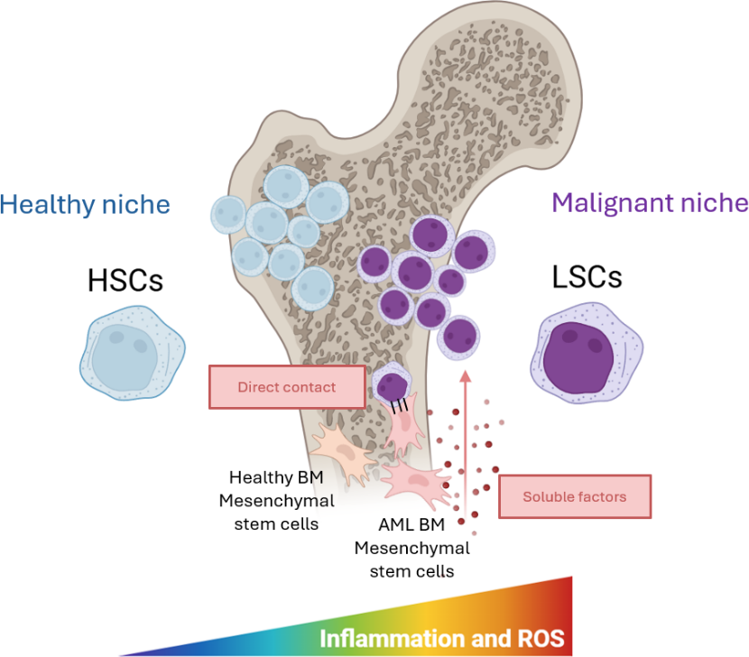
Indeed, such protective role of the BM niche towards AML cells has been strongly demonstrated. Particularly mesenchymal stem cells activate several pathways through direct contact or soluble factors which lead to the protection of AML cells from treatment. The BM niche is a key driver of cell adhesion-mediated drug resistance. In this process, leukemic cells exploit physical interactions with components of the BM microenvironment, including MSCs, ECs, and osteolineage cells, to evade apoptosis and withstand chemotherapy. Through adhesion molecules on their surface, leukemic cells activate signalling pathways that promote the establishment of a protective, pro-leukemic niche. At the same time, soluble mediators such as cytokines and chemokines, most notably CXCL12, further contribute to therapy resistance by reinforcing survival and resistance (Allert et al., 2024) (Fig. 1).
Among the different genetic alterations found in AML, mutations in the FMS-like tyrosine kinase 3 (FLT3) are the most common and with worse prognosis. Such mutations can occur as internal tandem duplication (FLT3-ITD) in the juxtamembrane domain or as a point mutation in the tyrosine kinase domain (FTL3-TKD), causing constitutive activation of the receptor and its downstream signalling pathways. Notably, FLT3-ITD+ AML cells undergo intrinsic endoplasmic reticulum (ER) stress due to the accumulation of FLT3-ITD misfolded protein in the ER (Takahashi S., 2020).
A recent study demonstrated how Retinoic acid and proteotoxic stress induce AML cell death overcoming stromal cell protection. Indeed, the combination of Retinoic Acid (R), Bortezomib (B) and Arsenic Trioxide (A) resulted strongly cytotoxic on FLT3-ITD+ AML cell lines and primary blasts isolated from patients due to ER homeostasis imbalance and generation of oxidative stress. However, as mentioned, the BM niche exerts a protective mechanism against treatment in vivo, rendering AML cells completely resistant to the RBA combination. For this reason, it was exploited a coculture system using a FLT3-ITD+ AML cell line (MOLM-13) and a murine stromal cell line (MS-5) to reproduce a more relatable BM microenvironment. As expected, MS-5 protected AML cells from the RBA combination. To overcome the protective mechanism, high doses of ascorbic acid (ASC) were added as an adjuvant to induce even more oxidative stress. The RBA + ASC resulted efficient in eliminating AML cells in vitro and in vivo (Liccardo et al., 2023).
Such evidence highlights the complex and necessary crosstalk between AML cells and MSCs, reinforcing the need to comprehend the molecular basis and interactions of the communications, rendering inevitable the study of AML as a complex and dynamic system involving more than one cell type. As previously discussed, there are many different strategies and pathways that MSCs activate to protect and support AML cells particularly during treatment. One of these is by enhancing antioxidant defences.
LSCs retain several properties of normal hematopoietic stem cells, yet their metabolism is profoundly reprogrammed to sustain the elevated energetic and biosynthetic demands of AML cells and notably, chemoresistant populations depend on mitochondrial oxidative phosphorylation (OxPhos) to meet their metabolic needs. Recent studies have shown that MSCs can transfer mitochondria to AML cells in vitro, supplying them with additional bioenergetic capacity. This transfer is further amplified following chemotherapy and is thought to promote resistance by preventing mitochondrial depolarization. Although AML cells already display elevated reactive oxygen species (ROS), leukemic stem cells maintain high yet non-toxic ROS levels through upregulated antioxidant pathways. In parallel, BMSC-driven increases in OXPHOS, TCA cycle activity, and glutathione (GSH)-mediated antioxidant defenses enable AML cells to meet their metabolic demands and withstand chemotherapy-induced stress. Together, these findings suggest that mitochondrial acquisition from the microenvironment is a key component of AML metabolic adaptation and therapy resistance (Forte et al., 2020).
In support of this, mitochondrial transfer has been experimentally demonstrated between the murine stromal cell line MS-5 and multiple AML cell lines. This process was quantified by flow cytometry, in which MS-5 cells were pre-labelled with MitoTracker and the subsequent uptake of MitoTracker-positive mitochondria by AML cells was measured. The phenomenon was independently validated by qPCR detection of mouse-derived mtDNA within human AML cells, providing molecular evidence of cross-species mitochondrial acquisition and reinforcing the functional relevance of stromal-to-leukemia mitochondrial transfer (Moschoi et al., 2016).
In fact, mitochondrial transfer has emerged as a relevant phenomenon within the tumour microenvironment, occurring not only among tumour cells but also between tumour cells and cells from the microenvironment including stromal cells, immune cells, endothelial cells, platelets, and even homologous cells. In most contexts, cancer cells acquire mitochondria from surrounding cells to enhance invasion, metastasis, chemotherapy resistance, and immune evasion (Brestoff, J. R. 2025).
Among the various mechanisms described, TNTs represent a common mechanism for intercellular mitochondrial transfer. TNTs are thin (50–1500 nm in diameter and 5–200 μm in length, with some reaching 700 nm thickness), F-actin–based membrane tubes that physically connect the plasma membranes of two cells, creating a direct bridge between the cells allowing exchange of cytoplasmic material including mitochondria. Two major models of TNT formation have been proposed. In the actin-driven mechanism, a cell extends an actin-rich protrusion that fuses with a neighbouring cell, enabling TNT formation independently of cell motility. In contrast, the cell-displacement mechanism occurs when two adjacent cells migrate in opposite directions, stretching membrane connections into TNTs; this process is highly dependent on cell movement (Guan et al., 2024).
Within the AML bone marrow niche, stromal–leukemic cell interactions create a supportive environment that promotes leukemic cell survival. AML blasts rely heavily on OxPhos and continuously adapt to fluctuations in nutrient and oxygen availability within the BM niche. Recent studies have shown that reactive oxygen species (ROS) stimulate MSCs to transfer mitochondria to leukaemia cells via TNTs, particularly under stress or chemotherapy. The acquisition of functional mitochondria increases mitochondrial respiration and ATP production in recipient AML cells, thus enhancing overall functional activity (Saito et al., 2021).
Taken together, these findings highlight the strong dependence of AML cells on MSCs, particularly under therapeutic stress. In this study we show that such crosstalk is rapid and dynamic, occurring in the first hours of direct contact and increases upon therapeutic stress.
Methods
Cell culture
The human AML cell line MOLM-13 was cultured in RPMI 1640 medium supplemented with 1% penicillin/streptomycin and 7% FBS. The murine BMSC line MS-5 was cultured in MEMα with 1% penicillin/streptomycin, 20% FBS and 100 μM β-mercaptoethanol. All the cultures were maintained at 37°C in a humified atmosphere containing 5% CO2.
Co-Cultures
50,000 MS-5 cells per well in an Ibidi µ-Slide 8 Well high ibiTreat (cat# 80806, Ibidi, Gräfelfing, Munich, Germany). After 72 hours, when cells reached approximately 80% confluency, the medium was replaced with 1 mL of RPMI 7% FBS 1%penstrep. The following day, MOLM13 cells were added at a concentration of 70,000 cells/mL, followed by treatment with 5-azacitidine (A) 100 nM and Venetoclax (V) 10 nM.
Live cell stain
MS5 cells were incubated with 1.67 µL of DiD in 500 µL of MEM Alpha medium (without serum) for 20 minutes at 37 °C in an incubator. After incubation, cells were washed twice with MEM Alpha containing serum to inactivate the dye, then resuspended and plated at 50,000 cells per well in an Ibidi µ-Slide 8 Well high ibiTreat (cat# 80806, Ibidi, Gräfelfing, Munich, Germany).
After 24 hours, MOLM13 cells were stained with DiI (1 µL/mL) for 20 minutes at 37 °C in serum-free RPMI medium, followed by three washes in RPMI supplemented with 7% FBS. The medium from the MSCs was then removed, and the DiI-labelled MOLM13 cells were added to the co-culture together with the respective treatments. After 6 hours, co-cultures (MOLM13-DiI / MS5-DiD) were analysed by confocal microscopy using a live-cell imaging chamber equipped with a heater and humidifier, maintaining stable conditions of 37 °C, 5–7% CO₂, and high humidity to preserve cell viability and physiological behaviour during imaging. DiD and DiI dyes were purchased from Thermo-Fisher Scientific (Vybrant™ Multicolor Cell-Labeling Kit, Cat# V22889).
For phalloidin staining, after fixation in 4% PFA and permeabilization with 0.1% TX100, cells were incubated with Rhodamine Phalloidin (R415, Thermo-Fisher Scientific) at a 1:50 dilution in 1% BSA-PBS for 30 minutes at room temperature. Nuclei were counterstained with DAPI (dilution 1:1000) for 8 minutes. Images were acquired and analysed by confocal microscopy by a Zeiss LSM 900 with Airyscan 2, equipped with ZEN 3.2 Blue Edition software.
TNTs quantification
TNTs were quantified based on established morphological criteria. Structures were classified as TNTs when they appeared as thin protrusions, contained F-actin as confirmed by phalloidin staining. For each biological replicate, 100 microscopic fields were analysed. Image evaluation was performed in a blinded manner to minimize observer bias during TNT identification.
Results
To highlight the interaction between AML cells and MSCs by TNTs, we differentially stained the murine stromal cell line MS-5 with the vital dye DiD (red) in coculture with the human AML cell line MOLM-13, stained with the vital dye DiI (yellow). Confocal microscopy revealed the presence of thin yellow nanotube-like structures extending from AML cells toward MS-5 cells, demonstrating direct crosstalk. These structures were detectable within the first hours of direct co-culture (Fig. 2A–B), revealing the rapid and dynamic nature of AML–MSCs communication.
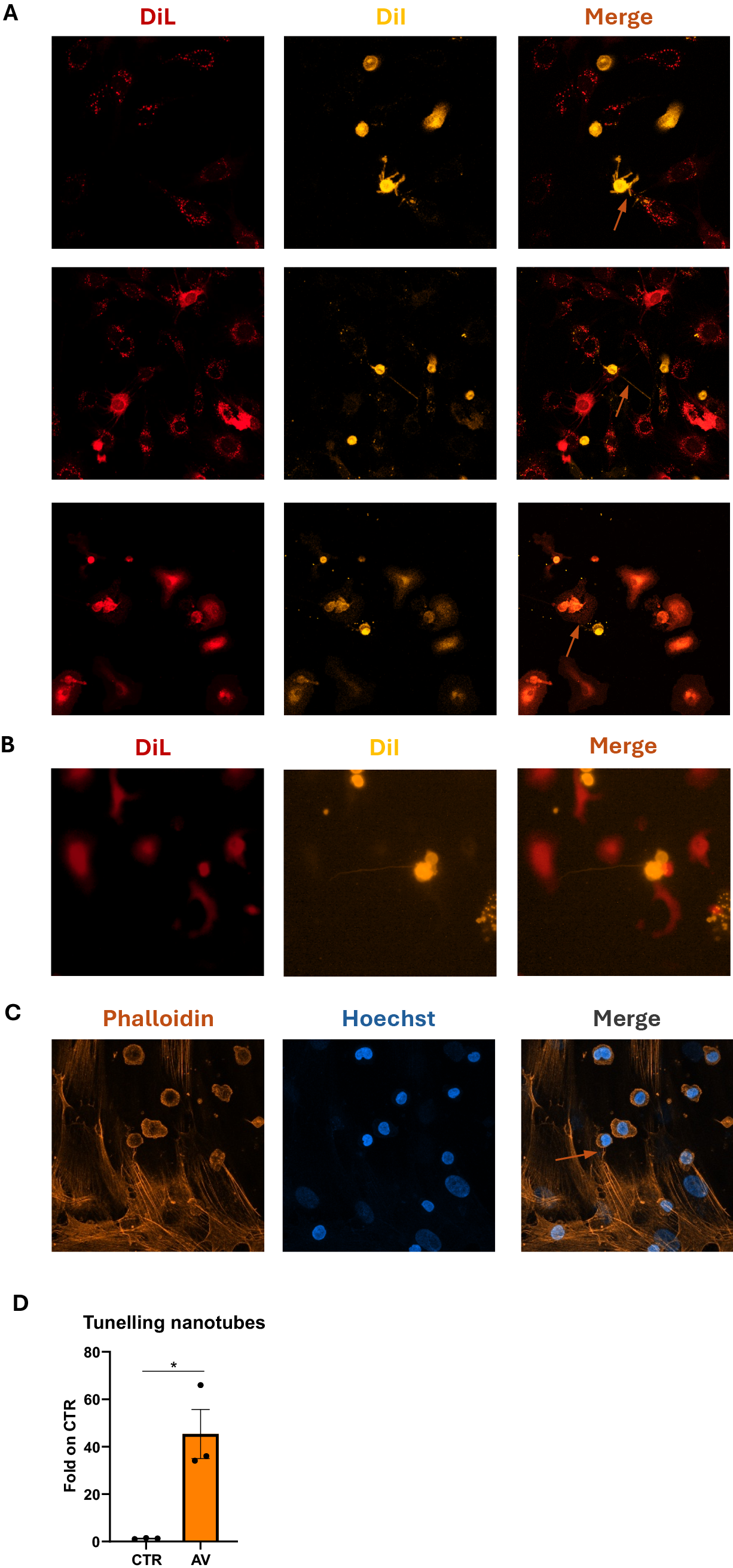
To confirm that the observed structures were consistent with TNTs features, both MS-5 and MOLM-13 cells were stained with rhodamine phalloidin to visualize F-actin. The connecting protrusion showed a continuous actin structure, in line with the canonical description of TNTs (Fig. 2C). Finally, given the central role of the bone marrow niche in promoting AML survival and therapy resistance, we assessed whether treatment influenced TNTs formation. Indeed, we observed an increase in TNTs frequency upon Azacytidine and Venetoclax (Aza-Ven) treatment, indicating that AML–MSCs communication is further enhanced under therapeutic pressure (Fig. 2D).
Discussion
AML is a heterogeneous and rapidly progressing blood cancer originating in the bone marrow. Many studies have shown that AML cells rely heavily on mesenchymal stromal cells for protection, metabolic support, and resistance to therapy. MSCs, endothelial cells, and osteolineage cells together create a pro-leukemic niche that impairs normal differentiation and favours leukemic expansion. Recent work has also highlighted how AML cells use tunnelling nanotubes and mitochondrial transfer to strengthen this interaction, especially under stress or chemotherapy. This makes the AML–MSCs relationship a key contributor to treatment resistance and an important target for new therapeutic strategies.
Our findings demonstrate that AML–MSCs communication is an early, rapid, and highly dynamic process, established within the first hours of direct contact and intensified under therapeutic pressure. By combining differential vital-dye labelling with confocal imaging, we show that AML blasts extend TNTs toward bone marrow stromal cells, forming thin, actin-rich cytoplasmic bridges that enable direct intercellular exchange. Although phalloidin staining confirms the presence of F-actin–containing protrusions, we acknowledge that TNTs identification is based primarily on morphological criteria. Nonetheless, the observed thin, actin-rich structures connecting distant cells are fully consistent with the canonical description of tunnelling nanotubes.
Importantly, we observed that TNTs formation is not static, but rather dynamic and amplified in response to treatment, suggesting that therapeutic stress increases the physical and metabolic crosstalk between AML cells and MSCs. This aligns with emerging evidence that the bone marrow niche actively contributes to leukemic resistance by enhancing metabolic support, antioxidant defences, and mitochondrial transfer during therapy.
Together, these results reinforce the concept that AML progression and therapy resistance cannot be understood solely through leukaemia-intrinsic mechanisms. Instead, AML must be considered as a multicellular system, in which stromal cells support and protect AML cells, enabling leukemic blasts to withstand cytotoxic stress. The rapid induction and treatment-driven amplification of TNT-mediated communication highlights TNTs as a functionally relevant component of AML–MSCs crosstalk and a potential therapeutic vulnerability. While we demonstrate that TNTs formation is rapidly induced and enhanced by treatment, future studies will be required to assess whether these protrusions support functional transfer of mitochondria or cytoplasmic material, thereby establishing a direct functional connection between TNTs formation and AML survival mechanisms within the stromal niche.
Overall, this study underscores the need for therapeutic strategies to focus not only on leukemic cells alone but on the entire niche, limiting TNT-mediated material exchange, and weakening the protective influence of the bone BM microenvironment. Targeting these microenvironment-dependent survival pathways may enhance the efficacy of existing treatments and improve outcomes for AML patients.
Funding
This work was supported by Fondazione GIMEMAonlus, bando “Fondo per le Idee” edizione 2022; Progetti “Ateneo” Sapienza University of Rome, 2025.
References
Allert, C., Müller-Tidow, C. & Blank, M. F. 2024. The relevance of the hematopoietic niche for therapy resistance in acute myeloid leukemia. Int. J. Cancer 154, 197–209. https://doi.org/10.1002/ijc.34684
Baccin, C. et al. (2020). Combined single-cell and spatial transcriptomics reveal the molecular, cellular and spatial bone marrow niche organization. Nat. Cell Biol. 22, 38–48. https://doi.org/10.1038/s41556-019-0439-6
Baryawno, N. et al. (2019). A cellular taxonomy of the bone marrow stroma in homeostasis and leukemia. Cell 177, 1915–1932.e16. https://doi.org/10.1016/j.cell.2019.04.040
Brestoff, J. R. (2025). Mitochondrial swap from cancer to immune cells thwarts anti-tumour defences. Nature 638, 42–43. https://doi.org/10.1038/d41586-025-00077-4
Forte, D. et al. (2020). Bone marrow mesenchymal stem cells support acute myeloid leukemia bioenergetics and enhance antioxidant defense and escape from chemotherapy. Cell Metab. 32, 829–843.e9. https://doi.org/10.1016/j.cmet.2020.09.001
Guan, F. et al. (2024). Mitochondrial transfer in tunneling nanotubes: a new target for cancer therapy. J. Exp. Clin. Cancer Res. 43, 147. https://doi.org/10.1186/s13046-024-03069-w
Kayser, S. & Levis, M. J. (2018). Advances in targeted therapy for acute myeloid leukaemia. Br. J. Haematol. 180, 484–500. https://doi.org/10.1111/bjh.15032
Liccardo, F. et al. (2023). Retinoic acid and proteotoxic stress induce AML cell death overcoming stromal cell protection. J. Exp. Clin. Cancer Res. 42, 223. https://doi.org/10.1186/s13046-023-02793-z
Moschoi, R. et al. (2016). Protective mitochondrial transfer from bone marrow stromal cells to acute myeloid leukemic cells during chemotherapy. Blood 128, 253–264. https://doi.org/10.1182/blood-2015-07-655860
Saito, K. et al. (2021). Exogenous mitochondrial transfer and endogenous mitochondrial fission facilitate AML resistance to OxPhos inhibition. Blood Adv. 5, 4233–4255. https://doi.org/10.1182/bloodadvances.2020003661
Schepers, K., Campbell, T. B. & Passegué, E. (2015). Normal and leukemic stem cell niches: insights and therapeutic opportunities. Cell Stem Cell 16, 254–267. https://doi.org/10.1016/j.stem.2015.02.014
Takahashi, S. (2020). Mutations of FLT3 receptor affect its surface glycosylation, intracellular localization, and downstream signaling. Leuk. Res. Rep. 13, 100187. https://doi.org/10.1016/j.lrr.2019.100187