https://doi.org/10.36253/ijae-17404
Variations in the dimensions of the sacrum and its implications for Forensic Anthropology
Anatomy and Histology Department, School of Medicine, Sefako Makgatho Health Sciences University, Gauteng, South Africa
*Corresponding author. E-mail:ymrtshedza@gmail.com
Abstract. With rising numbers of unidentified bodies in South Africa, the South African Police Service (SAPS) requires effective skeletal identification techniques. While the skull and pelvis are highly dimorphic, taphonomic fragmentation often renders them unavailable at forensic scenes. The sacrum is frequently well-preserved and exhibits population specific sexual dimorphism. This study aimed to evaluate the dimensions and three-dimensional (3D) shape of the ventral sacrum in a contemporary South African sample. Retrospective Computer Tomography (CT) scans of 51 black South African adults (26 females, 25 males) were obtained from Steve Biko Academic hospital. Utilising MeVisLab © (v.3.0.2), the sacra were isolated via threshold-based segmentation, and 3D landmarks were placed on the ventral sacral surface. Linear dimensions were calculated and evaluated using descriptive statistics and discriminant function analysis (DFA), while shape variations were assessed using a multivariate analysis and a two-group permutation test. Black South African males exhibited a significantly greater sacral height than females (p=0.004). The permutation test confirmed significant sex-based variation in overall ventral sacral shape (p= 0.005). DFA models predicted sex with a moderate 72.55% accuracy based on linear dimensions, but achieved an exceptional 94.12% accuracy utilising 3D shape coordinates. ROC analysis substantiated the predictive value of the ventral linear metrics (AUC = 0.73; 95% CI: 0.60 – 0.85; p = 0.004) for sacral height, yielding an optimal cut-off value of 103.41 mm (Sensitivity: 76 %, Specificity: 65.38 %). Sexual dimorphism significantly influences both the metrics and morphology of the ventral sacrum of black South Africans. Due to these distinct variations, the ventral sacral surface, particularly its 3D shape, provides a highly reliable alternative for accurate forensic sex estimation.
Keywords: sacrum, anatomical variation, biological profile, geometric morphometrics, Sexual dimorphism.
Contents
Congenital conditions associated with the sacrum
Sexual dimorphism of the sacrum
Population / ancestral variations
Introduction
With rising incidences of unsolved forensic cases, the South African Police Service (SAPS) needs reliable methods to determine the sex and population affinity of unknown victims. Forensic anthropologists assist the SAPS in their investigation by creating a biological profile using skeletal remains (L’Abbé and Steyn, 2012). The biological profile includes the sex, population affinity, age, stature and possible cause of death of the deceased (Krüger et al, 2018; Spradley, 2016).
Due to biological and geographical taphonomy, determining the biological profile of a victim may be challenging (Klales et al.,2009; Iscan and Steyn, 2013). Creating a biological profile for the deceased has proven to be a difficult task worldwide, as forensic anthropologists rarely receive a full skeleton from the police (Krüger et al, 2018). Often, the skull and limbs of the individual may not be present due to the harsh environmental conditions, scavenger activity or the nature of the crime (Krüger et al, 2018; Brits et al, 2020). This poses a challenge to investigators and forensic anthropologists alike, in trying to determine the sex, ancestry and age of the unknown individual in the attempt to solve a crime (Krogman and Iscan 1986). Due to these challenges, forensic anthropologists need to find additional, effective techniques to create reliable biological profiles of the deceased.
Numerous books and articles have been published based on metric and non-metric data of various bones of the human skeleton to assist forensic anthropologists in determining the sex and population affinity of the deceased (Flander, 1978, Iscan and Steyn 2013; Rusk and Ousley, 2016).
The sacrum is an imperative bone which can be useful in determining the ancestry and sex of an unknown victim, as it is often preserved well after death (Flander,1978, Rusk and Ousley, 2016). Due to this fact, the sacrum has captured the attention of forensic anthropologists in their quest to find reliable methods to determine the identity of the deceased (Flander, 1978; Rusk and Ousley, 2016).
Geometric morphometric techniques have been used to classify the human sacral landmarks based on their three dimensional shape (Passalacqua et al, 2010). Vollner and colleagues (2011) cross-validated sex and ancestry-sex classification accuracy measurements in a study performed on black and white Americans, which yielded an 89% and 65.6% accuracy for sex determination rate on the os coxae and sacrum respectively. Sexual dimorphism seems to be population specific as expected and therefore, population specific guidelines are needed (Iscan and Steyn, 2013).
Anatomy of the sacrum
The sacral bone is a large triangular bone situated at the base of the vertebral column where it contributes to the formation of the pelvis (Bogduk, 2005). The sacrum consists of five fused sacral vertebrae (Drake et al ,2009). It has a broad thick upper end, which articulates with L, and it then tapers to a blunt tip inferiorly where it articulates with the coccyx (Drake et al, 2009; Bogduk, 2005).
The sacrum has a smooth ventral surface (Fig 1A) that is concave and a rough dorsal surface which is convex, with both surfaces containing sacral foramina (Drake et al, 2009; Bogduk, 2005). The sacral promontory, which forms part of the pelvic inlet, is located on the ventral surface of the first sacral vertebra (Drake et al, 2009). The ala of the sacrum is located on its lateral surface, which articulates to the ilium of the os coxa to form the sacroiliac joint (Drake et al, 2009).
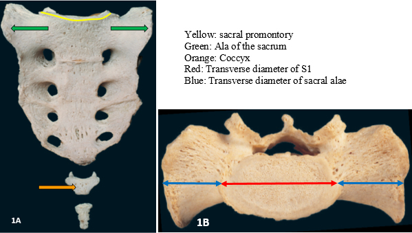
The ventral surface of the sacrum is of particular importance as it forms part of the birth canal, which is known to be sexually dimorphic and population specific (Betti, L., 2014).
Congenital conditions associated with the sacrum
Forensic anthropologists must be aware of congenital abnormalities of the skeleton when examining skeletal remains. Sacral defects commonly occur in three observed patterns: the absence of the vertebrae (for instance sacral agenesis), hemivertebrae (hemisacral), and defects of the neutral arch which results in sacral spina bifida (Stanley et al, 1979). A common congenital abnormality observed in the sacral region, includes the lumbosacral transitional vertebrae (Apazidis et al, 2011). The shape and size of the sacrum is affected by the presence of a lumbosacral transitional vertebra (LSTV), noted at the L5-S1 junction (Apazidis et al, 2011).
Lumbosacral transitional vertebra is associated with varied degrees of geometrical changes in different anatomical features of the human sacrum (Mahato, 2016). The LSTV is the most common congenital abnormality, such a transitional vertebra may present with abnormal variations in the normal lumbosacral articulations between the transverse process of the inferior lumbar vertebra and the sacrum (Mahato, 2016). When the L5 vertebra is fused to the sacrum it is referred to as sacralisation of L5, which results in the presence of only four lumbar vertebrae. When S1 is separated from the sacrum is referred to as lumbarisation of S1 (Connolly et al, 2003, Mahato, 2016).
Sexual dimorphism of the sacrum
Anatomical textbooks commonly describe the sacrum as “long and narrow” in males and “short and broad” in females (Benazzi et al.,2009; Iscan and Steyn 2013; Moore and Dalley, 2018). The sacrum is accepted to be sexually dimorphic due to its effect on the morphology of the shape of the pelvic inlet and outlet. The female sacrum is less curved, resulting in a larger, more oval shaped pelvic outlet (Flander, 1978). The discrete differences in the size, curvature, number of sacral vertebrae, extent of the articulation surfaces and the ratio of the first sacral vertebra to alae (S1/alae ratio) which is the ratio between the width of S1 and the width of the sacrum and the ratio between the width of the alae and the width of the sacrum, it plays a critical role in determining sex based on the sacrum (Iscan and Steyn, 2013)
With regard to the dimensions of the sacrum, it has been demonstrated to be an accurate indicator of sex differences using sacral breadth measurements (Trotter, 1926). According to Flander (1978), the anteroposterior dimensions and transverse length of the first sacral vertebral body are highly reliable indicators of sex (Flander,1978; Rusk and Ousley, 2016). In addition to these sexually dimorphic diameters, the S1/alae ratio, also referred to as the base-wing index, is an accurate metric for sex estimation (Kimura, 1982; Rusk and Ousley 2016).
A study performed by Rusk and Ousley (2016) used shape analysis based on multiple landmarks placed on the sacrum to determine variations among sexes between white and black Americans. They noted the sacrum had a classification accuracy ranging between 60.2 – 98.0% for sex determination and 60.0 – 95.8% for population affinity estimation.
Population / ancestral variations
Steyn and collegues (2016) explained the distinct morphology of South Africans’ bones due to the population originating from various locations with distinct climatic conditions. Tague (2007) discovered that white Americans have a larger sacrum as compared to that of black African-Americans. Furthermore, distinct sexual dimorphism exists across planes: males exhibited a greater S1 width in both the anteroposterior and transverse planes, whereas females displayed longer S1 transverse processes alongside larger transverse diameters and circumferences of the pelvic inlet (Tague, 2007). In contrast, Rusk and Ousley (2016) determined that the dorso-ventral curvature of the sacrum is more accurate in estimating population affinity (White vs Back Americans) than for sex estimation, despite the traditional reliance on this curve for sex determination.
Given the high preservation rate of the sacrum in skeletonised remains and the documented sexual dimorphism across diverse populations, this study aimed to evaluate variations in the dimensions and shape of the ventral surface of the sacrum within a black South African sample.
Material and methods
Material
A quantitative descriptive study was performed using Computed Tomography (CT) scans of 51 black South African adults (26 females, 25 males). Retrospective CT scans were selected from a repository at the Radiology Department of an academic hospital in Pretoria. Ethical approval (SMUREC/M/158/2022: PG) was obtained the research ethics committee at Sefako Makgatho Health Sciences University prior to data collection.
Patients were scanned in the supine position from the head to mid-thigh. slice thickness ranged between 2 to 2.5 mm. CT scanned data was collected in DICOM (Digital Imaging and Communications in Medicine) format which was imported into the fee medical image processing software, MeVisLab © v.3.0.2.
Methods
The collected DICOM files were imported into the MeVisLab © v.3.0.2 to visualise the skeletal structures of the articulated pelvis through threshold-based segmentation. Segmentation is an automated process that identifies the best limit between grey values of different densities, resulting in either a soft-tissue or hard-tissue 3D rendering (Spoor et al., 2000). For these CT scans, the attenuated threshold values typically ranged between 1100-1200 Hounsfield Units (HU) for bone tissue and 400 – 500 HU for soft tissue.
Ten landmarks were selected from literature to determine the dimensions and the shape of the sacrum (Table 1, Figs. 2 and 3). Table 2 defines the specific measurements calculated from the landmarks placed on the 3D sacral reconstruction.
| Landmark | Abbrev. | Definition |
|---|---|---|
| Sacral promontory | SPM | The most protruding point in the midline along the superior margin of S1 (referred to as the sacral promontory) |
| Right lateral S1 | RLS1 | The most lateral point along the right superior margin of the first sacral vertebra |
| Left lateral S1 | LLS1 | The most lateral point along the left superior margin of the first sacral vertebra |
| Right lateral ala | RLA | The most lateral point along the superior margin of the right ala |
| Left lateral ala | LLA | The most lateral point along the superior margin of the left ala |
| Sacral apex | SA | The tip of the sacrum, on the inferior margin of S5 |
| Ventral S2 | VS2 | The most protruding point along the antero-superior margin S2 in the midline |
| Ventral S3 | VS3 | The most protruding point along the antero-superior margin S3 in the midline |
| Ventral S4 | VS4 | The most protruding point along the antero-superior margin S4 in the midline |
| Ventral S5 | VS5 | The most protruding point along the antero-superior margin S5 in the midline |
| Measurements | Landmarks involved |
|---|---|
| Width of S1 | Between RLS1 and LLS1 |
| Length of the left ala | Between LLS1 and LLA |
| Length of the right ala | Between RLA and RLS1 |
| Width of the sacrum | Between LLA and RLA |
| S1/ala breath ratio | Ratio between the width of S1 and the width of the sacrum and the ratio between the width of the alae and the width of the sacrum |
| Anterior height of the sacrum | Between SPM and SA |
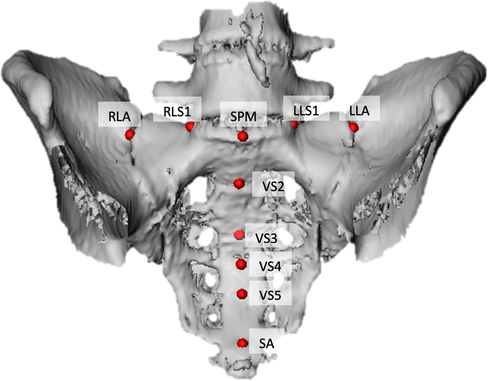
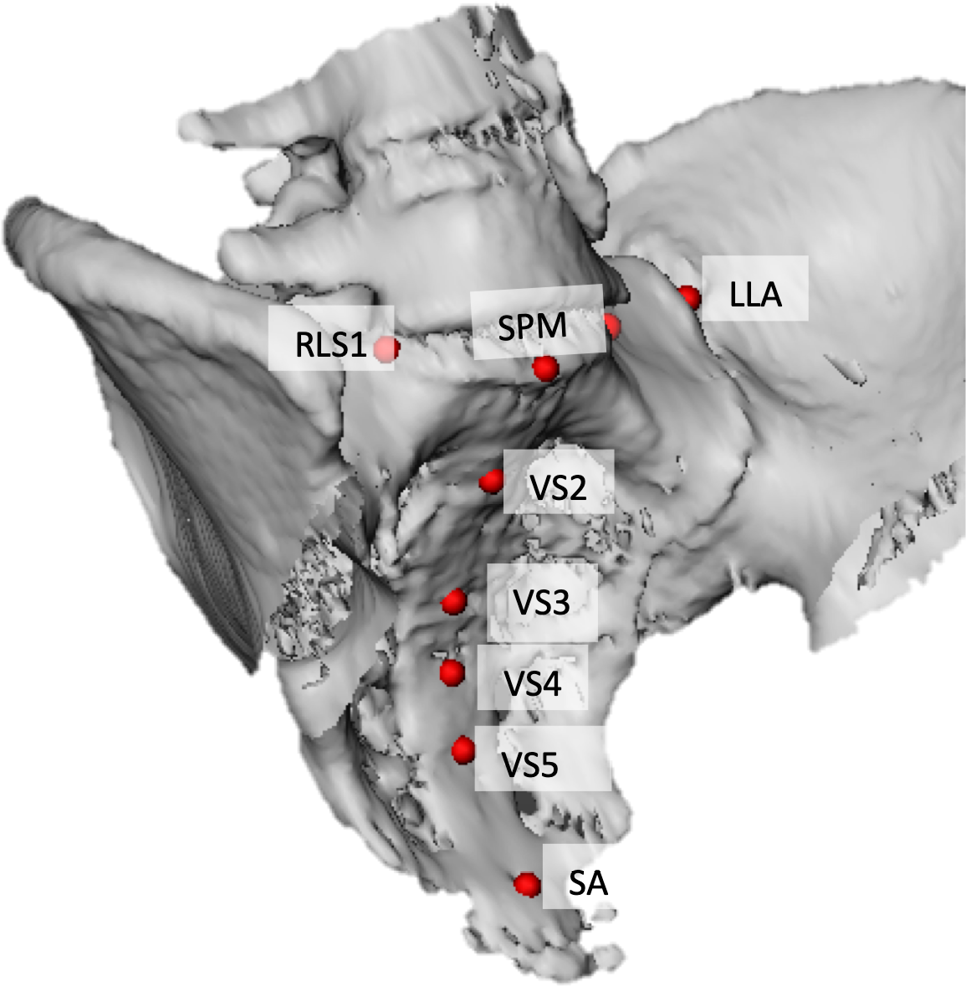
By placing the selected landmarks on the 3D ventral surface of the segmented sacrum, the landmarks were exported in XML format as x, y and z coordinates. The data was then imported into MS Excel to calculate the linear distances listed in Table 2. Normality testing and basic descriptive statistics were performed using the PAST v4.11 programme to determine the average dimensions of the ventral surface of the sacrum in black South African males and females. A Shapiro Wilk test were performed to evaluate data distribution and to select the appropriate statistical tests to assess significant difference between sexes.
To evaluate the effectiveness and discriminative ability of the sacral measurements in distinguishing between male and female individuals, a Receiver Operating Characteristic (ROC) curve analysis was conducted. The Area Under the Curve (AUC) was calculated for each measurement to evaluate its overall diagnostic accuracy, where an AUC of 0.5 indicates random guessing and 0.7 ≤ 0.9 indicates perfect discrimination, only applied to statistically significant (p ≤ 0.05) values. The optimal cut-off values maximising both sensitivity and specificity were determined. This ROC analysis protocol followed the methodology described by D’Arcangelo et al. (2019).
Results
The results of the Shapiro-Wilk test indicated that the S1/ala ratio was non-parametric in females, while the width of the ala was non-parametric in males. Independent t-tests were performed to determine if significant sex-based differences exist between black South Africans for parametric data, while Kruskal-Wallis tests were used for non-parametric data. Basic descriptive statistics and sex-specific comparisons are summarised in Table 3.
| Measurement (mm) | South African black females (n=26) |
South African black males (n=25) |
Differences (p-value) |
||||||||||||||||
|---|---|---|---|---|---|---|---|---|---|---|---|---|---|---|---|---|---|---|---|
| Width of S1 | 50.59 ± 7.34 (40.11–69.89) |
52.54 ± 5.10 (41.83–62.55) |
0.28 | ||||||||||||||||
| Width of the ala | 107.33 ± 6.87 (92.19–120.35) |
108.14 ± 8.06 (80.15–118.48) |
0.45 | ||||||||||||||||
| With of the right ala | 29.66 ± 3.76 (22.00–37.70) |
29.90 ± 3.48 (22.96–39.48) |
0.82 | ||||||||||||||||
| Width of the left ala | 30.25 ± 4.06 (21.89–37.54) |
30.83 ± 4.76 (22.53–40.16) |
0.65 | ||||||||||||||||
| Anterior height (height of the sacrum) | 99.88 ± 11.44 (79.13–126.32) |
108.49 ± 8.60 (89.81–123.20) |
0.004* | ||||||||||||||||
| S1/Ala ratio | 0.47 ± 0.06 (0.39–0.65) |
0.49 ± 0.04 (0.42–0.56) |
0.08 | ||||||||||||||||
| * statistical significant difference (p ≤ 0.05). | |||||||||||||||||||
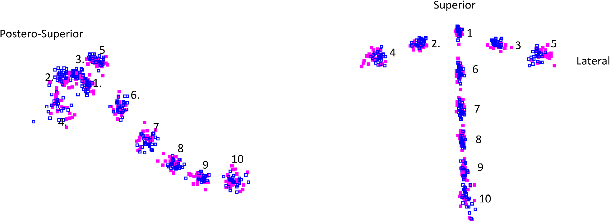
Sacral height was significantly greater in black South African males compared to their female counterparts. A shorter and wide sacrum was expected as it contributes to the oval shaped pelvic outlet of females which promote vaginal delivery. Sacral width was slightly greater in black South African males, but with further investigation, it was noted that the female sacral width was generally greater, but exhibited more variation resulting in a smaller mean value. This can be verified, by looking at the landmark distribution as indicated on Figure 7.
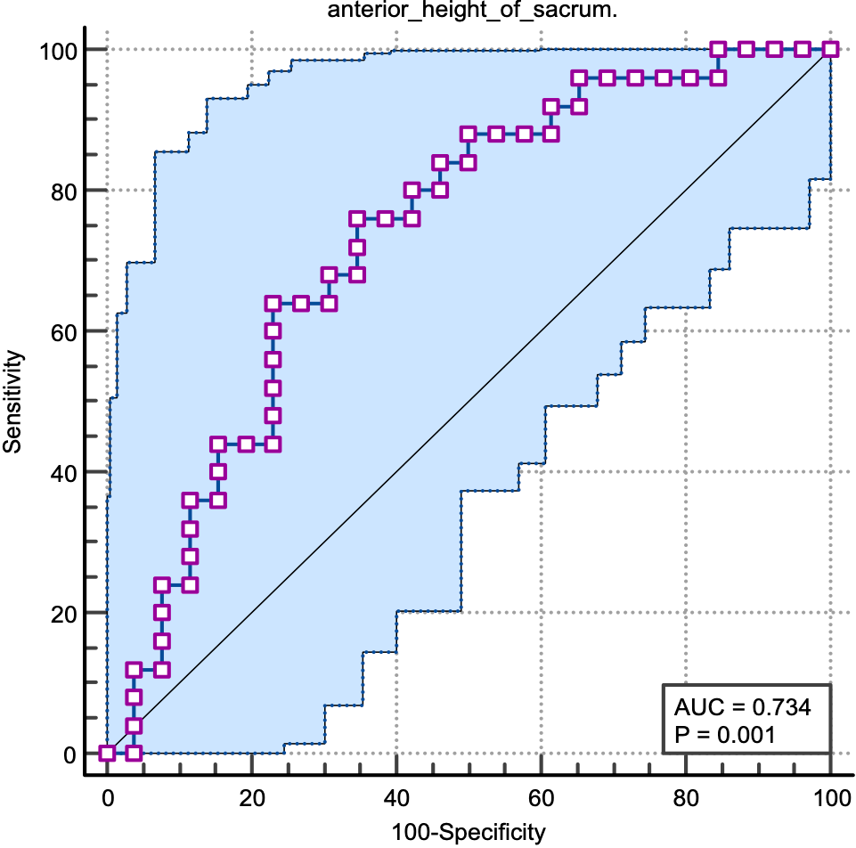
The sex discriminative capacity of the sacrum measurements was evaluated using ROC curve analysis, as shown in Table 4. Anterior height demonstrated the highest sex discrimination accuracy, yielding a statistically significant Area Under the Curve (AUC = 0.73; 95% CI: 0.60 – 0.85; p = 0.001), with an optimal cut-off value of 103.41 mm (Sensitivity: 76 %, Specificity: 65.38 %), as shown in Figure 4. In contrast, the remaining sacral dimensions (Width of S1, Width of the ala, Width of the right ala, Width of the left ala, and S1/Ala ratio) showed weak diagnostic performance, with AUC values ranging between 0.51 and 0.64, reflecting their lack of statistically significant differences between the sexes (p > 0.05).
| Measurement (mm) | Area under the curve (AUC) |
95% Confidence Interval (CI) |
Differences (p-value) |
||||||||||||||||
|---|---|---|---|---|---|---|---|---|---|---|---|---|---|---|---|---|---|---|---|
| Width of S1 | 0.61 | 0.47–0.75 | 0.28 | ||||||||||||||||
| Width of the ala | 0.56 | 0.42–0.70 | 0.45 | ||||||||||||||||
| With of the right ala | 0.51 | 0.37–0.66 | 0.82 | ||||||||||||||||
| Width of the left ala | 0.54 | 0.40–0.69 | 0.65 | ||||||||||||||||
| Anterior height (height of the sacrum) | 0.73 | 0.60–0.85 | 0.001* | ||||||||||||||||
| S1/Ala ratio | 0.64 | 0.50–0.78 | 0.08 | ||||||||||||||||
| * Statistical significant difference (p ≤ 0.05). | |||||||||||||||||||
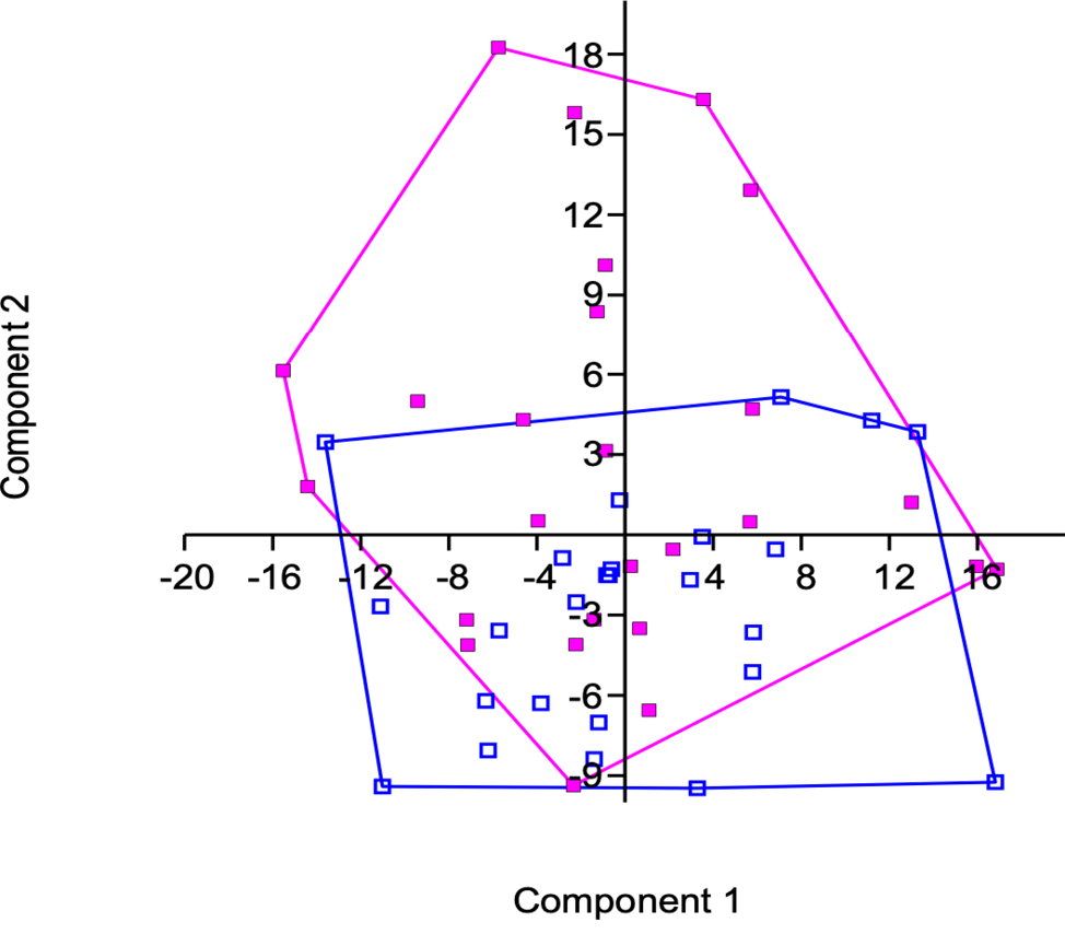
To evaluate 3D shape variations of the ventral surface of the sacrum between sexes, a multivariate Principal Component Analyses (PCA) was performed. Principal Component 1 (PC1) accounted for 28.97% of the total variance, while PCA 2 accounted for 21.73%. The distribution and clustering of these scores are illustrated in Figure 5. In addition, a two-group permutation test confirmed a statistically significant difference in the ventral sacral shape between black South African males and females (p= 0.005).
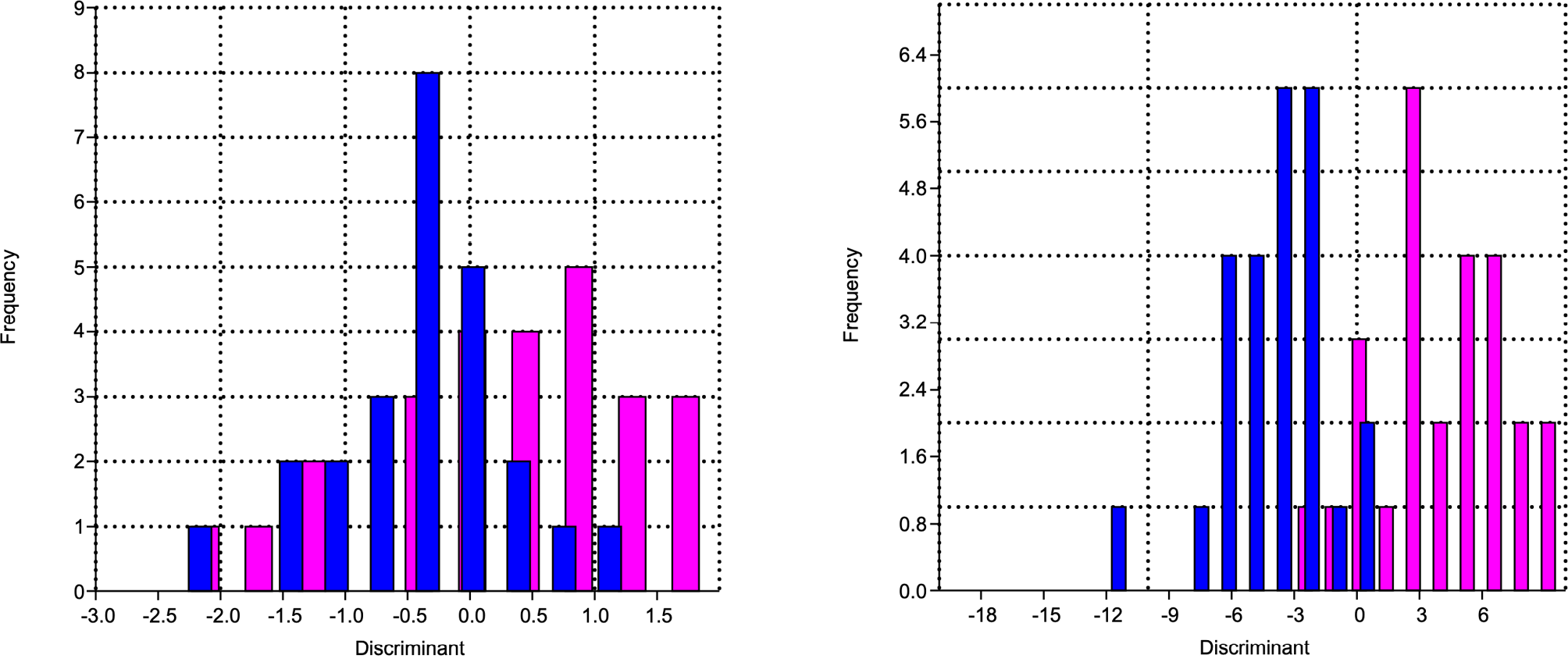
Discriminant function analyses were performed on the 3D coordinates and linear distances to evaluate the capacity of ventral sacral shape to estimate sex. The discriminant models achieved a 94.12% classification accuracy when using 3D shape data (Fig. 6a) and a 72.55% accuracy when based on linear dimensions (Fig 6b).
Discussion
In recent years, the sacrum has spiked the interest of scientist, with numerous studies demonstrating that its shape and dimensions are both sexually dimorphic and population-specific (Vollner et al., 2011; Iscan and Steyn, 2013; Rusk and Ousley, 2016; Lottering et al., 2022). Consequently, this study aimed to evaluate the efficacy of sex estimation within a black South African sample, using linear distances (measured between two 3D landmarks) and 3D shape coordinates on the ventral sacral surface.
Within this black South African sample, the sacral height varied significantly between the sexes (p = 0.004). Although the width of the of S1 was greater in males, it was not statistically significant. While the S1/Ala ratio (base-wing index) is widely documented as sexual dimorphic in the literature (Rusk and Ousley, 2016), it did not vary significantly between males and females in this sample
Cross-population comparisons highlight distinct geomorphological variations. In the literature, Chinese individuals present with the shortest sacral length compared to all studied population groups (Zhan et al., 2018). While sacral length is sexually dimorphic in the black South African and Turkish samples, it displays no significant dimorphism in Japanese or American populations (Table 5). Conversely, sacral width is dimorphic in those global populations, but lacks dimorphism among black South Africans. The width of the sacral alae (sacral breath) does not vary between sexes in any of the populations summarised in Table 5, confirming its limited utility for sex estimation.
| Study: | Population: | Modality: | S1 width | Alae width | Sacral height | S1/ala ratio | |||||||||||||
|---|---|---|---|---|---|---|---|---|---|---|---|---|---|---|---|---|---|---|---|
| This study | Black South Africans | CT | Female: 50.59±7.34 Male: 52.54 ± 5.1 |
Female: 108.14 ± 8.06 Male: 107.33 ± 6.87 |
Female: 99.88 ± 11.44* Male: 108.49 ± 8.6* |
Female: 0.47 ± 0.06 Male: 0.49 ± 0.04 |
|||||||||||||
| Zhan et al., 2018 | Chinese | Multidetector CT | Female: 82.0 ± 6.2 Male: 88.3 ± 5.4 |
||||||||||||||||
| Torimitsu et al., 2017 | Japanese | Cadaver | Female: 52.25 ± 5.11* Male: 57.41 ± 5.77* |
Female: 115.19 ± 5.60 Male: 116.98 ± 6.60 |
Female:102.58±10.52* Male:113.52±9.73* |
||||||||||||||
| Rusk and Ousley, 2016 | Black Americans | Sacral bone | Female:1/3:1/3;1/3 Males :1/4:1/2:1/4 |
||||||||||||||||
| Zech et al., 2012 | Swiss | Sacral bone | Female:47.6±4,2 Male: 55.5±6.2 |
Female:115.1±6.1 Male: 114.8±5.1 |
|||||||||||||||
| Plochocki, 2011 | Americans | Sacral bones | Female:98.3±10.03 Male:100.1±8.00 |
||||||||||||||||
| Pelin et al, 2005 | Turkish males | MRI | Male: 109.5±12.1 | ||||||||||||||||
| Flander, 1978 | Black and white Americans | Sacral bones | BF:47.44±4.15* BM:54.5±4.28* WF:46.56±3.94* WM:52.78±5.21* |
BF:111.36±7.23 BM:111.14±7.81 WF:117.62±7.08 WM:116.42±7.11 |
BF: 99.98±11.24 BM: 105.5±11.01 WF:109.64±12.56 WM:110.2±11.4 |
||||||||||||||
| * Statistically significant variations (p≤0.05), BF: Black female; BM: Black male; WF: White female; WM: White male. | |||||||||||||||||||
In this study, discriminant function analysis of the ventral sacral dimensions yielded a 72.55% accuracy for sex estimation, with males exhibiting a significantly greater sacral height than females. While this metric dimorphism is fully supported by Lottering and colleagues (2022), our classification accuracy is notably lower than the 88 - 94% achieved by Flander (1987). This discrepancy further underscores how linear skeletal dimensions can exhibit varying predictive reliability across different population groups. While ROC analysis substantiated the predictive value of these ventral linear dimensions with a significant Area Under the Curve (AUC: 0.76; CI 95%: 0.60 – 0.85; p = 0.004), the limitations of traditional linear metrics have increasingly driven researchers toward alternative methodologies.
Flander (1978) demonstrated that geometric morphometrics is a valuable method to determine sex from dry sacra after obtaining an 84% accuracy for white Americans and 91% accuracy for black Americans. In more recent years, Passalacqua and colleagues (2010) accurately determine sex from a black American sample, with 86% accuracy based on the shape of the sacrum defined by 3D landmarks. Notably, our results using3D coordinates of the ventral sacral surface yielded a 94,12 % accuracy in estimating sex of black South Africans, which demonstrates greater accuracy compared to the findings of Passalacqua and co-workers (2010).
Geometric morphometrics has been increasingly utilised for sex estimation in recent anthropological studies. Zech and colleagues (2012) achieved an 80 % sex prediction accuracy in a Swiss sample using CT scans. Conversely, the lowest sex classification accuracy, based on sacral shape was obtained by Franklin and co-workers (2014) who obtained a 70% classification accuracy using CT scans of an Australian sample.
Based on these studies conducted on across diverse population groups, sex can be estimated from the shape of the sacrum with an accuracy ranging between 70% - 94%. This overarching range demonstrates that shape-based analyses generally yield higher predictive rates than those based on traditional linear distances.
Conclusion
Knowledge of the anatomical variations of the sacrum between sex and population groups is integral in medical education, as it will influence the selection of operation techniques during surgical procedures and assist forensic scientists in human identification. This study demonstrates clear sexual dimorphism in the ventral surface of the sacrum, confirming its utility for accurately estimating sex in black South Africans.
The most effective method for sex estimation in this sample was based on 3D shape variation of the ventral surface of the sacrum, which has yielded an exceptional accuracy of 94,12 %. While linear distances measured across the ventral surface of the sacrum also successfully estimated sex with a 72,55 % accuracy rate, a finding supported by ROC analysis which confirmed a statistically significant Area Under the Curve (AUC = 0.73; 95% CI: 0.60 – 0.85; p = 0.004), shape analysis remains more accurate and reliable.
Consequently, it is recommended that forensic anthropologist and anatomical scientists prioritise 3D shape analysis of the ventral sacrum over traditional linear dimensions when estimating sex of skeletal remains within the black South African population.
References
Abrahams, P.H., Boon, J.M. and Spratt, J.D., 2008. McMinn’s clinical atlas of human anatomy. Elsevier Health Sciences.
Apazidis, A., Ricart, P.A., Diefenbach, C.M. and Spivak, J.M., 2011. The prevalence of transitional vertebrae in the lumbar spine. The Spine Journal, 11(9), pp.858-862.
Benazzi, S., Maestri, C., Parisini, S., Vecchi, F. and Gruppioni, G., 2009. Sex assessment from the sacral base by means of image processing. Journal of Forensic Sciences, 54(2), pp.249-254.
Betti, L., 2014. Sexual dimorphism in the size and shape of the os coxae and the effects of microevolutionary processes. American Journal of Physical Anthropology, 153(2), pp.167-177.
Bogduk, N., 2005. Clinical anatomy of the lumbar spine and sacrum. Elsevier Health Sciences, pp.51-54.
Brits, D.M., Steyn, M. and Hansmeyer, C., 2020. Identifying the unknown and the undocumented: The Johannesburg (South Africa) experience. Forensic science and humanitarian action: interacting with the dead and the living, pp.681-692.
Connolly, L.P., d’Hemecourt, P.A., Connolly, S.A., Drubach, L.A., Micheli, L.J. and Treves, S.T., 2003. Skeletal scintigraphy of young patients with low-back pain and a lumbosacral transitional vertebra. Journal of Nuclear Medicine, 44(6), pp.909-914.
D’Arcangelo, D., Scatozza, F., Giampietri, C., Marchetti, P., Facchiano, F. and Facchiano, A., 2019. Ion channel expression in human melanoma samples: in silico identification and experimental validation of molecular targets. Cancers, 11(4), p.446.
Drake, R., Vogl, A.W. and Mitchell, A.W., 2009. Dorland’s/Gray’s Pocket Atlas of Anatomy E-Book. Elsevier Health Sciences,pp.425.
Flander, L.B., 1978. Univariate and multivariate methods for sexing the sacrum. American journal of physical Anthropology, 49(1), pp.103-110.
Franklin, A. Cardini, A. Flavel, M.K. Marks.2014. Morphometric analysis of pelvic sexual dimorphism in a contemporary Western Australian population, Int. J. Leg. Med. 128
Iscan, M.Y. and Steyn, M., 2013. The human skeleton in forensic medicine. Charles C Thomas Publisher.
Kimura, K., 1982. A base-wing index for sexing the sacrum. Journal of the Anthropological Society of Nippon, 90(Supplement), pp.153-162.
Klales, A.R., Vollner, J.M. and Ousley, S.D., 2009. A new metric procedure for the estimation of sex and ancestry from the human innominate. Proc Am Acad Forensic Sci, 15, p.311.
Krogman, W.M. and Iscan, M.Y., 1986. The human skeleton in forensic medicine, Charles C. Thomas, Springfield, IL, 15(2), pp.202-08.
Krüger, G.C., Liebenberg, L., Myburgh, J., Meyer, A., Oettlé, A.C., Botha, D., Brits, D.M., Kenyhercz, M.W., Stull, K.E., Sutherland, C. and L’Abbé, E.N., 2018. Forensic anthropology and the biological profile in South Africa. In New Perspectives in Forensic Human Skeletal Identification (pp. 313-321). Academic Press.
Lottering, T., Hemingway, J. and Small, C., 2022. An exploration of sacral morphology using geometric morphometrics and three-dimensionally derived interlandmark distances. International Journal of Legal Medicine, 136(4), pp.1051-1065.
Mahato, N.K., 2016. Implications of structural variations in the human sacrum: why is an anatomical classification crucial?. Surgical and Radiologic Anatomy, 38(8), pp.947-954.
Moore, K.L. and Dalley, A.F., 2018. Clinically oriented anatomy. Wolters kluwer india Pvt Ltd.
Passalacqua NV, Vollner JM, Rainwater CW. 2010. Geometric morphometric analysis of the human sacrum and its utility in sex estimation. Am J Phys Anthropol 141(S50):185–186.
Pelin, C., Duyar, I., Kayahan, E.M., Zagyapan, R., Ağildere, A.M. and Erar, A., 2005. Body height estimation based on dimensions of sacral and coccygeal vertebrae. Journal of Forensic and Sciences, 50(2), pp.JFS2004010-4.
Plochocki, J.H., 2011. Sexual dimorphism of anterior sacral curvature. Journal of forensic sciences, 56(1), pp.161-164.
Rusk, K.M. and Ousley, S.D., 2016. An evaluation of sex‐and ancestry‐specific variation in sacral size and shape using geometric morphometrics. American Journal of Physical Anthropology, 159(4), pp.646-654.
Spoor, F., Jeffery, N. and Zonneveld, F., 2000, April. Imaging skeletal growth and evolution. In Linnean Society Symposium Series (Vol. 20, pp. 123-162).
Spradley K, M. and Jantz, R.L., 2016. Ancestry estimation in forensic anthropology: geometric morphometric versus standard and nonstandard interlandmark distances. Journal of forensic sciences, 61(4), pp.892-897.
Stanley, J.K., Owen, R.O.B.E.R.T. and Koff, S.T.E.V.E., 1979. Congenital sacral anomalies. The Journal of Bone & Joint Surgery British Volume, 61(4), pp.401-409.
Steyn, M., L’Abbé, E.N. and Myburgh, J., 2016. Forensic anthropology as practiced in South Africa. In Handbook of Forensic Anthropology and Archaeology (pp. 193-206). Routledge.
Tague, R.G., 2007. Costal process of the first sacral vertebra: sexual dimorphism and obstetrical adaptation. American Journal of Physical Anthropology: The Official Publication of the American Association of Physical Anthropologists, 132(3), pp.395-405.
Trotter M. 1926. The sacrum and sex. Am J Phys Anthropol 9:445–450.
Vollner JM, Passalacqua NV, Rusk K. 2011. A re-evaluation of the power of “standard” measurements in estimating sex and sex-specific ancestry from the innominate and sacrum. Am J Phys Anthropol 144(S52):301.
Zech, W.D., Hatch, G., Siegenthaler, L., Thali, M.J. and Lösch, S., 2012. Sex determination from os sacrum by postmortem CT. Forensic science international, 221(1-3), pp.39-43.
Zhan, M.J., Fan, F., Qiu, L.R., Peng, Z., Zhang, K. and Deng, Z.H., 2018. Estimation of stature and sex from sacrum and coccyx measurements by multidetector computed tomography in Chinese. Legal Medicine, 34, pp.21-26.