https://doi.org/10.36253/ijae-17382
Caffeine protects the blood brain barrier in type 2 diabetic rats, a histological and immunohistochemical study
1 Department of Anatomy, College of Medicine and Health Sciences, Arabian Gulf University, Manama, Bahrain
2 Department of Histology and Cell Biology, Faculty of Medicine, Assiut University, Assiut, Egypt
3 Department of Pharmacology and Therapeutics, College of Medicine and Health Sciences, Arabian Gulf University, Manama, Bahrain
*Corresponding author. E-mail: manalamo@agu.edu.bh
Abstract. Type 2 diabetes (T2D) is considered one of the metabolic conditions which may lead to degeneration of the central nervous system. The blood brain barrier (BBB) is recently recognized as a target that might be compromised in T2D. The pericyte and tight junction proteins, are major components of the BBB, that might be affected by diabetes. This may result in hyperpermeability of the BBB to toxic or hazardous substances, and this might affect its integrity. The current study addresses the effect of T2D on the BBB dysfunction and the role of caffeine to improve this condition. A rat model of T2D was used by feeding the rats with high calorie diet and injecting them with a single lower dose of streptozotocin. Diabetic rats were given caffeine orally for 5 weeks. At the end of the experiment (8 weeks), the rats were sacrificed, and their brains were processed for general histology and immunohistochemistry. The following markers were used: α-smooth muscle actin (α-SMA), occludin (tight junction protein) and transforming growth factor β1 (TGF-β1). There were degenerative changes in the cytoarchitecture of the CA1 area of the hippocampus. Immunohistochemistry revealed decrease in immunostaining of α-SMA and occludin and increase in TGF-β1 immunostaining, in diabetic rats. Caffeine intake resulted in improvement of the degenerative changes and reversal of the expression of the immunohistochemical markers. This study investigated the structural changes of the BBB in T2D, manifested by disruption of pericytes and tight junctions, with improvement of these findings after administration of caffeine.
Keywords: blood brain barrier, caffeine, occludin, pericyte, TGF-β1, Type 2 diabetes.
Contents
Induction of diabetes and animal groups
Measurements of blood glucose level
3.a. Caffeine reversed α SMA downregulation
3.b. Caffeine reversed occludin downregulation
3.c. Caffeine normalized TGF-β1 upregulation
3.d. Quantitative analysis of immunohistochemical marker expression
Introduction
Type 2 diabetes (T2D) is a common chronic condition that may lead to many health-related conditions (Garg and Duggal, 2022; Lenzi and Filardi, 2023). It may lead to several complications and is commonly associated with changes in lifestyle such as having food with high calories as well as adopting a sedentary life (Basak and Laskar, 2024). Insulin resistance is worsened with the intake of high calorie diets (Han et al., 2023). The use of high calorie diets with streptozotocin (STZ) at a lower dose, is generally accepted to produce a model of T2D and insulin resistance in rodents (Zhang et al., 2021). There is strong correlation of T2D with various neurodegenerative diseases including diabetic encephalopathy (Szablewski, 2025). Additionally, several health impairment conditions can occur, like cognitive dysfunction, memory loss, and several structural abnormalities (Moran et al., 2019). The hippocampus is an important area of the brain which is linked to cognitive behavior, and it is well known to be influenced by metabolic impairment related to diabetes (Aderinto et al., 2023; Zhang et al., 2023). Rats that received high caloric diet, were reported to display disorganization of the structure of the hippocampus (Mezo-González, 2022). The local microvascular failure is a common underlying mechanism in diabetic complications. An important component of the microvascular structure is the pericyte, a cell present within CNS endothelial microvasculature; its dysfunction can cause microvascular diabetic complications (El‐Ghazawi et al., 2024; Yu et al., 2025). Pericytes have important location in the CNS between neurons, endothelial cells, and astrocytes, and they are embedded within the basement membrane. They are crucial in controlling many important neurovascular functions; they participate in the BBB formation, vascular tone and remodeling (Chen et al., 2024). The transforming growth factor β (TGF-β) receptor signaling pathway modulates proliferation, differentiation, migration and attachment of pericytes and endothelial cells (Reyahi et al., 2015). However, during pathological conditions as diabetes, persistent elevation of TGF-β1 may result in dysregulation that promotes vascular remodeling, inflammatory response, and BBB dysfunction (Liu et al., 2021). The endothelial cells of the brain are specialized cells that make up one of the components of the BBB, characterized by high electric resistance, decreased transcytosis, and decreased paracellular permeability, which is mainly controlled by junctional molecules (Kadry et al., 2020). Tight junction is a type of these junctions that controls vascular paracellular transport by producing a strong electrical barrier. It is composed of some molecules as occludin, which may modulate paracellular vascular transport by making an electrical barrier that is highly resistant (Chen et al., 2024; Gopal et al., 2026). The BBB controls the homeostasis of the CNS by providing nutritional support and prevents the entry of neurotoxic substances from the circulation at the level of the microvascular endothelium (Rom et al., 2020). Caffeine is present in many food items and drinks, and it is well known for stimulation of the brain (Sharma et al., 2023) and has a high bioavailability and crosses lipid membranes quickly, therefore, it can quickly maintain blood levels (Song et al., 2024), and it can easily cross the BBB (Ikeda-Murakami et al., 2022). Caffeine has been suggested to ameliorate loss of memory associated with many neurodegenerative diseases (Ruggiero et al., 2022) and it is known for its anti-inflammatory and anti-oxidative properties (Ősz et al., 2022). Previously our lab reported an ameliorative role of caffeine on the brain of a rat model of type 2 diabetes, where it improved neurodegenerative changes of hippocampus and reduced inflammation (Othman et al., 2023). The aim of the present study is to use this animal model to investigate the disruption of pericytes, and tight junction protein of the brain, which might have a role in the integrity of the BBB in T2D. The possible role of caffeine as a therapeutic option is also addressed.
Materials and methods
Animals
The experiments were undertaken at the animal house of Arabian gulf university. Adult male Wistar rats of 250-300 g weight were used in the study. The rats were placed at 12/12 h light/dark cycle at room temperature of about 25 ºC. They were allowed access to tap water. The experimental design and protocols were performed after obtaining approval from the committee of Animal Care and Use of College of Medicine, Arabian Gulf University, Bahrain, Ethics approval #: E006-PI-04/17.
Induction of diabetes and animal groups
Forty rats were used in this study, and the procedures performed were following our previously established model of T2D with modifications (Othman et al., 2023). Briefly, rats were classified in 4 groups: 10 rats per group. The control group (group 1): were rats that had low calorie diet (LCD) (SF 18-050, Specialty Feeds, Australia) and were injected with citrate buffer and had oral saline. The diabetic group (group 2): had high calorie diet (HCD) (SF-04-001, Specialty Feeds, Australia) and 4 weeks later, they received a single injection of Streptozotocin (STZ) (SigmaTM, St. Louis, MO, USA) intraperitoneally at a dose of 35 mg/kg body weight (BW). The caffeine-treated diabetic group (group 3) received caffeine (Sigma TM, Saint Louis, MO, USA) orally through gavage at a dose of 100 mg/kg BW, for 5 weeks every day. Caffeine administration began at the start of week 4 (one week prior to STZ injection) and continued until the end of the study (week 8). Caffeine only group (group 4) were nondiabetic rats which had caffeine and LCD. By the end of the study (8 weeks), the animals were sacrificed, after inhalation of CO2, the brains were removed and put in formalin fixative and then processed for general histology and immunohistochemistry.
Measurements of blood glucose level
Evaluation of blood glucose levels was done to all rats at the start of the study (day 1), after 72 hours of STZ injection (the middle of week 4), and at the end of the study (week 8). A blood sample was withdrawn from distal ends of rat tails, after 12 h fasting, using a device for measurements of blood glucose (Accu-Check Active, Roche Diagnostics, Mannheim, Germany).
Histological evaluation
The brains were removed, from all groups, and placed in 10% neutral buffered formalin fixative. The area that contains the hippocampus was identified between 3-6 mm posterior to bregma (Paxinos and Watson, 2013). After processing, coronal sections were obtained from the paraffin blocks at 5 µm, and then processed for staining with hematoxylin and eosin (H&E) for general morphological evaluation. Photographs were obtained using 40X objective lens with emphasis to CA1 areas containing blood capillaries.
Immunohistochemistry
Five µm coronal sections, were processed for immunohistochemistry using (Vectastain Elite ABC Kit Universal, Vector laboratories, UK) according to the instructions of the manufacturer. The sections were incubated overnight in the refrigerator (4 °C) with the following antibodies, separately: anti-alpha-smooth muscle actin (α-SMA), a marker for pericytes, mouse monoclonal antibody, ab7817, Abcam, UK, anti-occludin (tight junction marker), ab216327, a rabbit monoclonal antibody, Abcam, UK, and anti-transforming growth factor-beta 1 (TGF-β1), ab215715, a rabbit monoclonal antibody, Abcam, UK. Visualization of the reaction was done using DAB (3,3-diaminobenzidine tetrahydrochloride, and counterstaining of the sections was performed by Harris hematoxylin. Slides were viewed and photographs captured by a light microscope with a digital camera (Axio Scope A1, Carl Zeiss, Microscopy, Germany). Immunohistochemistry photographs were analyzed quantitatively for percent area using ImageJ® software (Wayne Rasband NIH, Bethesda, MD, USA). Six sections were used for each animal and 6 non-overlapping fields per section were examined. All photographs were obtained using the objective lens 40X. All histological and immunohistochemistry procedures were performed according to (Bancroft et al., 2019).
Statistical analysis
Statistical evaluation was done by the software Statistical Package for Social Sciences (SPSS) version 27 for Windows (IBM Corp., Armonk, NY, USA). The variables were expressed as mean ± standard deviation (SD). Data were analyzed using one way analysis of variance (ANOVA) and Tukey post hoc test. P values < 0.05 are considered statistically significant.
Results
1. Blood glucose level
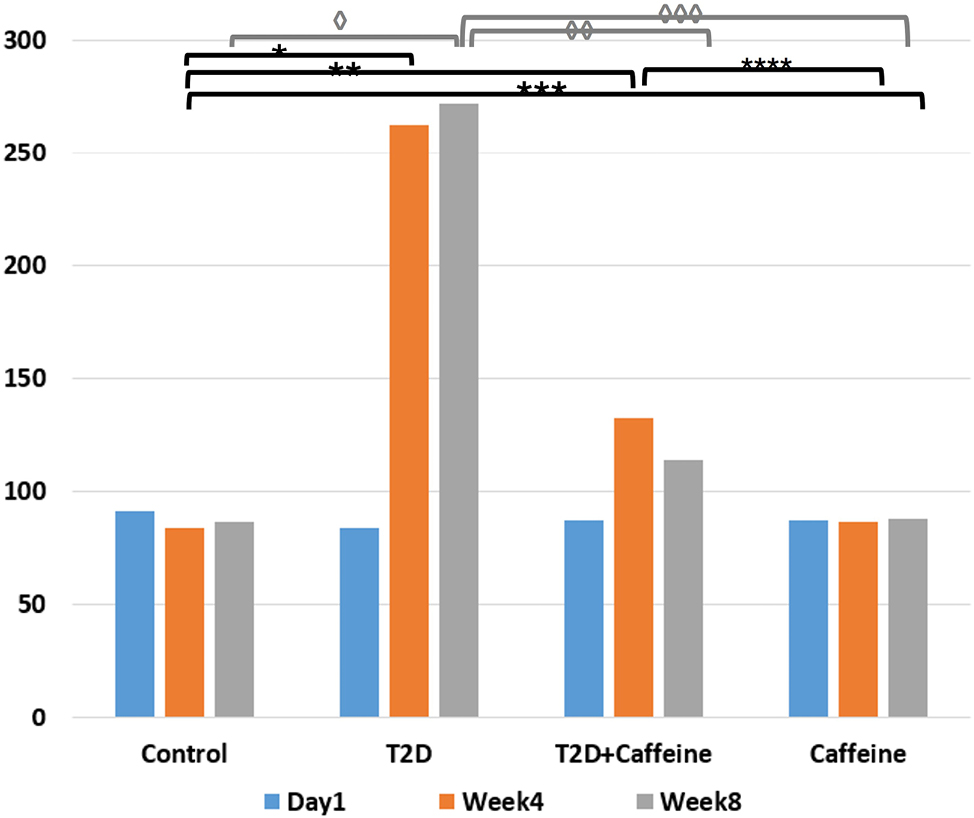
At day 1, blood glucose levels were within normal range with no statistically significant difference among the groups (ANOVA; F=0.668, P= 0.578). At week 4, significant difference was detected between the groups (ANOVA: F=121.87, P<0.0001). Group 2 demonstrated markedly elevated blood glucose levels compared with the other groups, while group 3 showed mildly increased levels. Groups 1 and 4 maintained relatively stable glucose levels. At week 8, significant intergroup differences persisted (ANOVA: F = 149, P <0.0001). Group 2 continued to exhibit the highest blood glucose levels, followed by group 3, which exhibited near normal values, whereas group 1 and 4 remained with comparable ranges (Figure 1).
2. Histological study
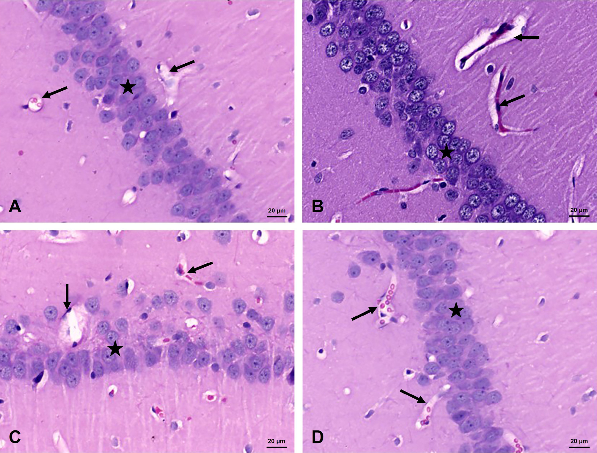
Evaluation of H&E sections of the CA1 (Cornu Ammonis 1) of the hippocampal region of the control group displayed normal structure with small pyramidal neurons that exhibited pale cytoplasm and nucleus. Blood capillaries appeared intact, together with other glial cells. The cells of the diabetic group appeared with dark cytoplasm and nucleus, and the blood capillaries looked disorganized. After caffeine administration, there was improvement of many cells and capillaries appeared intact. The histological sections of rats that received only caffeine, which serves as a control as well, revealed results similar to the control (Figure 2).
3. Immunohistochemistry
3.a. Caffeine reversed α SMA downregulation
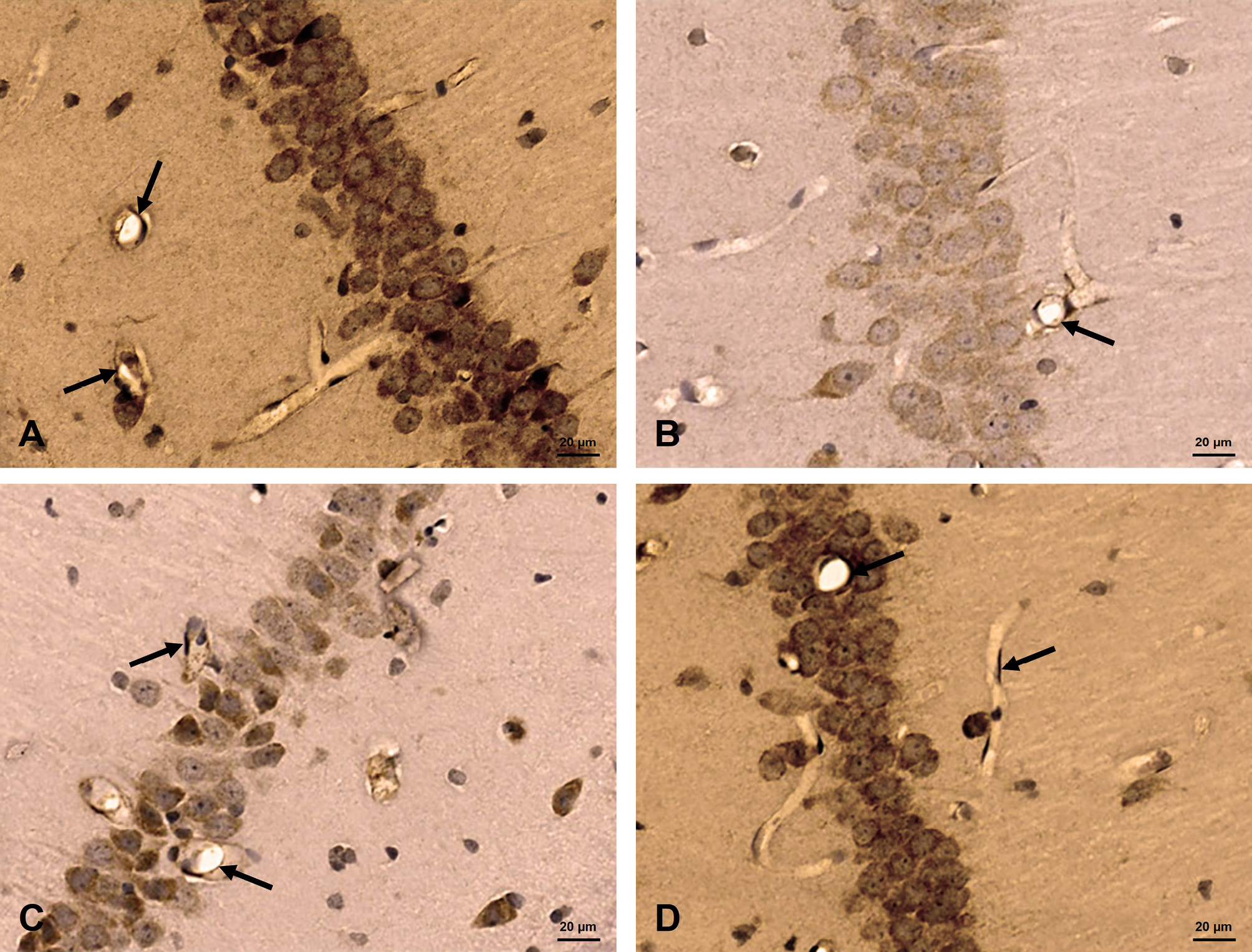
The α SMA immunohistochemistry of CA1area of the hippocampus of T2D rats showed reduction of immunoreactivity of the pericytes in the blood vessels, compared to the controls. Diabetic animals which were administered caffeine displayed upregulation of α SMA expression (Figure 3).
3.b. Caffeine reversed occludin downregulation
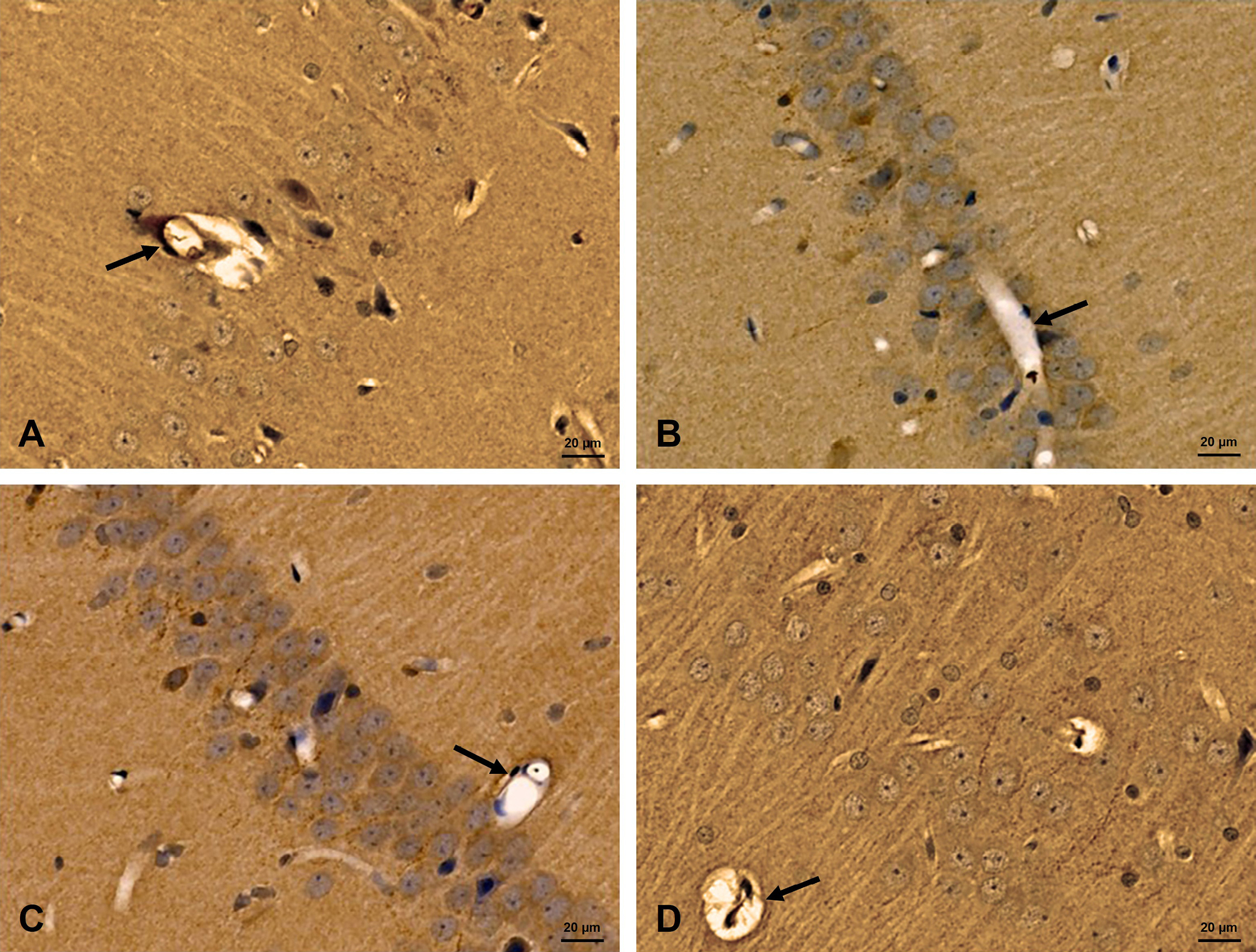
The occludin immunohistochemistry, which is a marker of tight junction, of CA1area of the hippocampus of T2D animals showed reduction of immunoexpression, compared to the controls. Diabetic animals which were administered caffeine displayed an increase of occludin immunoreactivity (Figure 4).
3.c. Caffeine normalized TGF-β1 upregulation
For the TGF-β1 immunohistochemistry, CA1area of the hippocampus of diabetic rats showed marked immunoreaction, compared to the controls. Diabetic rats that received caffeine revealed decrease of TGF-β1 immunoreactivity (Figure 5).
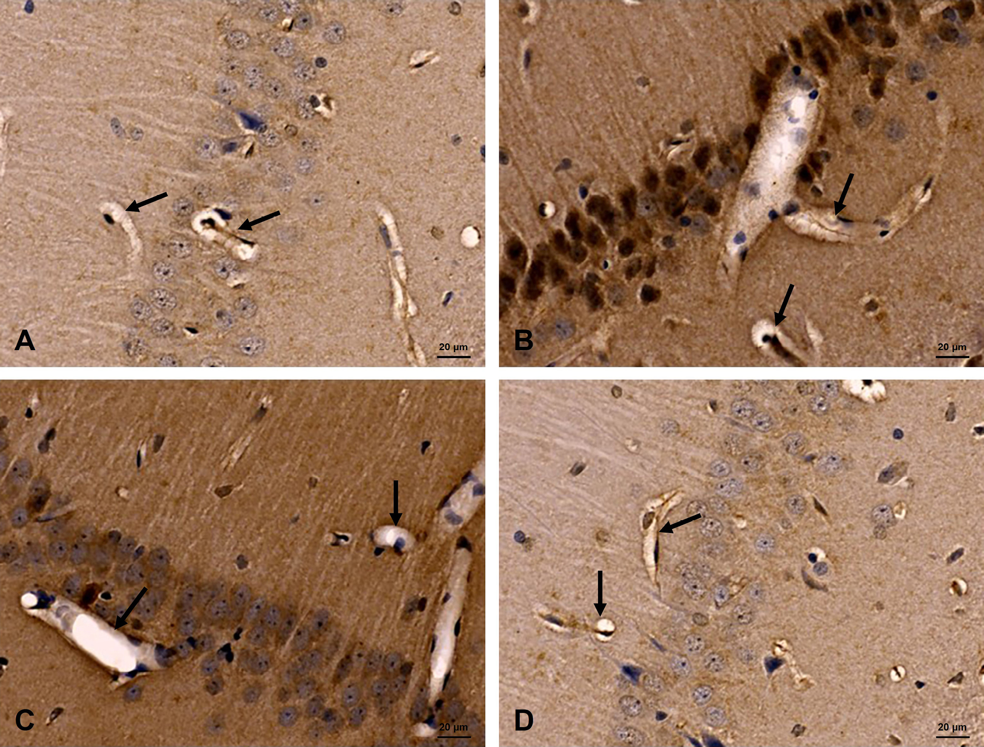
3.d. Quantitative analysis of immunohistochemical marker expression
Quantitative evaluation of the percent area of αSMA and occludin immunoreactivity showed a significant decrease in the immunoreaction in diabetic rats in relation to the controls, on the other hand, caffeine pretreatment to diabetic rats increased their reaction. For TGF-β1 expression, there was an increase in the reactivity in T2D animals in relation to the control, meanwhile caffeine administration to diabetic rats resulted in downregulation of its expression (Table 1).
| α SMA positive area Mean ± SD |
Occludin positive area Mean ± SD |
TFG β1 positive area Mean ± SD |
|||||||||||||||||
|---|---|---|---|---|---|---|---|---|---|---|---|---|---|---|---|---|---|---|---|
| Control | 11.05±0.9 | 14.36±0.831 | 7.17±0.7 | ||||||||||||||||
| D.M | 3.04 a,c,d ±0.557 | 6.25 a,c,d ±1.1 | 15.5 a,c,d ±1.04 | ||||||||||||||||
| D.M + Caffeine | 7.73 a,b,d ±0.775 | 10.45 a,b,d ±1.1 | 9.93 a,b,d ±0.973 | ||||||||||||||||
| Caffeine | 10.3 b,c ±0.654 | 13.43 b,c ±1.2 | 6.93 b,d ±1.401 | ||||||||||||||||
| Standard deviation (SD), one way analysis of variance (ANOVA) was used followed by Tukey post hoc test for multiple comparisons a significant compared to control group, b significant compared to DM group, c significant compared to DM treated with Caffein group, d significant compared to caffein only treated group. | |||||||||||||||||||
Discussion
Type 2 diabetes is becoming a serious metabolic disease with potential disabling complications. The pathophysiology of the disease is complicated and multifactorial that makes management a real challenge. In this study we are reporting the effects of caffeine as a treatment modality in a rat model of T2D by investigating the integrity of the BBB. Diabetes is a metabolic disease characterized by chronic hyperglycemia which may lead to injury to vital organs due to damage to microvasculature, which might affect neuronal function, and white matter disruption (Rom et al., 2020; Zhou et al., 2023). It was reported in the literature that the breakdown of BBB is linked to diabetes progression and the exact mechanisms for BBB permeability and therapeutic approaches, is not currently known (Rom et al., 2020). In this study we found that T2D disrupts the integrity of BBB through affecting pericytes and tight junction proteins. Staining with H & E detected degeneration of hippocampal neurons in diabetic rats, which might be related to BBB leakage and neurotoxic or vascular substances, which may exaggerate neuronal destruction (Lin et al., 2021). Leakage of BBB was evidenced in diabetic animal models as well as in human studies (Liu et al., 2021; Meijer and Gorter, 2024). The current study found downregulation of the αSMA, which is a marker of pericytes, in diabetic rats, a finding that signifies loss of the contractile ability of pericytes and pericyte dysfunction (Liu et al., 2021). The α-SMA was chosen as a marker for the pericytes, while it is not specific, and it stains other cell types and this presents a limitation to our study. In figure 3 we are illustrating the blood capillary with a position more suitable for pericytes. The pericytes can be stained with more specific markers, and this will be investigated in detail in our future studies. Diabetes is known to increase the risk of dementia and neurodegeneration, but the effects of hyperglycemia on pericyte/BBB function is controversial. Some studies proposed that the dysfunction of pericytes and BBB affection is related to oxidative stress and reduced ATP production resulting from prolonged hyperglycemia (Liu et al., 2021). Under physiological conditions, the pericytes are crucial for the formation of the BBB, regulation of neurovascular system, inflammatory response, and removal of toxic materials from the brain (Lin et al., 2021). In the current study, in diabetic rats, there was a decline in the occludin expression levels, which is a tight junction protein, suggesting impaired BBB as previously reported in mice, where they found degradation of TJ proteins, which can suggest it to be a mediator of BBB dysfunction (Machida et al., 2017). Immunohistochemical evaluation, in this study, revealed increased expression of TGF-β1 in diabetic rats, compared to controls, which decreased after caffeine administration. Physiological TGF-β is important for BBB maintenance, through regulation of endothelial-pericyte interactions. The increased TGF-β1 expression in diabetic animals may represent a compensatory protective mechanism, which may become maladaptive when chronically activated (Li et al., 2022). In this context, the caffeine induced reduction of TGF-β1 detected in this study may indicate normalization of the TGF-β1 overactivation rather than inhibition of its protective functions. Pericytes release certain factors, to improve the integrity of the tight junctions and enhance the stability of the BBB as TGF-β, and through this TGF-β signaling pathway, pericytes activate tight junction protein expression as occludin and others, which are important for formation of a high resistant barrier (Chaulagain et al., 2023). The release of TGF-β might result from thickening of the basement membrane, which may activate fibronectin release by pericytes (Li et al., 2022). In the absence of pericytes, endothelial cells display increased permeability allowing dysregulated paracellular transport and jeopardize the function of BBB (Kim et al., 2025). Previously, the expression of these proteins was detected in another study, in diabetic rats, which they may be associated with pericyte dysfunction, and loss of tight junction integrity of endothelial cells (Fan et al., 2015). In the current study caffeine was administered to the diabetic animals, which led to improvement of the BBB parameters tested and structure of hippocampal neurons. When caffeine is consumed on a regular basis, it enhances adaptive mechanisms in the hippocampus, which may affect the primary metabolism and modulate synaptic plasticity (Lopes et al., 2023). Additionally, this role of caffeine in neuroprotection might be due to anti-inflammatory and glial cell modulation (da Silva Neves et al., 2025). This study is focusing on the role played by caffeine to enhance BBB function, by reversing pericyte loss and enhancing tight junction protein efficiency. Caffeine can provide neuroprotection which is related to its ability to block adenosine receptors (Lopes et al., 2023). Caffeine has both hydrophilic and lipophilic nature, it can cross biological membranes, including BBB, thus having stimulatory effects in the central nervous system. Additionally, caffeine has anti-inflammatory, and antioxidative effects and neuroprotective effects of caffeine were reported by previous studies in some neurodegenerative conditions and stroke (Sharma et al., 2025). A meta-analysis in 2020 observed that regular intake of caffeine revealed a lowered risk of Parkinson disease and its progression (Hong et al., 2020). The relation between caffeine intake and the risk of dementia and AD was also investigated in another meta-analysis in 2024; they detected lowered risk of dementia and AD, with caffeine intake (Li et al., 2024). Caffeine has been shown to have a significant role in protecting against damage of the brain such as neurodegeneration, and cognitive impairment. Caffeine has a role in blocking oxidative stress and controlling inflammatory pathways in brain injuries. The anti-inflammatory and antioxidant roles are not through blocking of adenosine receptors, indicating alternative pathways (Vargas-Pozada et al., 2022; Awashra et al., 2026). A role for activated microglia and release of certain cytokines such as IL-6 and TNF-α, can mediate the neurotoxicity. These anti-inflammatory effects of caffeine can be the mechanism of the neuroprotection via minimizing damage to tissues and improving survival of neurons (Othman et al., 2023). An in vivo and invitro study, investigating the protective role of caffeine in lipopolysaccharide-induced injury to retinal cells; they found decrease in inflammatory expression of IL6, TNFα, IL-β1. Additionally, caffeine upregulated the lowered tight junction protein (ZO-1) expression (Conti et al., 2022). This aligns nicely with the findings in this study regarding the effect of caffeine in enhancing BBB integrity and occludin upregulation. It was reported in the literature that the breakdown of the BBB is one of the mechanisms of dementia and Alzheimer’s disease (AD) (Rom et al., 2020; Kim et al., 2025). The effects of caffeine in protecting against AD can be due to blocking of adenosine receptors, which are important in the formation of amyloid beta, which mediates neurotoxicity in neuronal culture models of AD (Stockwell et al., 2017). Caffeine can also be considered a neuroprotective drug in cases of PD for delaying the progress of the disease, which is mediated by blocking adenosine receptors, that suppresses dopamine 2 receptor, and via antioxidant effect (reviewed in: Song et al., 2024).
Limitations of the study
“A limitation of the current study is the use of relatively high caffeine dose (100 mg/kg body weight). This dose was chosen based on our previous studies using the same diabetic rat model, in which the same dose revealed neuroprotective effects to the brain by ameliorating inflammation and gliosis (Othman et al., 2023). Additionally, a study from other researchers, used several doses of caffeine (10-120 mg/kg body weight), in a mouse model of sleep deprivation (Onaolapo et al., 2015). Although this dose exceeds typical human caffeine consumption values, there is probably a species-specific difference in metabolism and pharmacokinetics, which may make this dose achieve pharmacological effects in the animal model, rather than mimic typical human dietary consumption. Future studies using lower doses of caffeine are warranted”. This study did not include electron microscopic evaluation, which could have addressed detailed structure of the BBB. Additionally, behavioral testing was not performed, which might have highlighted the cognitive function. These studies are among our future plans to have more insights about the BBB, and the functional impact of its compromise.
Conclusion
The current study showed structural changes of different components of the BBB in the CA1 area of hippocampus in a rat model of T2D. Additionally, it highlighted the ameliorative effects of caffeine against these changes that could be related to its ability to enhance the BBB. Future studies are in progress to investigate the role of caffeine in such protective roles.
Author contributions
Conceptualization: MA Othman, YI Tayem. Laboratory work: A Rashid, N Bakhit. Manuscript drafting: MA Othman, N Bakhit. Data analysis: A Rashid, N Bakhit. Critical review of the manuscript: MA. Othman, YI Tayem. Final manuscript approval: All authors.
Acknowledgements
The authors declare no conflicts of financial or non-financial interests. Data are available with the corresponding author and can be shared upon request. The authors thank Mrs. Vengilyn Garcia, the secretary of Anatomy department for administrative and processing support.
Funding
This study was fully funded by a grant from the Arabian Gulf University, grant # E006-I-04/17.
References
Aderinto N., Olatunji G., Abdulbasit M., Ashinze P., Faturoti O., Ajagbe A., Ukoaka B., Aboderin G. (2023) The impact of diabetes in cognitive impairment: A review of current evidence and prospects for future investigations. Medicine. Oct 27;102(43):e35557. https://doi.org/10.1097/MD.0000000000035557.
Awashra A., AbuBaha M., Mahafdah B., Sarama A., Alattar D., AbuBaha B., Abu-Khazneh O., Emara A, Milhem A., Elgendy M.S., Shubietah A. (2026) Caffeine as a cardiovascular protective agent: a comprehensive review of mechanisms and outcomes. European journal of medical research. Mar 4. https://doi.org/10.1186/s40001-026-04089-2.
Bancroft J.D., Gamble M., Suvarna S.K., Layton C. (2019) Theory and practice of histological techniques. 8th ed. Philadelphia: Elsevier.
Basak M., Laskar M.A. (2024) Pathophysiology, lifestyle intervention and complications of Type-2 diabetes: A review. Journal of Applied Pharmaceutical Research. Jun 30;12(3):01-10. https://doi.org/10.69857/joapr.v12i3.482.
Chaulagain B., Gothwal A., Lamptey R.N., Trivedi R., Mahanta A.K., Layek B., Singh J. (2023) Experimental models of in vitro blood–brain barrier for CNS drug delivery: an evolutionary perspective. International journal of molecular sciences. Jan 31;24(3):2710. https://doi.org/10.3390/ijms24032710.
Chen T., Dai Y., Hu C., Lin Z., Wang S., Yang J., Zeng L., Li S., Li W. (2024) Cellular and molecular mechanisms of the blood-brain barrier dysfunction in neurodegenerative diseases. Fluids Barriers CNS. Jul 19;21(1):60. https://doi.org/10.1186/s12987-024-00557-1.
Conti F., Lazzara F., Romano G.L., Platania C.B.M., Drago F., Bucolo C. (2022) Caffeine protects against retinal inflammation. Front Pharmacol. Jan 6;12:824885. https://doi.org/10.3389/fphar.2021.824885.
da Silva Neves, L., de Mattos, G.V.R.M., Oliveira-Nazareth, Y., da Silva, R.S. and Campello-Costa, P. (2025) Effects of caffeine on neuroinflammation in anxiety and depression: a systematic review of rodent studies. Translational Psychiatry, 15(1), p.477. https://doi.org/10.1038/s41398-025-03668-x.
El‐Ghazawi K., Eyo U.B., Peirce S.M. (2024) Brain microvascular pericyte pathology linking Alzheimer’s disease to diabetes. Microcirculation. Oct;31(7):e12877. https://doi.org/10.1111/micc.12877.
Fan F., Geurts A.M., Murphy S.R., Pabbidi M.R., Jacob H.J., Roman R.J. (2015) Impaired myogenic response and autoregulation of cerebral blood flow is rescued in CYP4A1 transgenic Dahl salt-sensitive rat. Am J Physiol Regul Integr Comp Physiol 308: R379–R390. https://doi.org/10.1152/ajpregu.00256.2014.
Garg P., Duggal N. (2022) Type 2 diabetes mellitus, its impact on quality of life and how the disease can be managed-a review, Obesity Medicine, Volume 35, 100459, https://doi.org/10.1016/j.obmed.2022.100459.
Gopal S.S., Kaur M., Lanzkron S., Srivastava A.K. (2026) The breakdown of neurovascular barriers: molecular mechanisms of tight junction dysfunction. Molecular Neurobiology. Jan;63(1):273. https://doi.org/10.1007/s12035-025-05592-z.
Han Y., Wu H., Sun S., Zhao R., Deng Y., Zeng S., Chen J. (2023) Effect of high fat diet on disease development of polycystic ovary syndrome and lifestyle intervention strategies. Nutrients. May 8;15(9):2230. https://doi.org/10.3390/nu15092230.
Hong C.T., Chan L., Bai C.H. (2023) The Effect of Caffeine on the Risk and Progression of Parkinson’s Disease: A Meta-Analysis. Nutrients. 2020;12:1860. https://doi.org/10.3390/nu12061860. Erratum in Nutrients, 15, 699.
Ikeda-Murakami K., Tani N., Ikeda T., Aoki Y., Ishikawa T. (2022) Central Nervous System Stimulants Limit Caffeine Transport at the Blood-Cerebrospinal Fluid Barrier. Int J Mol Sci. Feb 7;23(3):1862. https://doi.org/10.3390/ijms23031862.
Kadry H., Noorani B., Cucullo L. (2020) A blood-brain barrier overview on structure, function, impairment, and biomarkers of integrity. Fluids Barriers CNS. Nov 18;17(1):69. https://doi.org/10.1186/s12987-020-00230-3.
Kim S., Jung U.J., Kim S.R. (2025) The Crucial Role of the Blood-Brain Barrier in Neurodegenerative Diseases: Mechanisms of Disruption and Therapeutic Implications. J Clin Med. Jan 9;14(2):386. https://doi.org/10.3390/jcm14020386.
Lenzi F.R., Filardi T. (2023) Social determinants of vulnerabilities in type 2 diabetes: a call to action. Journal of Endocrinological Investigation. Apr;46(4):841-4. https://doi.org/10.1007/s40618-022-01952-x.
Li F., Liu X., Jiang B., Li X., Wang Y., Chen X., Su Y., Wang X., Luo J., Chen L., et al. (2024) Tea, coffee, and caffeine intake and risk of dementia and Alzheimer’s disease: A systematic review and meta-analysis of cohort studies. Food Funct. 15:8330–8344. https://doi.org/10.1039/D4FO01750A.
Li X., Cai Y., Zhang Z., Zhou J. (2022) Glial and Vascular Cell Regulation of the Blood-Brain Barrier in Diabetes. Diabetes \& Metabolism Journal. 46: 222-238. https://api.semanticscholar.org/CorpusID:247521156}
Lin L., Wu Y., Chen Z., Huang L., Wang L., Liu L. (2021) Severe Hypoglycemia contributing to cognitive dysfunction in diabetic mice is associated with pericyte and blood-brain barrier dysfunction. Front Aging Neurosci. Nov 26;13:775244. https://doi.org/10.3389/fnagi.2021.775244.
Liu Y., Zhang H., Wang S., Guo Y., Fang X., Zheng B., Gao W., Yu H., Chen Z., Roman R.J., Fan F. (2021) Reduced pericyte and tight junction coverage in old diabetic rats are associated with hyperglycemia-induced cerebrovascular pericyte dysfunction. Am J Physiol Heart Circ Physiol. Feb 1;320(2):H549-H562. https://doi.org/10.1152/ajpheart.00726.2020.
Lopes C.R., Oliveira A., Gaspar I., Rodrigues M.S., Santos J., Szabó E., Silva H.B., Tomé Â.R., Canas P.M., Agostinho P., Carvalho R.A., Cunha R.A., Simões A.P., Lopes J.P., Ferreira S.G. (2023) Effects of Chronic Caffeine Consumption on Synaptic Function, Metabolism and Adenosine Modulation in Different Brain Areas. Biomolecules. Jan 4;13(1):106. https://doi.org/10.3390/biom13010106.
Machida T., Dohgu S., Takata F., Matsumoto J., Kimura I., Koga M., Nakamoto K., Yamauchi A., Kataoka Y. (2017) Role of thrombin-PAR1-PKCθ/δ axis in brain pericytes in thrombin-induced MMP-9 production and blood-brain barrier dysfunction in vitro. Neuroscience. 2017 May 14;350:146-157. https://doi.org/10.1016/j.neuroscience.2017.03.026.
Meijer W.C., Gorter J.A. (2024) Role of blood–brain barrier dysfunction in the development of poststroke epilepsy. Epilepsia. 65:2519–2536. https://doi.org/10.1111/epi.18072.
Mezo-González C.E., Daher Abdi A., Reyes-Castro L.A., Olvera Hernández S., Almeida C., Croyal M., Aguesse A., Gavioli E.C., Zambrano E., Bolaños-Jiménez F. (2022) Learning deficits induced by high-calorie feeding in the rat are associated with impaired brain kynurenine pathway metabolism. International Journal of Tryptophan Research. Jul;15:11786469221111116. https://doi.org/10.1177/11786469221111116.
Moran C., Beare R., Wang W., Callisaya M., Srikanth V. (2019) Alzheimer’s Disease Neuroimaging Initiative (ADNI), Alzheimer’s Disease Neuroimaging Initiative (ADNI). Type 2 diabetes mellitus, brain atrophy, and cognitive decline. Neurology. Feb 19;92(8):e823-30. https://doi.org/10.1212/WNL.0000000000006955.
Onaolapo O.J., Onaolapo A.Y., Akanmu M.A., Olayiwola G. (2015) Caffeine/sleep-deprivation interaction in mice produces complex memory effects. Ann Neurosci 22(3):139–149. https://doi.org/10.5214/ans.0972.7531.220304.
Othman M.A., Fadel R., Tayem Y., Jaradat A., Rashid A., Fatima A., Al-Mahameed A.E., Nasr El-Din W.A. (2023). Caffeine protects against hippocampal alterations in type 2 diabetic rats via modulation of gliosis, inflammation and apoptosis. Cell Tissue Res 392, 443–466 https://doi.org/10.1007/s00441-022-03735-5
Ősz B.E., Jîtcă G., Ștefănescu R.E., Pușcaș A., Tero-Vescan A., Vari C.E. (2022) Caffeine and its antioxidant properties—it is all about dose and source. International journal of molecular sciences. Oct 28;23(21):13074. https://doi.org/10.3390/ijms232113074.
Paxinos G., Watson C. (2013) The rat brain in stereotaxic coordinates, 7th edn. Academic Press, Amsterdam.
Reyahi A., Nik A.M., Ghiami M., Gritli-Linde A., Pontén F., Johansson B.R., Carlsson P. (2015) Foxf2 Is Required for Brain Pericyte Differentiation and Development and Maintenance of the Blood-Brain Barrier. Dev Cell. Jul 6;34(1):19-32. https://doi.org/10.1016/j.devcel.2015.05.008.
Rom S., Heldt N.A., Gajghate S., Seliga A., Reichenbach N.L., Persidsky Y. (2020) Author Correction: Hyperglycemia and advanced glycation end products disrupt BBB and promote occludin and claudin-5 protein secretion on extracellular microvesicles. Sci Rep. 2020 Oct 28;10(1):18828. https://doi.org/10.1038/s41598-020-75940-7. Erratum for: Sci Rep. 2020 Apr 29;10(1):7274. https://doi.org/10.1038/s41598-020-64349-x.
Ruggiero M., Calvello R., Porro C., Messina G., Cianciulli A., Panaro M.A. (2022) Neurodegenerative diseases: Can caffeine be a powerful ally to weaken neuroinflammation?. International Journal of Molecular Sciences. Oct 26;23(21):12958. https://doi.org/10.3390/ijms232112958.
Sharma B., Agriantonis G., Dawson-Moroz S., Brown R., Simon W., Ebelle D., Chapelet J., Cardona A., Soni A., Siddiqui M., Patel B., Cheerasarn S., Chang J., Cobb L., John F., Hasan M.M., Garcia C., Shaefee Z., Twelker K.D., Bhatia N., Whittington J. (2025) Caffeine: A Neuroprotectant and Neurotoxin in Traumatic Brain Injury (TBI). Nutrients. Jun 4;17(11):1925. https://doi.org/10.3390/nu17111925.
Sharma V.K., Sharma A., Verma K.K., Gaur P.K., Kaushik R., Abdali B. (2023) A comprehensive review on pharmacological potentials of caffeine. Journal of Applied Pharmaceutical Sciences and Research. Dec 14;6(3):16-26. https://doi.org/10.31069/japsr.v6i3.04.
Song X., Singh M., Lee K.E., Vinayagam R., Kang S.G. (2024) Caffeine: A multifunctional efficacious molecule with diverse health implications and emerging delivery systems. Int J Mol Sci. Nov 8;25(22):12003. https://doi.org/10.3390/ijms252212003.
Stockwell J., Jakova E., Cayabyab F.S. (2017) Adenosine A1 and A2A Receptors in the Brain: Current Research and Their Role in Neurodegeneration. Molecules. 22:676. https://doi.org/10.3390/molecules22040676.
Szablewski L. (2025) Associations Between Diabetes Mellitus and Neurodegenerative Diseases. International Journal of Molecular Sciences. 26(2):542. https://doi.org/10.3390/ijms26020542.
Yu T., Wang Z., Chen Y., Xiang Y., Wu M., Zhang M., Yin X., Chen Z. (2025) Blood–brain barrier (BBB) dysfunction in CNS diseases: paying attention to pericytes. CNS Neuroscience & Therapeutics. May;31(5):e70422. https://doi.org/10.1111/cns.70422.
Vargas-Pozada E.E., Ramos-Tovar E., Rodriguez-Callejas J.D., Cardoso-Lezama I., Galindo-Gómez S., Talamás-Lara D., Vásquez-Garzón V.R., Arellanes-Robledo J., Tsutsumi V., Villa-Treviño S., et al. (2022) Caffeine Inhibits NLRP3 Inflammasome Activation by Downregulating TLR4/MAPK/NF-ΚB Signaling Pathway in an Experimental NASH Model. Int. J. Mol. Sci. 23:9954. https://doi.org/10.3390/ijms23179954.
Zhang L., Huang Y.J., Sun J.P., Zhang T.Y., Liu T.L., Ke B., Shi X.F., Li H., Zhang G.P., Ye Z.Y., Hu J. (2021) Protective effects of calorie restriction on insulin resistance and islet function in STZ-induced type 2 diabetes rats. Nutrition & metabolism. May 5;18(1):48. https://doi.org/10.1186/s12986-021-00575-y.
Zhang S., Zhang Y., Wen Z., Yang Y., Bu T., Bu X., Ni Q. (2023) Cognitive dysfunction in diabetes: abnormal glucose metabolic regulation in the brain. Frontiers in endocrinology. Jun 16;14:1192602. https://doi.org/10.3389/fendo.2023.1192602.
Zhou Y., Wei L., Gao S., Wang J., Hu Z. (2023) Characterization of diffusion magnetic resonance imaging revealing relationships between white matter disconnection and behavioral disturbances in mild cognitive impairment: a systematic review. Front. Neurosci. 17:1209378. https://doi.org/10.3389/fnins.2023.1209378.