https://doi.org/10.36253/ijae-17095
Embedding virtual tools and innovative approaches in neuroanatomy teachings: A single center pilot and feasibility experience
Experimental Neurology Unit, School of Medicine and Surgery, University of Milano-Bicocca, Monza, Italy
NeuroMI (Milan Center for Neuroscience), Milan, Italy
E-mail: paola.alberti@unimib.it
Abstract. A solid knowledge of neuroanatomy is essential given the rapidly increasing use of neuroimaging in clinical practice. As a result, developing innovative and engaging methods to support students throughout the learning process has become a central topic of interest. Implementing digital tools and interactive approaches represents an elegant and promising solution. At our University, the international degree program in Medicine and Surgery (IMS)—taught entirely in English and welcoming students from all over the world—was established in the 2017–2018 academic year. IMS follows a specific framework for teaching human anatomy: during the first year, students attend a course entitled Fundamentals of Human Morphology, which provides the foundational knowledge needed for their training. In subsequent years, anatomy is revisited through the so-called vertical tracks, in which it is presented from a clinically oriented perspective. Moreover, the number of IMS students is smaller than in the Italian course (n < 50), making IMS an ideal environment in which to introduce and implement innovative strategies relying heavily on virtual dissections, gamification, and flipped-classroom approaches in a pilot and feasibility phase. In this paper, we describe the implementation of the advanced neuroanatomy course, designed to achieve a seamless integration of frontal lectures and interactive teaching methods. Survey results from 30 voluntary participants demonstrated high satisfaction, with median scores of 9.5/10 for the use of the virtual dissection table compared to textbooks, and 10/10 for the gamification tournament. Knowledge retention improved from a mean baseline score of 64% correct answers in the pre-test to over 85% of students answering all questions correctly in the post-test, confirming the feasibility and potential efficacy of this interactive curriculum.
Keywords: virtual dissection, innovative teaching methods, gamification, flipped classroom, human anatomy.
Contents
Introduction
A comprehensive and rigorous understanding of neuroanatomy is essential for the management of disorders involving both the central and peripheral nervous system. Such knowledge is fundamental for the correct interpretation of the neurological examination and for the initiation of an appropriate diagnostic workup, which relies extensively on neuroanatomically based investigations including computed tomography (CT), magnetic resonance imaging (MRI), nuclear medicine techniques, and ultrasound (US) [1-5], as well as neurophysiological assessment [6]. Despite often being regarded as a domain primarily relevant to specialists in neuro-psychiatric disciplines – such as neurology, neurosurgery, psychiatry, child neurology/psychiatry, neuroradiology, and physiotherapy – neuroanatomy now plays a crucial role across a broader spectrum of healthcare professionals.
In the emergency setting, recent advancements in the treatment of acute ischemic stroke [7-10] and the incorporation of endovascular therapy into standard clinical practice [11] have reshaped organizational models [11,12], thereby requiring multidisciplinary teams to possess adequate neuroanatomical competence. Similarly, US has gained prominence beyond its traditional application in the diagnosis of peripheral neuropathies [3,4], becoming an essential tool for guiding procedures such as regional anesthesia and pain management [13-16]. These developments collectively underscore how neuroanatomy has evolved into an indispensable component of contemporary clinical practice, extending well beyond the boundaries of classical neuro specialties.
Stemming from these considerations, we introduced a tentative approach aimed at developing novel strategies to empower students and actively engage them in the learning process, with the goal of fostering a solid, clinically oriented neuroanatomical foundation that would remain stable over time. At our University, the international degree program in Medicine and Surgery (IMS) is structured in a way that is particularly suitable for this purpose. During the first year, students attend the Fundamentals of Human Morphology course, which provides the essential basis for their anatomical training. In the following years, anatomy is revisited within the so-called vertical tracks, where it is taught from a clinically oriented perspective. As an example, we describe here the implementation of this framework in the context of neuroanatomy teaching.
The IMS course was established in the 2017–2018 academic year (AY) and was structured differently from the Italian course (IC), not only because all classes are delivered in English – thus accommodating an international cohort of students from around the world – but also in its overall educational design.
During the first two years, the curriculum is intended to build a solid foundation in the basic (preclinical) disciplines, including human anatomy. To this end, as mentioned, students attend the course Fundamentals of Human Morphology, which provides a comprehensive introduction to human anatomy across all organs, systems, and regions. However, this represents only the initial phase of their training.
In the subsequent years, teaching is organized into the so-called vertical tracks, a system of integrated courses in which each medical topic is explored in depth, beginning with clinically oriented anatomy, physiology, and pathology, and progressing toward semiotics, diagnostics, and the nosographic classification and treatment of diseases related to that system.
With regard to neuroanatomy, students in the fourth year (of the six-year program) attend an advanced course embedded within the Neuroscience vertical track. This course is designed to establish a rapid and explicit link with clinical applications. By this stage, students already possess a foundational understanding of neuroanatomy, which they further consolidate to the point of integrating anatomical knowledge seamlessly into clinical reasoning.
Additionally, the relatively small class size – compared with the IC – makes IMS an ideal environment for introducing innovative, interactive teaching strategies. In 2017–2018 AY, the program offered 36 seats, a number that progressively increased to 48 seats by 2023–2024 AY.
Here, we describe how the advanced neuroanatomy course was progressively refined starting in 2021–2022 AY and subsequently consolidated into the format presented in this paper for the most recent academic year (2024–2025 AY, syllabus available here: Summary of Neuroanatomy I | e-Learning - UNIMIB; https://elearning.unimib.it/course/info.php?id=56249).
The primary objective was to assess the feasibility, student acceptance, and preliminary educational impact of a multifaceted, interactive neuroanatomy curriculum within an international medical program. While individual digital tools are increasingly common, the novelty of our approach lies in the seamless, structured integration of continuous virtual dissection, peer-to-peer gamification tournaments, and a flipped-classroom model, tightly aligned with clinical applications in a vertical-track curriculum
Methods
All lessons for this course are delivered in the Anatomy Room, which is equipped with 3D plastic models and a virtual dissection tool, the Anatomage Table™ (Anatomage Inc., Santa Clara, California, US). This device provides 3D reconstructions of five segmented cadavers, enabling hands-on virtual dissections. It also includes its own case library and allows the upload of Digital Imaging and Communication in Medicine (DICOM) data, which can be rendered with photorealistic filters. In addition, the software supports the upload and easy visualization of DICOM datasets originating from users’ own clinical practice, facilitating in-depth discussion of real clinical cases with strong anatomical relevance.
Furthermore, each student received an individual license (purchased by the University) for the companion e-book/platform associated with the Anatomage system, Anatomage Lessons™. Paper-based exercises (primarily structure coloring and labelling activities) were also provided.
During the 2024-2025 AY, each lesson was delivered in a 4-hour session combining both lecture-based and interactive activities. Students (n = 36) were divided into six teams of six members each, with team names chosen by the students themselves. These groups collaborated during the interactive breakout sessions.
At the beginning of the very first lesson, students anonymously completed a self-assessment multiple-choice questionnaire to gauge how much knowledge they had retained from the first-year course. The test was accessed anonymously via mobile phone through a Google form linked to a QR code.
At the start of all subsequent lessons, the class was divided into groups and each session opened with an assignment. The Anatomage Table™ quiz mode enabled the creation of a tournament: each group, one at a time, performed an 8-minute dissection exercise, generating a score that was recorded. Each quiz was designed to review and reinforce the topic covered in the previous lesson. While one group was engaged in this activity, the others worked collaboratively on exercises using the Anatomage Lessons™ platform and paper-based materials.
The lecture-based portion of the lesson then followed. Topics were presented through a combination of virtual dissections and minimal use of traditional slide decks, which were reserved primarily for diagrams and summaries. After each topic was introduced, students again worked in teams: one team at the Table and the others completing paper-based or Anatomage Lessons™ activities.
Considering the available literature on the topic [17-20], a gamification approach was tentatively integrated into the course. During the final lesson, the Table’s quiz mode was used once again to assess all teams on the entire syllabus. By pooling the scores accumulated across all lessons, one team was ultimately declared the winner of the tournament. Finally, students were given the opportunity to retake the self-assessment multiple-choice questionnaire.
Students who attended the neuroanatomy course in the 2024–2025 AY were the same cohort that had received human anatomy instruction exclusively online in 2019–2020 AY due to the COVID-19 pandemic. This made them an ideal group for evaluating whether a more engaging, interactive approach would be perceived as beneficial.
At the end of the course, students were invited to anonymously rate the activities conducted throughout the program using an ad hoc evaluation questionnaire developed via a Google form accessed via a QR code; completing the survey was not mandatory. The evaluation followed the standard 0–10 grading system used by our university for assessing students’ opinions on teaching activities (https://opinionistudenti.unimib.it/valdid/): low (0-4), intermediate (5-7), and high score (8-10).
Results
Thirty students voluntarily and anonimously provided their feedback. Figure 1 summarizes the perceived usefulness of the live dissections performed by the professor during the lecture-based portion of the lessons, showing overall highly satisfactory results.
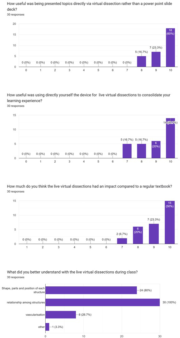
Figure 2 presents students’ ratings of the activities they felt were most empowering. The online materials – offered through Anatomage Lessons™ – were particularly appreciated, also supporting a flipped-classroom approach that is gaining increasing relevance in anatomy education. Since the platform was accessible at all times, students could review topics in advance, as all materials were made available from the very first lesson. All learning modalities (i.e., online materials, paper-based exercises, and the gamification approach) received more than satisfactory scores, although paper-based exercises were rated slightly lower overall.
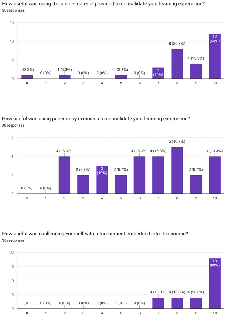
The following median scores were recorded for the most relevant items:
a) use of the Table compared with a regular textbook: 9.5 (Q1: 8.25; Q3: 10);
b) importance of students directly using the Table: 9 (Q1: 8; Q3: 10);
c) use of Anatomage Lessons™: 9 (Q1: 8; Q3: 10);
d) completion of paper-based exercises: 6.5 (Q1: 4; Q3: 8);
e) Anatomage tournament integrated into the lessons: 10 (Q1: 8.25; Q3: 10).
To tentatively assess the effectiveness of the teaching method, performance on the preliminary multiple-choice test was compared with that on the final test. At the beginning of the course, students achieved a mean score of 64% correct answers overall, with only 25% of questions answered correctly by fewer than 45% of students. After completing the course, more than 85% of students answered all questions correctly; moreover, 25% of the questions received correct answers from the entire cohort, and even the question with the lowest success rate was answered correctly by 83% of students. It should be noted, however, that the same questions were used in both tests and, as these were not formal examinations, students may have had access to study materials on both occasions.
Discussion
Our data, although preliminary and primarily intended as a feasibility assessment, were consistent with the published literature, which highlights the importance of virtual and 3D approaches in learning anatomy compared to traditional 2D tools [21-24]. Furthermore, the survey results were once again consistent with previously published approaches to anatomy teaching, highlighting both the relevance of gamification and the value of a student-centered learning environment [19,24-26].
The evaluation of our educational experience inevitably intersects with the global and ongoing debate regarding the role of traditional cadaveric dissection versus digital innovations in modern medical curricula. As recently highlighted in an interactive consensus feature in the New England Journal of Medicine [27], medical schools face critical policy decisions regarding whether to maintain resource-intensive body-bequest programs or fully embrace advanced technological learning tools. Proponents of traditional dissection argue that working with human donor bodies provides irreplaceable, transformative opportunities, acting as a ‘first clinical discipline’ where students acquire emotional competencies, teamwork, and a profound awareness of anatomical variation. Conversely, a recent debate by Shastri and colleagues [28] emphasizes that modern undergraduate medical teaching has shifted toward multimodal, educationally principled curricular designs. They point out that traditional dissection is heavily resource-laden, requiring specialized laboratories and embalming chemicals, and is exceptionally time-intensive within an increasingly packed undergraduate medical curriculum; furthermore, they note that disproportionate student time is often spent removing fat or connective tissue rather than conceptualizing functional anatomy, whereas digital 3D anatomy tools and interactive applications facilitate inclusive, self-paced, and flexible learning with precise structural visualization.
In this context, advanced digital platforms provide an elegant, effective bridge. They allow students to engage with photorealistic 3D anatomical networks in ways that go beyond traditional methods, effectively preparing them for a healthcare environment increasingly dominated by radiologic imaging and digital interfaces. Rather than viewing these modalities as mutually exclusive, our pilot experience suggests that virtual tools can successfully act as powerful pedagogical enhancements that respect the tight time constraints of modern undergraduate training while actively empowering students through interactive, student-centered frameworks
Overall, this experience proved highly promising, and further developments are already underway. During the first year, one of the modules of the Fundamentals of Human Morphology course, Regional Anatomy, has already been delivered using an approach similar to the one described in this paper, starting from 2023–2024 AY. The same applies to a course on the female reproductive system for sixth-year students (implementation initiated in 2024–2025 AY) within the Woman and Child vertical track. Likewise, the Anatomy of the Digestive System course (part of the Digestive Health vertical track) for fifth-year students is currently being revised following the same framework, with implementation beginning in 2025–2026 AY.
In addition to updating the curricula of formal courses, we are also promoting a peer-to-peer tutoring program, whose positive impact was documented in a publication describing its pilot phase; the initiative was highly appreciated by students in both the IMS and IC degree programs [29]. This activity will be further expanded to include peer-to-peer tutors specifically trained to support younger students in the independent use of the Table, as a second Table is being made available in the library and will be accessible to all trained students through a dedicated booking system.
The approaches described above constitute a preliminary pilot phase during which the teaching methods were cautiously upgraded in response to students’ emerging needs. A formal research protocol is being developed to systematically determine the effectiveness and long-term impact of this educational trajectory. Several limitations, in fact, must be acknowledged in this pilot study. First, it is a single-center experience with a relatively small sample size (n = 36, with 30 survey respondents), which may limit the generalizability of our findings to larger student cohorts or different institutional settings. Second, the study lacks a parallel control group (e.g., students undergoing traditional lectures only), preventing a definitive causal conclusion regarding the superior educational efficacy of the digital approach. Third, the evaluation of student satisfaction relies on self-reported outcomes, which are inherently subject to subjective bias. Lastly, because the pre- and post-tests utilized the identical multiple-choice questions in a non-formal examination environment, the observed performance improvement must be interpreted with caution, as it might partly reflect test familiarity or access to study resources rather than long-term knowledge retention. These limitations will be addressed in our upcoming formal research protocol involving a controlled, longitudinal evaluation.
References
[1] Yen C, Lin CL, Chiang MC. Exploring the Frontiers of Neuroimaging: A Review of Recent Advances in Understanding Brain Functioning and Disorders. Life (Basel). 2023;13(7).
[2] Skadorwa T, Kunicki J, Nauman P, Ciszek B. Image-guided dissection of human white matter tracts as a new method of modern neuroanatomical training. Folia Morphol (Warsz). 2009;68(3):135-139.
[3] Coraci D, Masiero S, Padua L. Ultrasound advanced techniques for nerve assessment: One of the fires of PrometheUS. Clin Neurophysiol. 2022;135:164-165.
[4] Coraci D, Loreti C, Fusco A, Giovannini S, Padua L. Peripheral Neuropathies Seen by Ultrasound: A Literature Analysis through Lexical Evaluation, Geographical Assessment and Graph Theory. Brain Sci. 2021;11(1).
[5] Fritz JV. The practice of neuroimaging within a neurology office setting. Neurol Clin Pract. 2013;3(6):501-509.
[6] Kędzia A, Derkowski W. Modern methods of neuroanatomical and neurophysiological research. MethodsX. 2024;13:102881.
[7] Lens C, Demeestere J, Casolla B, et al. From guidelines to clinical practice in care for ischaemic stroke patients: A systematic review and expert opinion. Eur J Neurol. 2024:e16417.
[8] Shakir M, Irshad HA, Lodhi BA, et al. Endovascular thrombectomy after 24 hours for patients with acute ischemic stroke due to large vessel occlusion: A systematic review and meta-analysis of outcomes. Clin Neurol Neurosurg. 2024;247:108610.
[9] Orscelik A, Bilgin C, Cortese J, et al. Comparative analysis of single plane and biplane angiography systems for mechanical thrombectomy for acute ischemic stroke: a systematic review and meta-analysis. J Neurointerv Surg. 2024.
[10] Salim HA, Pulli B, Yedavalli V, et al. Endovascular therapy versus medical management in isolated posterior cerebral artery acute ischemic stroke: A multinational multicenter propensity score-weighted study. Eur Stroke J. 2024:23969873241291465.
[11] Détraz L, Ernst M, Bourcier R. Stroke Transfer and its Organizational Paradigm : Review of Organizational Paradigms and the Impact on Outcome. Clin Neuroradiol. 2018;28(4):473-480.
[12] Ciccone A, Berge E, Fischer U. Systematic review of organizational models for intra-arterial treatment of acute ischemic stroke. Int J Stroke. 2019;14(1):12-22.
[13] Am S, Patel N, Kumar R, et al. Medial versus lateral approach in ultrasound-guided costoclavicular brachial plexus block for upper limb surgery: a randomized control trial. Anaesthesiol Intensive Ther. 2024;56(3):199-205.
[14] Wilcox CM, Bang JY, Buxbaum J, et al. Effect of endoscopic ultrasound guided celiac plexus block on the palliation of pain in chronic pancreatitis (EPOCH Trial): study protocol for a randomized multicenter sham-controlled trial {1}. Trials. 2024;25(1):676.
[15] De Cassai A, Behr A, Bugada D, et al. Anesthesiologists ultrasound-guided regional anesthesia core curriculum: a Delphi consensus from Italian regional anesthesia experts. J Anesth Analg Crit Care. 2024;4(1):54.
[16] Torrano V, Zadek F, Bugada D, et al. Simulation-Based Medical Education and Training Enhance Anesthesia Residents’ Proficiency in Erector Spinae Plane Block. Front Med (Lausanne). 2022;9:870372.
[17] Krishnamurthy K, Selvaraj N, Gupta P, et al. Benefits of gamification in medical education. Clin Anat. 2022;35(6):795-807.
[18] van Gaalen AEJ, Brouwer J, Schönrock-Adema J, Bouwkamp-Timmer T, Jaarsma ADC, Georgiadis JR. Gamification of health professions education: a systematic review. Adv Health Sci Educ Theory Pract. 2021;26(2):683-711.
[19] Ang ET, Chan JM, Gopal V, Li Shia N. Gamifying anatomy education. Clin Anat. 2018;31(7):997-1005.
[20] Perumal V, Dash S, Mishra S, Techataweewan N. Clinical anatomy through gamification: a learning journey. N Z Med J. 2022;135(1548):19-30.
[21] Wesencraft KM, Clancy JA. Using Photogrammetry to Create a Realistic 3D Anatomy Learning Aid with Unity Game Engine. Adv Exp Med Biol. 2019;1205:93-104.
[22] Al-Khalili SM, Coppoc GL. 2D and 3D stereoscopic videos used as pre-anatomy lab tools improve students’ examination performance in a veterinary gross anatomy course. J Vet Med Educ. 2014;41(1):68-76.
[23] Hoyek N, Collet C, Di Rienzo F, De Almeida M, Guillot A. Effectiveness of three-dimensional digital animation in teaching human anatomy in an authentic classroom context. Anat Sci Educ. 2014;7(6):430-437.
[24] Allen LK, Ren HZ, Eagleson R, de Ribaupierre S. Development of a Web-Based 3D Module for Enhanced Neuroanatomy Education. Stud Health Technol Inform. 2016;220:5-8.
[25] Van Nuland SE, Roach VA, Wilson TD, Belliveau DJ. Head to head: The role of academic competition in undergraduate anatomical education. Anat Sci Educ. 2015;8(5):404-412.
[26] Felszeghy S, Pasonen-Seppänen S, Koskela A, et al. Using online game-based platforms to improve student performance and engagement in histology teaching. BMC Med Educ. 2019;19(1):273.
[27] Lin K, Hildebrandt S, McMenamin P. Teaching Anatomy - To Dissect or Not to Dissect? N Engl J Med. 2026;394(18):1862-1864.
[28] Shastri R, Wiles C, Sweeney O, Easton L, Jacques S. Is the dissection of cadavers a necessary part of medical education? BMJ. 2025;388:q2829.
[29] Alberti P. 3D Digital tools in neuro-anatomy teaching: from peer-to-peer tutoring to clinically oriented approaches Italian Journal of Anatomy and Embriology. 2024;128(2):125-134.