https://doi.org/10.36253/ijae-17036
Unusual pulmonary vein variations: A new case report and its clinical significance
1 Department of Experimental and Clinical Medicine, Human Anatomy Section, University of Florence, Florence, Italy
2 Department of Medicine, Surgery and Neuroscience, University of Siena, Siena, Italy
3 UniCamillus - Saint Camillus International University of Health Sciences, Rome, Italy
# These authors share first authorship
*Corresponding author. E-mail: ferdinando.paternostro@unifi.it
Abstract. Anatomical variations in pulmonary circulation, especially in the number and drainage patterns of pulmonary veins, have significant clinical implications. The typical anatomy includes four pulmonary veins, which transport oxygenated blood to the left atrium through distinct ostia. However, variations have been reported in up to 36% of cases, a discovery facilitated by advances in imaging and cadaveric dissection. This study presents the anatomical findings of supernumerary pulmonary veins observed during routine thoracic dissection of a 75-year-old female cadaver. The implications of such variations are critical in cardiothoracic and pulmonary surgery, where unrecognized anatomical differences may lead to complications, including intraoperative bleeding and incomplete resections. Knowledge of these variations is also pivotal to radiological interpretation, as misinterpretation can lead to diagnostic errors. This study emphasizes the need for clinicians to be aware of pulmonary venous variability to enhance surgical planning, reduce procedural risks, and improve patient outcomes.
Keywords: pulmonary veins, anatomical variation, left atrium, supernumerary pulmonary vein, cadaveric dissection, clinical anatomy.
Contents
Institutional review board statement
Introduction
It is widely known that the human vasculature commonly exhibits variable patterns in many organs, such as the kidneys [1], aortic arch [2], and thyroid gland [3]; these are therefore regarded as “normal” variations [4].
Regarding anatomical variations in the pulmonary circulation, variability in pulmonary venous ostial anatomy was previously considered rare and documented only in a few case reports [5].
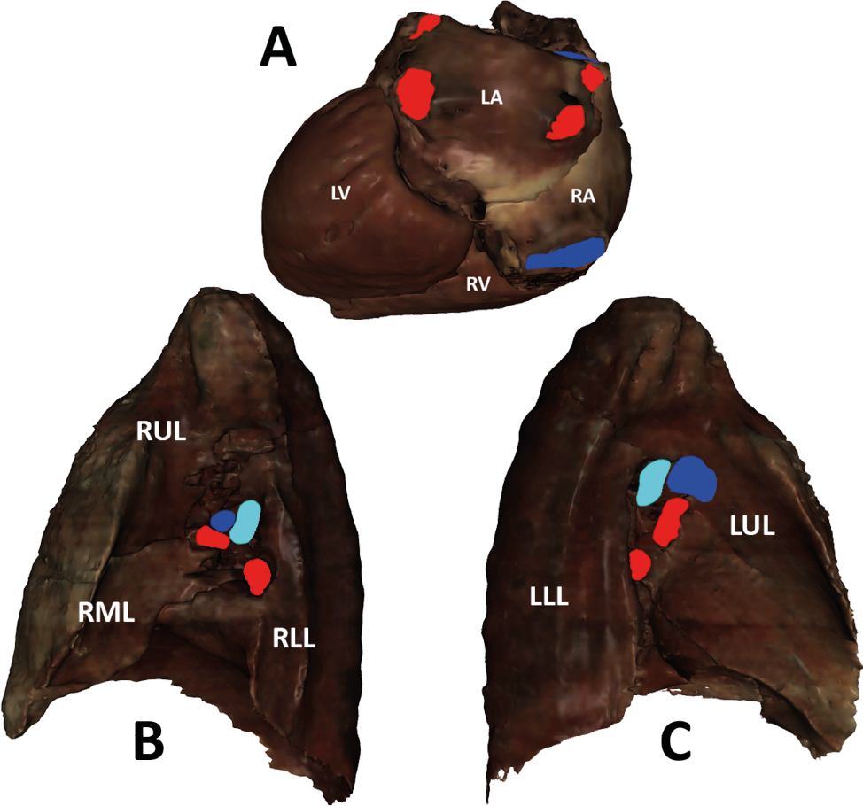
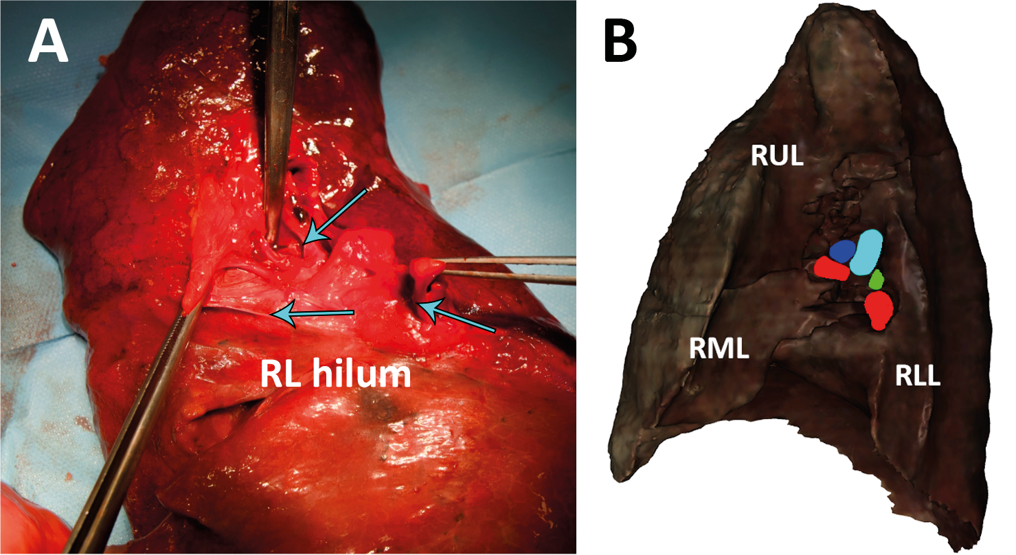
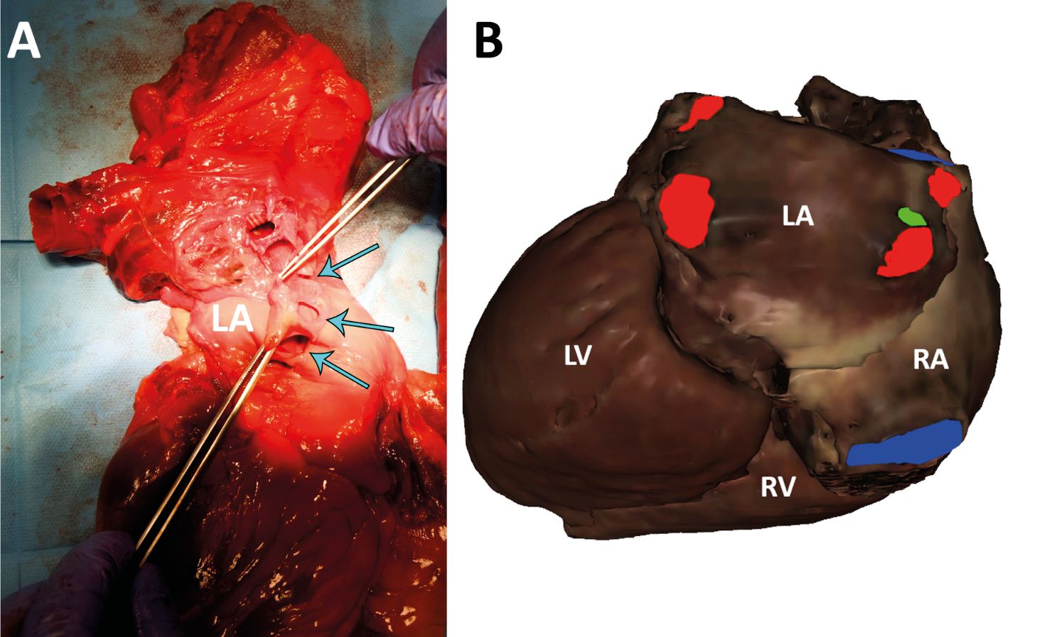
The “normal” anatomy comprises four pulmonary veins, two from each lung, which return oxygenated blood to the left atrium, with each vein opening through a separate ostium (Figure 1). Since the second half of the 20th century, different patterns in the number of pulmonary veins and left atrial ostia have been reported, and these numbers do not always correspond [5]. Accordingly, contrary to earlier assumptions, recent studies have reported variations in pulmonary venous anatomy in approximately 36% of cases. This increased recognition has been facilitated by advances in imaging, non-invasive diagnostic techniques, conventional angiography, and cadaveric dissection [6]. Variations in number and drainage patterns have recently gained increasing attention not only in cardiothoracic surgery and pulmonary lobectomy, because pulmonary veins play an important role in determining segmental lung anatomy [7], but also in interventional procedures such as radiofrequency ablation for atrial fibrillation. Indeed, pulmonary venous variability has been associated with ectopic cardiac activity [8]. The present study aims to highlight the importance of knowledge of pulmonary venous anatomy, focusing on the presence of supernumerary pulmonary veins and their drainage into the left atrial ostia (Figures 2 and 3).
Materials and methods
The anatomical variability of the pulmonary veins was observed in a 75-year-old female cadaver. The specimen was obtained during routine undergraduate thoracic dissection classes held at the ICLO Teaching and Research Center (Verona, Italy), under the supervision of dissection technicians, clinicians, and professors.
The unfixed lung was resected at the hilum, close to the vascular and bronchial pedicles. Any adhesions between the parietal and visceral pleura were removed by hand.
Consent for the use of the self-donated cadaver in this study was obtained from the donor before death.
The study protocol conformed to the guidelines set out by the Declaration of Helsinki.
Discussion
Four pulmonary veins, two superior and two inferior, typically drain oxygenated blood from the lungs into the left atrium through distinct ostia. However, anatomical variations in the number of pulmonary veins can occur. While these variations are often clinically silent, an in-depth understanding of pulmonary venous anatomy and its relationship to the left atrium is essential. This knowledge is critical not only for pre-procedural planning in thoracic and cardiac surgery [10], but also for understanding the association between pulmonary vein anomalies and atrial fibrillation [9].
In addition to radiofrequency ablation for atrial fibrillation, other clinical scenarios demonstrate the significance of pulmonary vein variations. For instance, during pulmonary lobectomies, where the aim is to remove lung segments affected by cancer, knowledge of supernumerary pulmonary veins can help avoid intraoperative complications, such as inadvertent venous injury or excessive bleeding. Misidentification of these vessels may lead to incomplete resections or compromised postoperative lung function. Similarly, in lung transplantation procedures, surgeons must be aware of variations in venous anatomy to ensure successful anastomosis and proper venous drainage, minimizing the risk of graft failure or postoperative complications such as pulmonary vein thrombosis.
Furthermore, variations in pulmonary venous anatomy can affect the interpretation of diagnostic imaging, such as in computed tomography (CT), angiography or magnetic resonance imaging (MRI). Failure to recognize supernumerary veins or abnormal drainage patterns may lead to misdiagnosis of conditions such as pulmonary embolism or to misinterpretation of vascular masses. Radiologists must be familiar with potential anatomical variations to ensure accurate diagnoses and avoid unnecessary interventions.
Advanced diagnostic imaging and cadaveric dissections offer clearer insights into such variations. Arrhythmias such as atrial fibrillation, can be effectively treated through radiofrequency ablation targeting ectopic foci. The success of this procedure relies on precise knowledge of normal pulmonary venous anatomy and its variations [5].
In conclusion, a comprehensive understanding of the anatomical features, including the number of pulmonary veins and left atrial ostia, is invaluable for clinicians. It facilitates accurate diagnosis and better planning and execution of surgical procedures, while reducing the risk of adverse events. As more cases of pulmonary vein variability come to light, clinicians must be well-equipped with this knowledge to optimize patient outcomes in diagnostic and therapeutic contexts.
Author contributions
Conceptualization: J.J.V.B., N.F., F.P.; Methodology: J.J.V.B., N.F., A.M.M., A.P., I.B.; Investigation: J.J.V.B., N.F., A.M.M., G.G., F.P.; Data curation: J.J.V.B., N.F., A.P.; Writing – original draft: J.J.V.B., N.F.; Writing – review & editing: I.B., A.M.M., G.G., A.P., F.P.; Visualization and figure supervision: I.B.; Supervision: F.P.
Institutional review board statement
The study was conducted in accordance with the Declaration of Helsinki.
Informed consent statement
Informed consent was obtained from the body donor prior to death for the use of donated tissues for educational and research purposes.
Acknowledgments
The authors sincerely thank the body donors whose generosity made this anatomical research possible. The authors also thank the ICLO Teaching and Research Center (Verona, Italy) for providing the necessary support. The authors gratefully acknowledge the invaluable help and guidance of Dr. Gianni Sereni and Riccardo Cominotti.
References
[1] Recto, C.; Pilia, A.M.; Campi, R.; Branca, J.J.V.; Pacini, A.; Paternostro, F. Renal Artery Variations: A 20.782 Kidneys Review. Ital J Anat Embryol 2019, 124, 153–163, https://doi.org/10.13128/ijae-10776.
[2] Recto, C.; Boddi, M.; Branca, J.J.V.; Morucci, G.; Pacini, A.; Gulisano, M.; Paternostro, F. Aortic Arch Branching Pattern Variation: Its Incidence on a 20030 Cases Review. Ital J Anat Embryol 2019, 124, 5–15, https://doi.org/10.13128/ijae-25245.
[3] Branca, J.J.V.; Lascialfari Bruschi, A.; Pilia, A.M.; Carrino, D.; Guarnieri, G.; Gulisano, M.; Pacini, A.; Paternostro, F. The Thyroid Gland: A Revision Study on Its Vascularization and Surgical Implications. Medicina 2022, 58, 137, https://doi.org/10.3390/medicina58010137.
[4] Wannasopha, Y.; Oilmungmool, N.; Euathrongchit, J. Anatomical Variations of Pulmonary Venous Drainage in Thai People: Multidetector CT Study. Biomed Imaging Interv J 2012, 8, e4, https://doi.org/10.2349/biij.8.1.e4.
[5] Prasanna, L.; Praveena, R.; D’Souza, A.; Bath, K. Variations in the pulmonary venous ostium in the left atrium and its clinical importance. J Clin Diagn Res 2014, 8, AC01–AC03, https://doi.org/10.7860/JCDR/2014/7649.3992.
[6] Harbi, A.; Mhish, H.; AlShehri, H.Z.M.; Das, K.M. Anatomical Variation of Pulmonary Venous Ostium and Its Relationship with Atrial Arrhythmia in the Saudi Population. J Saudi Heart Assoc 2014, 26, 81–85, https://doi.org/10.1016/j.jsha.2013.11.007.
[7] Polaczek, M.; Szaro, P.; Jakubowska, L.; Zych, J.; Religioni, J.; Orlowski, T.M. Pulmonary Veins Variations with Potential Impact in Thoracic Surgery: A Computed-Tomography-Based Atlas. J Thorac Dis 2020, 12, 383–393, https://doi.org/10.21037/jtd.2020.01.34.
[8] Shukla, L.; Gaur, N.; Soni, G.; Dhall, U. Variation in Number and Drainage Pattern of Pulmonary Veins Draining into the Left Atrium. J Anat Soc India 2012, 61, 5–8, https://doi.org/10.1016/S0003-2778(12)80003-7.
[9] Pandey, N.N.; Taxak, A.; Kumar, S. Normal Pulmonary Venous Anatomy and Non-Anomalous Variations Demonstrated on CT Angiography: What the Radiologist Needs to Know? BJR 2020, 93, 20200595, https://doi.org/10.1259/bjr.20200595.
[10] Skowerski, M.; Wozniak-Skowerska, I.; Hoffmann, A.; Nowak, S.; Skowerski, T.; Sosnowski, M.; Wnuk-Wojnar, A.M.; Mizia-Stec, K. Pulmonary Vein Anatomy Variants as a Biomarker of Atrial Fibrillation – CT Angiography Evaluation. BMC Cardiovasc Disord 2018, 18, 146, https://doi.org/10.1186/s12872-018-0884-3.