https://doi.org/10.36253/ijae-16839
3-dimensional scanning of the fresh cadaver brains for anatomy education
1 Department of Anatomy, Institute of Medical Sciences, University of Opole, 45-040 Opole, Poland
2 Department of Forensic Medicine, Institute of Medical Sciences, University of Opole, 45-040 Opole, Poland
*Corresponding author. E-mail: msimka@uni.opole.pl
Abstract. Background. Nowadays, anatomy education use 3D digital models, which are particularly helpful when a cadaveric material is scarce or absent. Such models can be created using the photogrammetry, which consists of multiple digital photographs taken from various angles and then are used for constructing a virtual anatomical model. With the advance of novel smartphones and applications, a high-quality and easy-to-perform photogrammetry became user-friendly. Methods. In this study we assessed the feasibility and technique of creating 3D digital models of the fresh cadaver brains. For this purpose we applied the KIRI Engine application installed on the iPhone smartphone. With this method, five brains of recently deceased patients were scanned and 3D virtual models of these organs were created. Results. We did not encounter major problems with scanning of the brain with the above-described method. After completion of the scanning, models of the brain could be downloaded and either displayed as the virtual reality objects, or as isolated 3D objects. These virtual brains could be moved or rotated or enlarged in order to see details. Also, it was possible to transfer the file containing virtual model the scanned organ to another phone or computer, in order to be handled by another user. Conclusions. We found 3D scanning of the brains with the use of a smartphone application feasible and highly valuable from the educational perspective, especially considering visual appearance of 3D virtual models of the brains, which were very similar to the alive organs.
Keywords: anatomy, brain, medical education, photogrammetry.
Contents
Introduction
Although anatomy based on cadaveric dissections is regarded the backbone of undergraduate medical education, nowadays such dissections are increasingly augmentted or even displaced by other educational tools. This shift in the approach to teaching anatomy was not exclusively associated with the limited access to cadaver bodies. It has rather been associated with the shift toward clinically oriented undergraduate medical education, which is not easy to achieve when teaching is solely based on cadaver dissections. Besides, new attractive educational tools, such as virtual immersive reality or “living anatomy” with the use of ultrasonography, were introduced to the anatomical education, which made the traditional approach to teaching anatomy debatable.
There is also another problem with the education based on cadaver dissections. For the purpose of such a teaching, the bodies should be embalmed with chemicals. Traditionally, formaldehyde had been used. Nowadays, since this chemical substance is highly toxic and carcinogenic, it is displaced by novel chemical preservatives, for example Thiel fluid [1-10]. However, chemical embalming of the cadaver bodies, irrespective of the chemical compound used, is associated with good preservation of some tissues, while other tissues and organs easily disintegrate. Moreover, embalmed cadaver bodies, especially when formaldehyde is used, poorly resemble living tissues and organs. Consequently, the undergraduate anatomical education quiet often focuses at these structures that are well preserved and are easily recognizible on embalmed bodies, such as bones, muscles and ligaments. By contrast, organs and structures poorly preserved during embalming, are often neglected during anatomical courses, if other educational methods are not used. Besides, quite often in embalmed organs details of negligible clinical relevance are well preserved, while those of clinical importance are not well visible in these speciments.
All these problems concern teaching of neuroanatomy. Although the brain can be quite well preserved if formaldehyde embalming is used, such an embalmed organ changes its color from whitish to grey, blood vessels are no longer visible and only superficial macroscopic structures are recognizible [11-13]. Besides, a free use of formaldehyde is no longer possible in the European Union. Due to toxic and carcinegenic activity of this chemical compound, its use during anatomical education is strictly limited and requires a costly equipment in the lab to minimize side effects related to its toxicity [14,15]. Unfortunatelly, the brain is poorly preserved following less toxic methods of the embalming, such as the Thiel fluid. In addition, handling with the nervous tissue is associated with potential risk of prion infection [16,17].
Although contemporary clinical imaging, particularly magnetic resonance, offers a valuable alternative for the brain dissection speciemens, especially regarding the subcortical structures, still such a teaching, which is exclusively based on radiological images cannot replace traditional methods completely. Importantly, magnetic resonance or computed tomography scans are 2-dimensional (2D). Unlike graduated doctors, students during their first years at the medical university are usually incapable of mental transformation of the 2D scans into 3-dimensional (3D) structures. This skill is steadily developed during subsequent medical education but is typically undeveloped when the anatomy courses are performed [18]. Hence, need of a good quality brain specimens at anatomical classes.
This conudrum could be partially solved if 3D digital brains were used, instead of the real embalmed organs. Until recently, it was not easy to do and was not economically affordable. Still, with the advance of novel smartphones and applications, a high-quality and easy-to-perform photogrammetry became assessible and cheap. In this study, we assessed the feasibility and technical challenges associated with the creation of 3D digital models of the fresh cadaver brains.
Description
For the purpose of this study, we applied the KIRI Engine application (KIRI Innovations, Shenzhen, China) installed on the iPhone 13Pro Max (Apple Inc., Cupertino, CA, USA) smartphone. With this method, five brains of recently deceased patients were scanned. The brains were scanned using the Light Detection and Ranging (LiDAR) Object Capture tool of the KIRI Engine application. This particular tool utilizes the algorithm that captures and processes high-quality 3D models, using a combination of the LiDAR’s light-detection technology and the photogrammetry. After switching on the Object Capture tool of the application, the phone was moved around the brain, following the instructions given by the application. The pictures of the target object were taken automatically, finally creating highly detailed 3D virtual models of the brain.
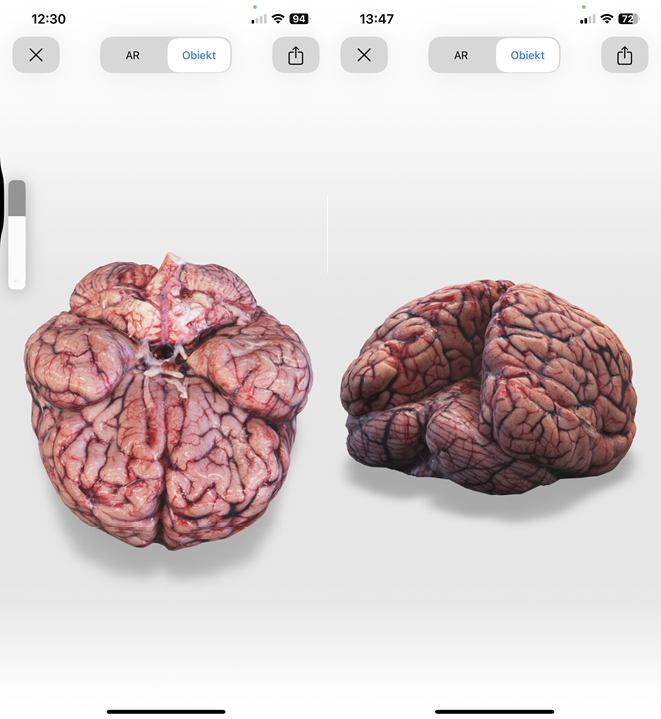
We did not encounter major problems with scanning of the brain with the above-described method. There were, however, some technical issues that should be solved in the future, and these will be further discussed. After completion of the scanning, models of the brain could be downloaded from the phone and either displayed as virtual reality objects (Figure 1), or as isolated 3D objects (Figure 2). These virtual brains could be moved or rotated or enlarged in order to see details (Supplementary File). Also, it was possible to transfer the file containing virtual model the scanned organ to another phone or computer, in order to be handled by another user.
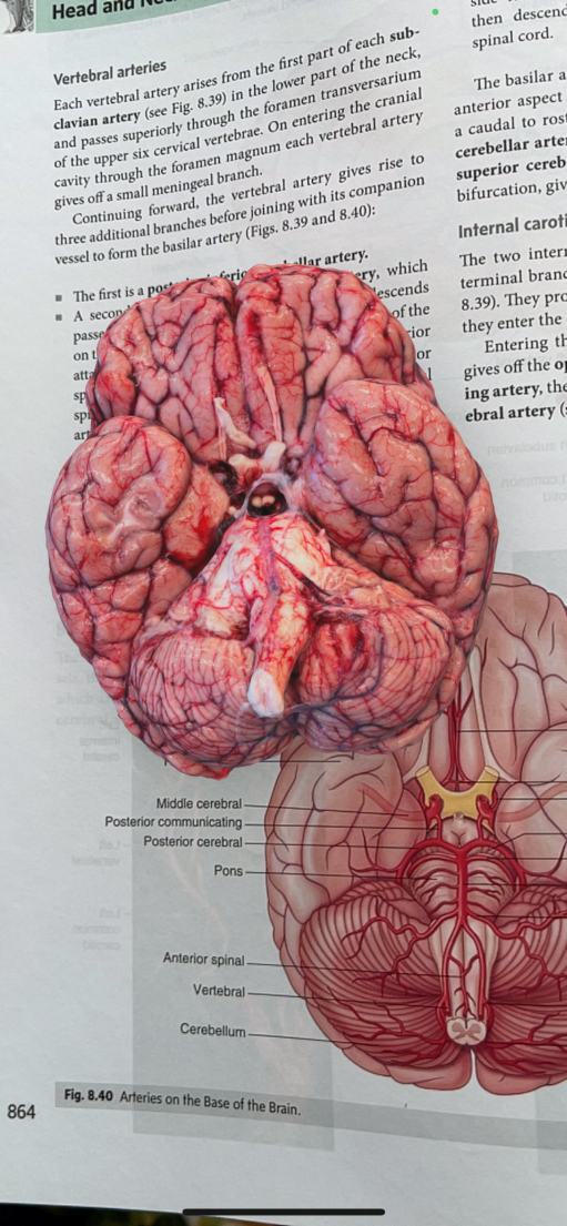
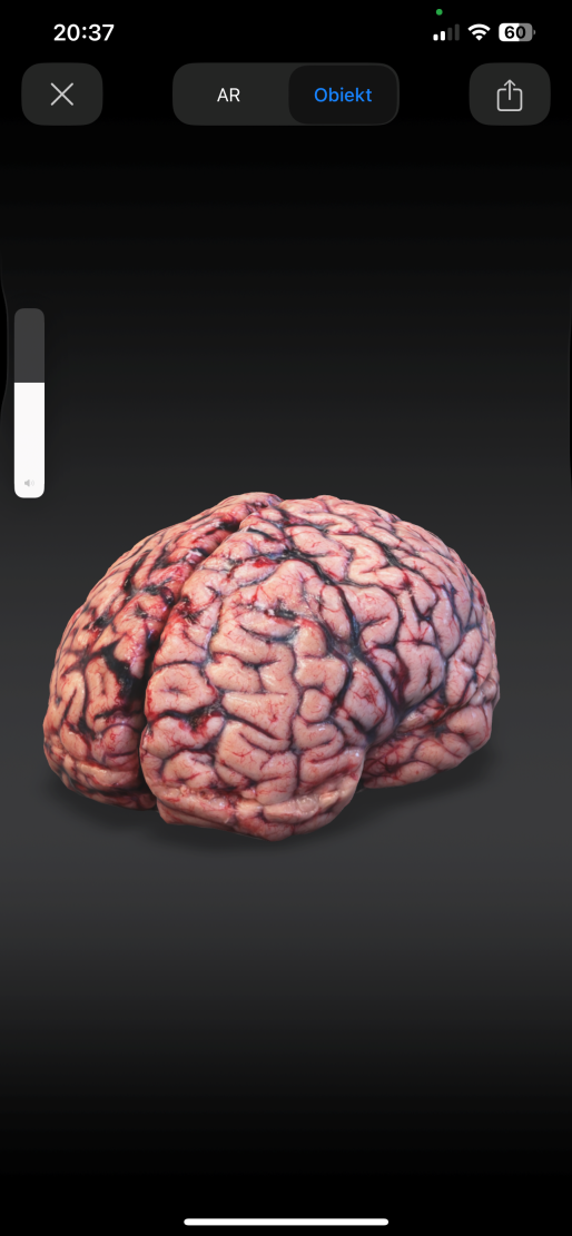
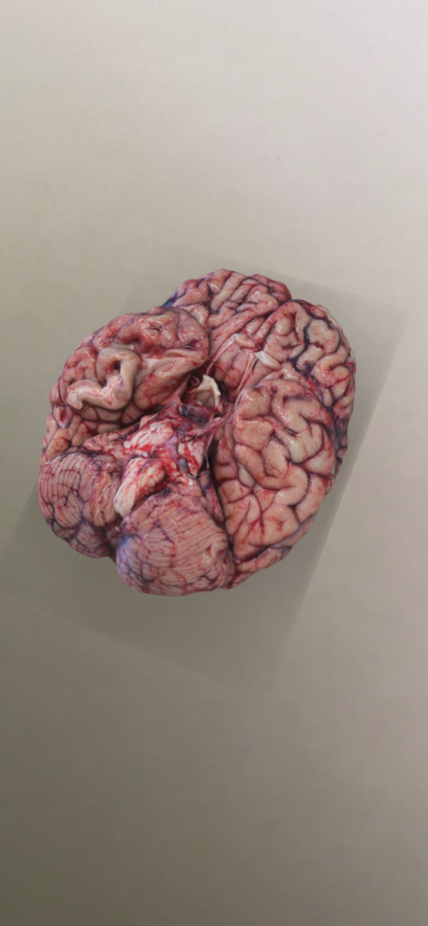
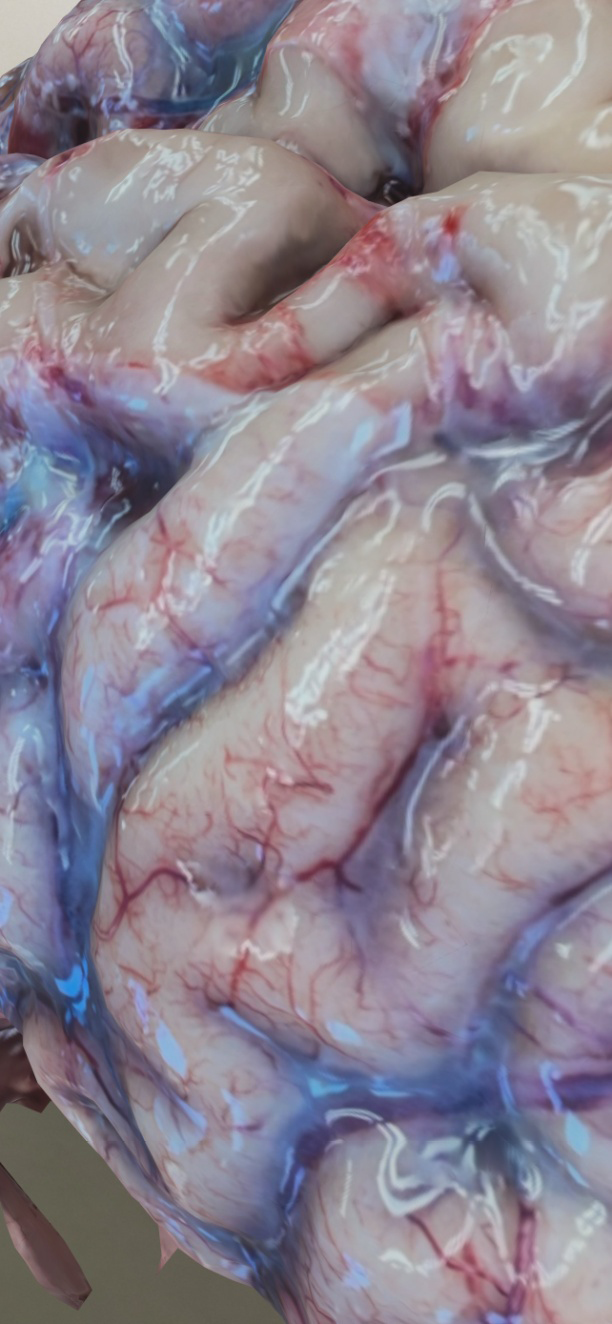
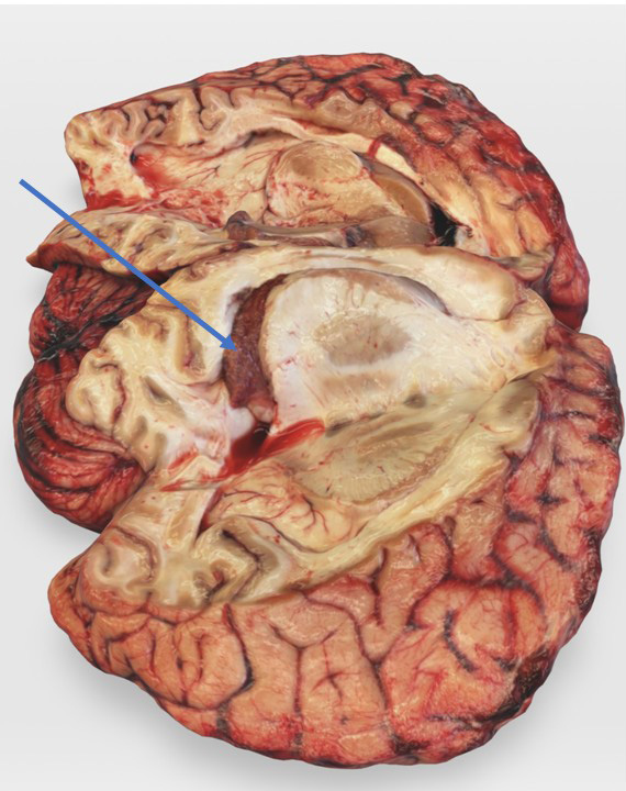
We found this method feasible and highly valuable from the educational perspective, especially considering visual appearance of these virtual organs, which were very similar to the alive brains (Figure 3). Since these virtual objects were created using the high-resolution photographs, after augmentation of a model, the details were clearly visible (Figures 4,5,6). Still, at this stage of research, we encountered some problems with creation of the 3D models. Unlike embalmed brains, fresh brains are quite soft and prone to gravitational distortions. Consequently, some parts of this organ that topographically are close together, after removal from the cranial cavity moved aside (Figure 7). However, although the virtual brain was somewhat distorted, on the other hand this distortion enabled the view of the medial surface of the brain with its important anatomical structures (Figure 8).
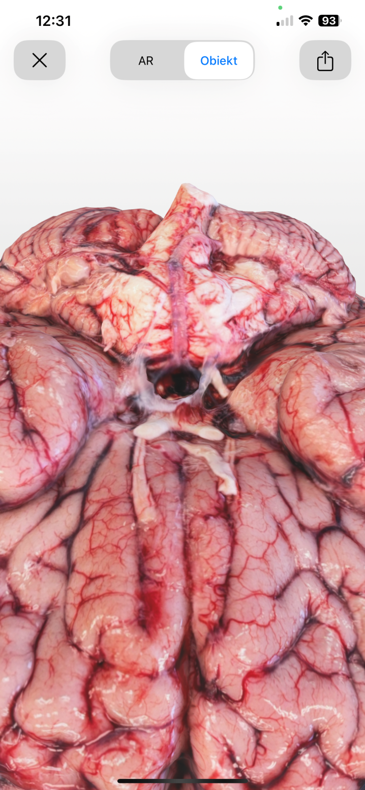
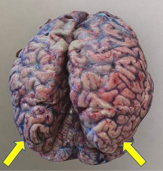
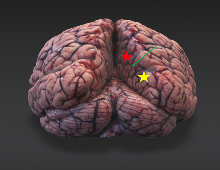
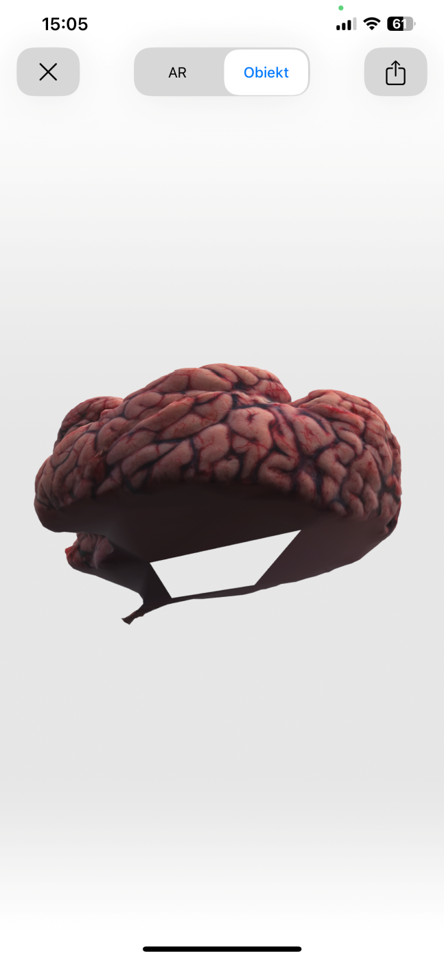
Until now, we were unable to build a virtual model of the whole brain. It was possible to scan this organ from above, with the brain lying either on its lower or upper surface. Unfortunately, unlike other more rigid anatomical specimens that could either be suspended in the air or steadily rotated during scanning, a fresh brain was too soft and prone to distortion. Consequently, it was possible to get good quality 3D virtual brains (Figure 9), either seen from below or above (Figure 8), but actually these models lacked the other surface (Figure 10) when rotated. Still, even considering these shortcomings, which probably can be solved in the future with more experience in the scanning, 3D virtual models of the fresh brains seem to be a promising educational tool for undergraduate medical students.
Discussion
Our preliminary observations demonstrate that scanning of the fresh cadaver brains with the KIRI Engine application is easy, feasible and of potentially high educational value, especially considering the visual appearance of these models, which were very similar to alive brains. Several anatomical features, such as the tiny blood vessels of the pia mater, a translucent arachnoid mater, or the choroid plexus, unlike brains preserved by chemical solutions, are quite similar to those structures of the alive organ, seen for example during the neurosurgical procedures. Although we have encountered several technical problems during scanning (discussed in the Results section of this paper), they seem to be of minor educational importance and most likely can be solved in the future. Besides, during this feasibility study we scanned only superficial aspects of the whole brains. In the future, we plan to scan also the internal structures of brain. Of note, since the brains are scanned during the autopsies, such a scanning should not interfere with this examination and, in general, with the work of a hospital morgue or forensic pathology center. Alternatively, more detailed and focused scanning of a brain of recently deceased donor could be performed, still it should be remembered that an opening of the skull may disturb the process of embalming of the rest of cadaver’s body.
Over the last decade, the photogrammetry (the term refers to a technology that measures something using the recording of light) has been steadily introduced to the medical education, including anatomy [19-23]. However, until recently, its use has been limited by high costs of the scanners and an IT-expertise needed to operate the models acquired after the scanning. Recent advance in technology, especially development of the operator-friendly applications, and the fact that some modern smartphones are equipped with the LiDAR device (LiDAR is a remote sensing technology that uses the laser light to measure distances) made possible a creation of highly accurate, 3D representations of the target object. Consequently, it became possible to scan anatomical specimens and create their 3D virtual representations. In the recent publications, a shift from expensive commercial scanners to the free smartphone applications is notable. Several different smartphone applications were used [24,25]. Currently, it remains unclear which of them is of the best educational value, especially because they are being constantly updated and improved. In addition to the KIRI Engine, applications that may be potentially used for anatomical scanning comprise: the Qlone, the Polycam, the Scandy Pro, the Trnio 3D Scanner, the SCANN3D, and the RealityScan. Probably, those basing on the LiDAR technology should be chosen, since this method provides better quality pictures.
In this feasibility study we have chosen the KIRI Engine application, since for the purpose of this research it was possible to use the non-commercial version of this application, with no associated costs for our university. Besides, in the not yet published study (Wawer, J.; et al. Evaluation of the commercial applications for mobile devices for the scanning of 3D anatomical model. Presented at the 3rd Conference on Anatomical Didactics; 08-09.05.2024, Łódź, Poland), it was reported that for anatomical scanning, this particular application is of the best usefulness. Another advantage of the KIRI Engine is the fact that photogrammetry requires a high computing power. The KIRI Engine effectively utilizes cloud-processing and storage to combat this limitation.
Importantly, until now, 3D anatomical virtual models were created from the embalmed prosection specimens [25,26-30]. By contrast, in our study we used fresh brains of the recently deceased individuals. The scanning was performed during the post-mortem, not during dissecting the body of a cadaver. It seems that the use of fresh organs can overcome limitations associated with the use of embalmed brains for anatomy education. Although embalmed brains can be used for a longer period of time, yet after the embalming they change their color and structure, while some important anatomical structures, particularly blood vessels, are no longer well visible. Some authors tried to solve the problems of poorly visible cerebral vessels in brain specimens by injecting the cerebral vessels with a colored silicone or another filling material [31-33]. Still, such injected specimens, even if valuable from educational point of view, lack the appearance of an alive brain. Besides, a wider use of virtual models instead of embalmed cadaver specimens would reduce the risk associated with exposure of the students and academic teachers to formaldehyde and other toxic and carcinogenic chemicals, as well as to a potential exposure to the brain-residing prions.
We found 3D scanning of the brains with the use of a smartphone application feasible and highly valuable from the educational perspective, especially considering visual appearance of 3D virtual models of the brains, which were very similar to the alive organs. Such a 3D scanning of the organs of fresh cadavers seems to be a new and promising educational tool for undergraduate medical students.
Ethics statement
This study was conducted in accordance with the Declaration of Helsinki and has been approved by the Bioethical Committee of the University of Opole; approval No UO/0015/KB/2024.
This research received no external funding. The authors declare no conflicts of interest.
There are no conflicts of interest associated with this submission. No AI tools were used in the preparation of this manuscript.
Author contributions
Conceptualization, M.S.; methodology, P.G.; validation, P.G. and M.S.; investigation, P.G.,J.M. and W.R.; data curation, P.G.; writing – original draft preparation, M.S.; writing – review and editing, P.G.,J.M. and W.R.; visualization, P.G.; supervision, M.S.; project administration, M.S. All authors have read and agreed to the published version of the manuscript.
References
[1] Benet, A.; Rincon-Torroella, J.; Lawton, M.T.; González Sánchez, J.J. Novel embalming solution for neurosurgical simulation in cadavers. J Neurosurg 2014, 120, 1229-37.
[2] Hammer, N.; Löffler, S.; Bechmann, I.; Steinke, H.; Hädrich, C.; Feja, C. Comparison of modified Thiel embalming and ethanol-glycerin fixation in an anatomy environment: Potentials and limitations of two complementary techniques. Anat Sci Educ 2015, 8, 74-85.
[3] Hammer, N. Thirty years of Thiel embalming-A systematic review on its utility in medical research. Clin Anat 2022, 35, 987-997.
[4] Antipova, V.; Niedermair, J.F.; Siwetz, M.; Fellner, F.A.; Löffler, S.; Manhal, S.; Ondruschka, B.; Pietras, S.M.; Poilliot, A.J.; Pretterklieber, M.L.; Wree, A.; Hammer, N. Undergraduate medical student perceptions and learning outcomes related to anatomy training using Thiel- and ethanol-glycerin-embalmed tissues. Anat Sci Educ 2023, 16, 1144-1157.
[5] Thompson, B.; Green, E.; Scotcher, K.; Keenan, I.D. A Novel cadaveric embalming technique for enhancing visualisation of human anatomy. Adv Exp Med Biol 2022, 1356, 299-317.
[6] Haizuka, Y.; Nagase, M.; Takashino, S.; Kobayashi, Y.; Fujikura, Y.; Matsumura, G. A new substitute for formalin: Application to embalming cadavers. Clin Anat 2018, 31, 90-98.
[7] Nagase, M.; Nagase, T.; Tokumine, J.; Saito, K.; Sunami, E.; Shiokawa, Y.; Matsumura, G. Formalin-free soft embalming of human cadavers using N-vinyl-2-pyrrolidone: perspectives for cadaver surgical training and medical device development. Anat Sci Int 2022, 97, 273-282.
[8] Waerlop, F.; Rashidian, N.; Marrannes, S.; D’Herde, K.; Willaert, W. Thiel embalmed human cadavers in surgical education: Optimizing realism and long-term application. Am J Surg 2021, 221,1300-1302.
[9] Balta, J.Y.; Cronin, M.; Cryan, J.F.; O’Mahony, S.M. Human preservation techniques in anatomy: A 21st century medical education perspective. Clin Anat 2015, 28, 725-34.
[10] Hachabizwa, C.; Banda, M.; Hainza, J.; Mutemwa, S.; Erzingastian, K.; Kafumukache, E. Cadaveric embalming using a modified Thiel method as an alternative to the formalin method. Anat J Africa 2020, 9, 1797-1806.
[11] Miyake, S.; Suenaga, J.; Miyazaki, R.; Sasame, J.; Akimot,o T.; Tanaka, T.; Ohtake, M.; Takase, H.; Tateishi, K.; Shimizu, N.; Murata, H.; Funakoshi, K.; Yamamoto, T. Thiel’s embalming method with additional intra-cerebral ventricular formalin injection (TEIF) for cadaver training of head and brain surgery. Anat Sci Int 2020, 95, 564-570.
[12] Tomalty, D.; Pang, S.C.; Ellis, R.E. Preservation of neural tissue with a formaldehyde-free phenol-based embalming protocol. Clin Anat 2019, 32, 224-230.
[13] Nardi, L.; Schmeisser, M.J.; Schumann, S. Fixation and staining methods for macroscopical investigation of the brain. Front Neuroanat 2023, 17, 1200196.
[14] Hauptmann, M.; Stewart, P.A.; Lubin, J.H.; Beane Freeman, L.E.; Hornung, R.W.; Herrick, R.F.; Hoover, R.N.;, Fraumeni, J.F. Jr.; Blair, A.; Hayes, R.B. Mortality from lymphohematopoietic malignancies and brain cancer among embalmers exposed to formaldehyde. J Natl Cancer Inst 2009, 101, 1696-708.
[15] Soonklang, N.; Saowakon, N. Evaluation of formaldehyde exposure among gross dissection after modified embalming solution and health assessment. Environ Sci Pollut Res Int 2022, 29, 65642-65654.
[16] Ogami-Takamura, K.; Saiki, K.; Endo, D.; Murai, K.; Tsurumoto, T. The risk of Creutzfeldt-Jakob disease infection in cadaveric surgical training. Anat Sci Int 2022, 97, 297-302.
[17] Koyama, S.; Yagita, K.; Hamasaki, H.; Noguchi, H.; Shijo, M.; Matsuzono, K.; Takase, K.I.; Kai, K.; Aishima, S.I.; Itoh, K.; Ninomiya, T.; Sasagasako, N.; Honda, H. Novel method for classification of prion diseases by detecting PrPres signal patterns from formalin-fixed paraffin-embedded samples. Prion 2024, 18, 40-53.
[18] Simka, M. Teaching anatomy of the lower extremity veins: Educational challenges.Vasc Invest Ther 2025, 8, 1-6.
[19] Chytas, D.; Paraskevas, G.; Noussios, G.; Demesticha, T.; Salmas, M.; Vlachou, C.; Vasiliadis, A.V.; Troupis, T. Use of photogrammetry-based digital models in anatomy education: An overview. Morphologie 2024, 108, 100792.
[20] de Oliveira, A.S.; Leonel, L.C.; LaHood, E.R.; Nguyen, B.T.; Ehtemami, A.; Graepel, S.P.; Link, M.J.; Pinheiro-Neto, C.D.; Lachman, N.; Morris, J.M.; Peris-Celda, M. Projection of realistic three-dimensional photogrammetry models using stereoscopic display: A technical note. Anat Sci Educ 2024 , 17, 39-46.
[21] Struck, R.; Cordoni, S.; Aliotta, S.; Pérez-Pachón, L.; Gröning, F. Application of photogrammetry in biomedical science. Adv Exp Med Biol 2019, 1120, 121-130.
[22] Krause, K.J.; Mullins, D.D.; Kist, M.N.; Goldman, E.M. Developing 3D models using photogrammetry for virtual reality training in anatomy. Anat Sci Educ 2023, 16, 1033-1040.
[23] Tóth, D.; Petrus, K.; Heckmann, V.; Simon, G.; Poór, V.S. Application of photogrammetry in forensic pathology education of medical students in response to COVID-19. J Forensic Sci 2021, 66, 1533-1537.
[24] Piazza, A.; Corvino, S.; Ballesteros, D.; Campeggi, A.; Agosti, E.; Serioli, S.; Corrivetti, F.; Bortolotti, C.; De Notaris, M. Neuroanatomical photogrammetric models using smartphones: a comparison of apps. Acta Neurochir (Wien) 2024, 166, 378.
[25] de Oliveira Manduca Palmiero, H.; Ribas, E.C.; Carlotti, C.G. Jr.; Figueiredo, E.G. Anatomical assessment of cerebral sulci and gyri for neuroanatomy education using photogrammetry technique to generate 3D-tridimensional models. Neurosurg Rev 2025, 48, 172.
[26] Petriceks, A.H.; Peterson, A.S.; Angeles, M.; Brown, W.P.; Srivastava, S. Photogrammetry of human specimens: An innovation in anatomy education. J Med Educ Curric Dev 2018, 5, 2382120518799356.
[27] Van Vlasselaer, N.; Keelson, B.; Scafoglieri, A.; Cattrysse, E. Exploring reliable photogrammetry techniques for 3D modeling in anatomical research and education. Anat Sci Educ 2024, 17, 674-682.
[28] Morichon, A.; Dannhoff, G.; Barantin, L.; Destrieux, C.; Maldonado, I.L. Doing more with less: Realistic stereoscopic three-dimensional anatomical modeling from smartphone photogrammetry. Anat Sci Educ 2024,17, 864-877.
[29] de Oliveira, A.S.; Leonel, L.C.; LaHood, E.R.; Hallak, H.; Link, M.J.; Maleszewski, J.J.; Pinheiro-Neto, C.D.; Morris, J.M.; Peris-Celda, M. Foundations and guidelines for high-quality three-dimensional models using photogrammetry: A technical note on the future of neuroanatomy education. Anat Sci Educ 2023, 16, 870-883.
[30] de Oliveira, A.S.; Leonel, L.C.; Bauman, M.M.; De Bonis, A.; LaHood, E.R.; Graepel, S.; Link, M.J.; Pinheiro-Neto, C.D.; Lachman, N.; Morris, J.M.; Peris-Celda, M. Photogrammetry scans for neuroanatomy education - a new multi-camera system: technical note. Neuroinformatics 2024, 22, 317-327.
[31] Smith, K.; Ventre, G.J.; Palmisciano, P.; Hussein, A.E.; Hoz, S.S.; Forbes, J.A.; Lowrie, D.J. Jr.; Zuccarello, M.; Andaluz, N.; Prestigiacomo, C.J. Brain vasculature color-labeling using the triple-injection method in cadaveric heads: A technical note for improved teaching and research in neurovascular anatomy. Oper Neurosurg (Hagerstown) 2023, 24, 291-300.
[32] Çırak, M.; Yağmurlu, K.; Soldozy, S.; Norat, P.; Shaffrey, M.E.; Kalani, M.Y.S. Common challenges and solutions associated with the preparation of silicone-injected human head and neck vessels for anatomical study. Brain Sci 2020, 11, 32.
[33] Sanan, A.; Abdel Aziz, K.M.; Janjua, R.M.; van Loveren, H.R.; Keller, J.T. Colored silicone injection for use in neurosurgical dissections: anatomic technical note. Neurosurgery 1999, 45, 1267-71; discussion 1271-4.