https://doi.org/10.36253/ijae-16769
Cardiac anatomy in the ‘Madonna of the Carnation’ attributed to Leonardo da Vinci
Department of Vascular Surgery; Helsicore; Tevdore Mgvdeli st. 13; Tbilisi, Georgia
E-mail: gagakeshelava@gmail.com
Abstract. Around 1478-1480, the ‘Madonna of the Carnation’, also known as the ‘Madonna with Vase’, the ‘Madonna with Child’, or the ‘Virgin with Flower’, was produced. Using the program Paint X, we moved one detail (indicated by a bird-like shape drawn by Leonardo himself) ob Mary’s left breast and we obtained an image of an ox heart with its aortic arch. The image of the heart obtained by us is identical to the ox’s heart described by Leonardo in his anatomical drawing. Comparison reveals a striking resemblence to the heart configuration and arrangement of the branches of an aortic arch. Based on the fact that the heart is symbol of love, in this artwork Leonardo symbolically depicted a mother’s love for her son and at the same time he described the anatomy of the heart.
Keywords: Leonardo da Vinci, Madonna of the Carnation, cardiac anatomy, aortic arch.
Contents
Interpretation of the painting
Leonardo’s innovations in the cardiovascular anatomy
Introduction
The aim of this study is to interpret ‘The Madonna of the Carnation’ (Madonna with a Vase of Flowers) in anatomical aspect. This study provides insight into how Leonardo translated his innovative anatomical knowledge into art.
The ‘Madonna of the Carnation’ was produced around 1472-1478, (Fig. 1). The thorough study of the painting conducted by Moller eventually allowed the panel to be confidently attributed to Leonardo (Moller, 1937), an attribution that remained unchallenged during the last 30 years and was substantiated afresh by the study conducted by Brown (Brown, 1998). Although some historians believe that Verrocchio produced the original design for the ‘Madonna of the Carnation’ (Dunkerton, 2011).
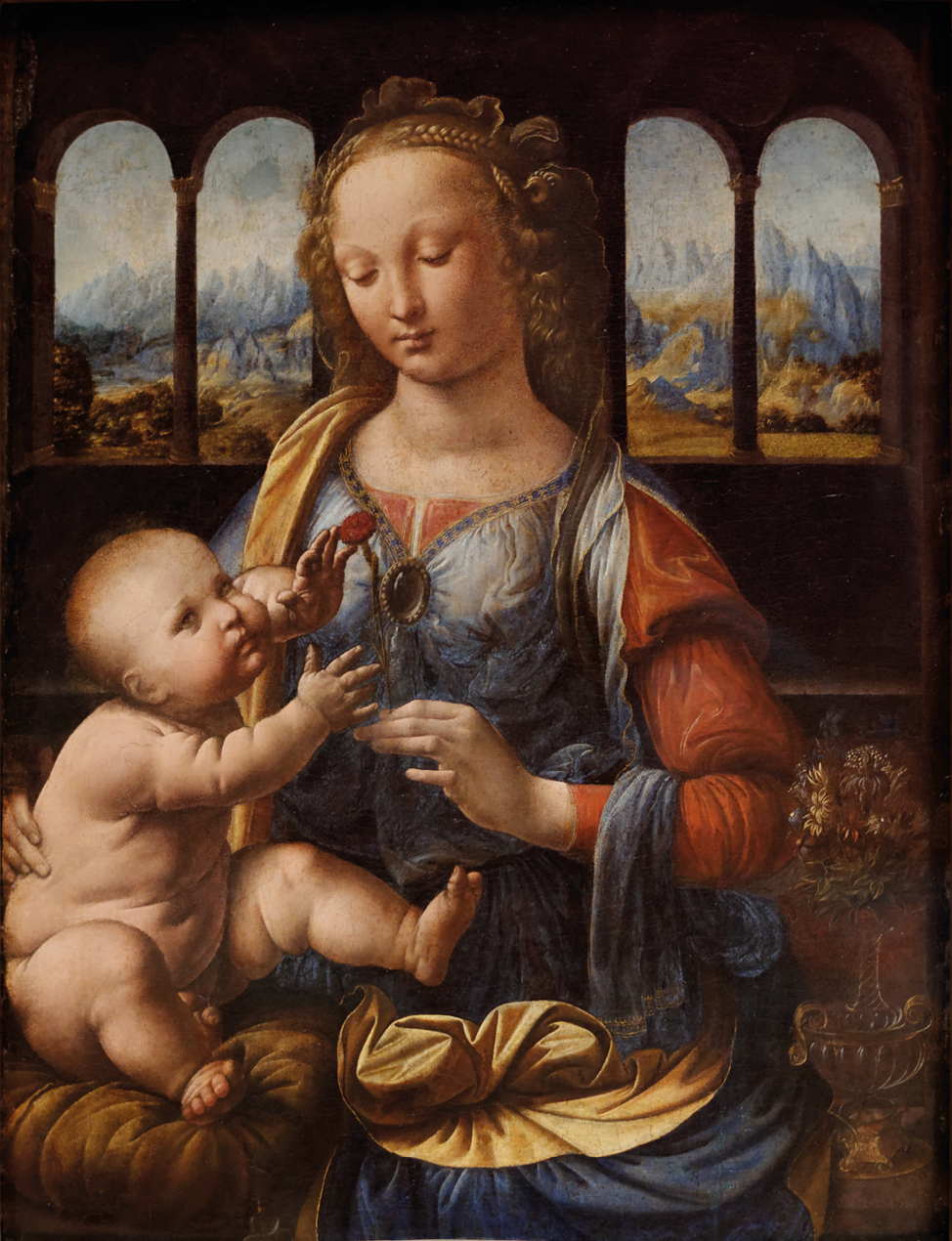
The iconography of the Madonna handing a flower to her son is hardly original. The flower can be the symbol of the virtue of the virgin but also that of the death of Jesus. The carnation, by its shape associated with that of a nail and its blood-red color, announces the future of Christ.
The work is stored in the Alte Pinakothek Gallery in Munich, Germany.
Interpretation of the painting
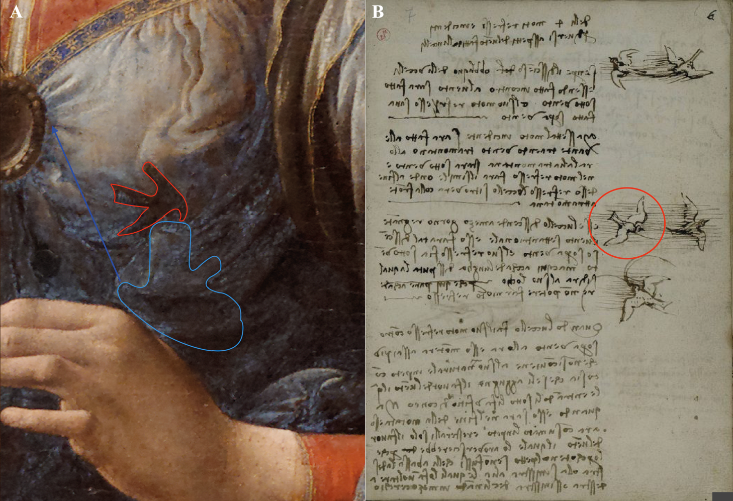
In the ‘Madonna of the Carnation’, our attention was drawn to the shadow drawn by Leonardo himself on Mary’s left breast (Fig. 2A – bounded by a red contour). This shadow resembles an arrow indicating the direction of something. But in Leonardo’s era, the arrow was not yet used as a direction indicator. The second thing this shadow resembles is a bird depicted in a drawing by Leonardo (Fig. 2B). The ‘bird’ indicates the detail bordered by author of a painting with a faint contour (Fig. 2 A- blue marked contour).
Using the program Paint X, we moved the detail in the direction of the blue arrow to final location and we got an image of the heart (Fig. 3 A, B). It depicts the left and right subclavian arteries, common carotid arteries, and ventricles (Fig. 3 C). The image of the heart obtained by us is strikingly similar to the ox’s heart described by Leonardo in his anatomical drawing (RL 19073-74v; K/P 1 66v) (Fig. 3 D). By comparing them we see a sharp resemblance to the heart configuration and arrangement of the branches of an aortic arch (Fig. 3 C, D).
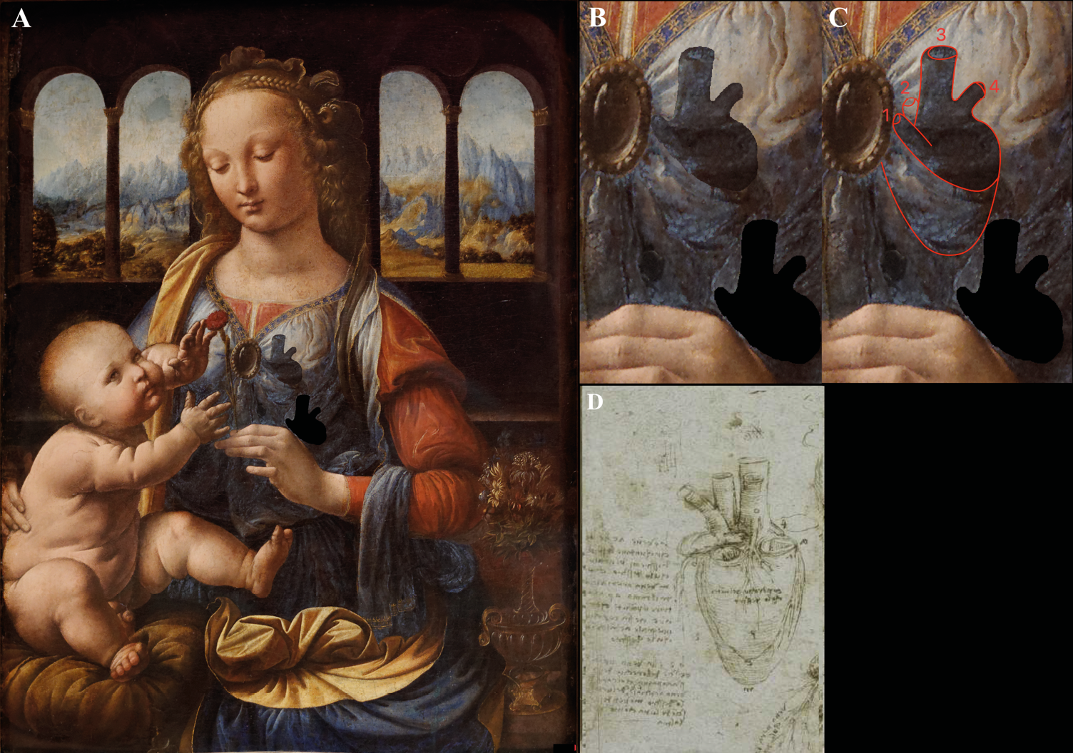
It is noteworthy that after placing the detail in the correct place, the insignificant contour in the painting acquired the meaning of the second branch of the aortic arch (Fig. 3 B, C).
A similar concept is found in the ‘Dreyfus Madonna’ attributed to Leonardo da Vinci. Also, by moving one detail in this artwork an image of an ox’s heart is obtained. The difference is that in the second painting, the detail is emphasized by the Madonna’s hand. In this case too, the obtained image of the heart resembles the heart depicted in Leonardo’s anatomical sketch (Keshelava, 2021). Based on the fact that the heart is symbol of love, in the both artwork Leonardo originally depicted the feeling of mother towards her son and at the same time he described the anatomy of the heart.
Peter Paul Rubens’s copy of ‘The Battle of Anghiari’ by Leonardo da Vinci is also an example of the transfer of anatomical knowledge into art (Keshelava, 2023).
It should be noted that there is a discrepancy between the periods of creation of the painting and Leonardo’s anatomical studies. It was speculated that Leonardo created this work in 1472-1478 and an important contribution to Leonardo’s approach to anatomy was provided after 1510, which meant dissection of animal and human cadavers. The anatomical drawing (RL 19073-74v; K/P 1 66v) similar to image obtained by us is created in 1513. This discrepancy can be explained in only one way: the master could have made changes to the painting later. The fact that Leonardo made changes in his work is confirmed by researchers (Cotte, 2005).
Leonardo’s innovations in the cardiovascular anatomy
Leonardo da Vinci’s study in anatomy can be divided into three periods: an early (from c. 1487), a middle (1506-1510) and a late phase (after 1510). The drawings from early phase was not based on the dissection of bodies and meant simply upon information that could be deduced from the surface of the human corps, from the bodies of animals and from the human skeleton. During this period, animal examination, documents from ancient Greece and medieval Italy, and the occasional autopsy of a condemned criminals were the main source of anatomical knowledge in Europe (Park, 1994; Olry, 1997).
Marco Antonio Della Torre, an anatomist and a medical doctor, known mainly for his collaboration with Leonardo da Vinci, made a significant contribution to Leonardo’s knowledge and approach in the anatomy (Picardi et al., 2010)
At the time of Leonardo, ideas about the anatomy and physiology of the heart were based on the views of the authorities (Cambiagi, 2019). At the same time, the heart had an almost spiritual role (Heater, 2010).
In order to examine the hemodynamic properties of blood flow through the heart and its valve, the master created multiple wax castings of the bull heart. From these casts he built glass models. Leonardo described the characteristic of the sinus of Valsalva. He also studied the dynamics of water flow in rivers, using colors to show the flow patterns. He stated: “…the water is continuously bouncing against the banks… and as time passes the course of the river gets more and more tortuous…” Leonardo da Vinci translated those findings to blood flow in vessels (Boon, 2010; Webb, 2010) and described turbulence formation in the aortic sinus (Keele, 1979).
His first achievement was to describe the heart as a muscle, constantly contracting and expanding. He also noted that this muscle is much stronger than other muscles (Keele, 1952). Leonardo believed that the contraction of the heart causes a wave to propagate along the vessels and concluded that this is the reason for the existence of a pulse in a living organism (Pasipoularides, 2014).
The master also deduced that the blood is supplied to every part of the body by the aorta. He proposed that new blood is received by the bronchial arteries from the bronchi, and that venous blood is freshened in the lungs before to returning to the heart. He came to the notion that the heart feeds itself by looking at the coronary arteries (Keele, 1973; Sterpetti, 2019). “The heart is a vessel made of thick muscle, vivified and nourished by artery and vein as are other muscles” - he made such a conclusion on the background of the dissection of the coronary arteries, which are clearly described in the anatomical sketches. Leonardo da Vinci used his understanding of hydrodynamics and anatomy to highlight the atherosclerotic alterations in arteries.18 He was also interesting in the tortuosity and elongation of the vessels in old people.
He distinguished between the atria and ventricles based on their function and physical characteristics, and he thought that the heart was composed of four chambers (Sterpetti, 2019). Leonardo adopted Galenic beliefs, unaware of the circulatory system’s relationship, which William Harvey would not completely establish until 1628.
Da Vinci’s most important findings in cardiovascular anatomy and physiology were as follow (Wells, 2007): the view of the cardiac cycle and asynchronous contraction and dilation of the atria and ventricles; he noted that the heart is a muscle; Leonardo rejected the idea that the heart receives air from the lungs; He solved the mechanism of valves function; His drawings describe the coronary arteries in detail; Leonardo’s study on old bodies provided showed the atherosclerotic changes in vessels; da Vinci was the first to describe the trabecula septomarginalis or moderator band of the right ventricle (this anatomical detail is also known as Reil’s band, but some scholars proposed the term “Leonardo’s band” (Shoja, 2013).
Leonardo da Vinci’s own studies in anatomy and physiology were mainly based on empirical observation. As mentioned above, he used simple and reasonable experiments, and based on them, he obtained important conclusions. His achievement are a good example of how logical thinking can be used to understand the physiology of life. It is regrettable that although his art was appreciated by his contemporaries, but he was not known as scientist. After his death the sketches and drawings remained hidden for a century (Dorland, 1922). His vision of the world was logical, using an empiric and unusual method of study. He wrote,”…nature is driven by the intelligence of its law”.
During the Renaissance, the artists already saw the heart as a symbol of romantic love and the love of God (Figueredo, 2024). The fact that the heart was a symbol of love in that era logically explains Leonardo’s idea and the meaning of hidden heart images in his paintings. The images of the heart in the “Madonna of the Carnation” depict a mother’s love for her child.
Authorship
Grigol Keshelava has made substantial contribution to all of the following: 1) the conception and design of the study, acquisition of data and interpretation of data; 2) drafting the article and revising it critically for intellectual content; 3) final approval of the version to be submitted.
References
Boon B. (2010) Leonardo da Vinci on atherosclerosis and function of the sinuses of Valsalva. Netherlands Heart Journal. 17: 496-499.
Brown D.A. (1998) Leonardo da Vinci. Origins of a Genius. New Haven, London.
Cambiagi M., Hausse H. (2019) Leonardo da Vinci and his study of the heart. European Heart Journal. 40: 1823-1831.
Cotte P. (2005) Lumiere on the Lady with an Ermine. Vinci Edition.
Dorland W.A. (1922) The American illustrated medical dictionary. Philadelphia, Saunders.
Dunkerton J. (2011) Leonardo in Verrocchio’s workshop: re-examining the technical evidence. National Gallery Technical Bulletin. 32: 1-31.
Figueredo V.M. (2024) The heart Renaissance. Reviews in Cardiovascular Medicine. 25(3): 91.
Heater W. (2010) The Medieval Heart. New Haven, London: Yale University Press.
Keele K.D. (1952) Leonardo da Vinci on movement of the heart and blood. Philadelphia: Lippincott.
Keele K.D (1973) Leonardo da Vinci’s views on atherosclerosis. Medical History. 17(3): 304-308.
Keele K.D., Pedretti C. (1979) Leonardo da Vinci. Corpus of anatomical studies in the Collection of Her Majestry the Queen at Windsor Castle. Harcourt Brace Jovanovich, New York.
Keshelava G. (2021) Cardiac anatomy in the ‘Dreyfus Madonna’ by Leonardo da Vinci. Interact Cardiovascular and Thoracic Surgery. 32(4): 582-584.
Keshelava G. (2023) Hidden brain anatomy in Peter Paul Rubens’s copy of ‘The battle of Anghiari’ by Leonardo da Vinci. The Neuroscientist. 29(6): 676-680.
Moller E. (1937) Leonardos Madonna mit der Nelke in der Alteren Pinakothek. Muchner Jahrbuck der Bildenden Kunst. 12: 5-40.
Olry R. (1997) Medieval neuroanatomy: the text of Mondino dei Luzzi and the plates of Guido da Vigevano. Journal of the History of the Neurosciences. 6(2): 113-123.
Park K. (1994) The criminal and the sainthly body: autopsy and dissection in Renaissance Italy. Renaissance Quarterly. 47(1): 1-33.
Pasipoularides A. (2014) Historical continuity in the methodology of modern medical science: Leonardo leads the way. International Journal of Cardiology. 171: 103-115.
Picardi E.E., Macchi V., Porzionat A., Boscolo-Berto R., Loukas M., Tubbs S., De Caro R. (2010) Marco Antonio Della Torre and Leonardo da Vinci. Clinical Anatomy. 2(6): 744-748.
Shoja M.M., Agutter P.S., Loukas M., Benninger B., Shokouhi G., Namdar H., Ghabili K., Khalili M., Tubbs R.S (2013) Leonardo da Vini’s studies of the heart. International Journal of Cardiology. 167(4): 1126-1138.
Sterpetti A.V. (2019) Cardiovascular research by Leonardo da Vinci (1452-1519). Circulation Research. 124: 189-191.
Wells F. (2007) The Renaissance heart: the drawings of Leonardo da Vinci. In: Peto, J. The Heart. London/New Haven: Yale University Press: 70-94.