https://doi.org/10.36253/ijae-16696
Embryologic layers in dermatology: Developmental checkpoint disorders, diagnostic insight, and regenerative futures
¹ Department of Dermatology, Sivas Medicana Hospital, Sivas, Türkiye
² Department of Histology and Embryology, Faculty of Medicine, Erzincan Binali Yıldırım University, Erzincan, Türkiye
³ Department of Medical Genetics, Istanbul Faculty of Medicine, Istanbul University, Istanbul, Türkiye
⁴ Department of Dermatology, Faculty of Medicine, Bezmiâlem Vakıf University, Istanbul, Türkiye
*Corresponding Author. E-mail: gkhnkya@gmail.com
Abstract. Objectives: This review aims to present a developmental framework linking embryonic lineage with non-heritable cutaneous anomalies to improve diagnostic precision and educational approaches in dermatology. Materials and Methods: A narrative literature review was conducted using PubMed, Scopus, and Web of Science databases covering the years 2000–2025. Keywords included “skin development,” “embryology,” “developmental checkpoint disorders,” and “non-genetic congenital disorders.” Data on morphogenesis, embryologic signaling pathways, and representative disorders were synthesized into a layer-based model. Results: Disorders such as self-healing collodion baby (periderm retention anomaly), pigmentary mosaicism (postzygotic melanocyte patterning defect), and focal dermal hypoplasia (connective tissue maldevelopment) reflect disruptions at specific morphogenetic checkpoints. Mapping these conditions to their embryonic origins revealed layer-specific vulnerability windows and facilitated differential diagnosis from inherited disorders. Understanding these embryologic principles supports earlier diagnosis, informed prenatal counseling, and structured integration into dermatology curricula. Advances in regenerative medicine, particularly stem cell–based strategies, highlight the translational potential of dermatoembryology in developing targeted therapies. Conclusion: A layer-oriented dermatoembryological perspective enhances recognition of developmental skin disorders, especially when genetic analyses are inconclusive. Incorporating embryologic concepts into clinical reasoning not only improves diagnostic accuracy but also fosters regenerative therapeutic innovations and enriches dermatology education.
Keywords: embryonic development, skin abnormalities, congenital disorders, dermatology, regenerative medicine.
Contents
2. Overview of skin development
2.2. Epidermal and dermal layer formation
2.4. Adnexal structure formation
2.5. Molecular signaling pathways
3. Developmental defects and related non-genetic disorders
3.2. Basal Layer and stratification defects
3.3. Granular layer and cornification abnormalities
3.4. Embryological pigment patterning defects
3.6. Dermal–epidermal junction defects
3.7. Adnexal development defects
3.8. Embryological patterning lines
3.9. Externally induced developmental defects
4.1. Diagnostic utility of dermatoembryology
4.2. Educational integration: teaching dermatologic development
4.3. Future directions: a stem cell perspective
1. Introduction
Human skin is a complex, multilayered organ essential for homeostasis, immune defense, and environmental interaction. Structurally, it comprises three principal layers: epidermis, dermis, and hypodermis. Each derives from distinct embryonic sources – surface ectoderm gives rise to the epidermis, mesoderm to the dermis and hypodermis, and neural crest cells to melanocytes, vascular elements, and select sensory structures [1–3]. While epidermal and dermal development has been extensively characterized, the hypodermis remains comparatively underexplored, despite its key functions in mechanical cushioning, endocrine signaling, and immune regulation. Recent advances in tri-layered skin modeling demonstrate that inclusion of adipose tissue enhances both structural fidelity and physiological relevance in engineered constructs [4].
Skin morphogenesis begins in the third gestational week, encompassing sequential processes such as epidermal stratification, melanoblast migration, adnexal morphogenesis, and maturation of the dermoepidermal junction [2]. Single-cell transcriptomic and spatial analyses have revealed dynamic interactions between immune and nonimmune populations during this period; notably, macrophages actively shape angiogenesis, neurogenesis, and hair follicle formation beyond their classical immunologic roles [5]. Keratinocyte differentiation proceeds from basal progenitors upward through spinous and granular layers, with lineage specification orchestrated by conserved signaling pathways – Wnt, Notch, Hedgehog, FGF, and MAPK/ERK – operating in precise spatiotemporal patterns [6–8]. Although epithelial cells are embryologically committed, regenerative studies highlight latent plasticity that links embryogenesis with postnatal repair, bridging developmental and regenerative dermatology [9].
Errors in these morphogenetic checkpoints may result in clinically significant anomalies, some without defined genetic etiologies. Conditions such as self-healing collodion membrane, pigmentary mosaicism, and focal dermal hypoplasia illustrate how disruption of critical embryonic stages can yield cutaneous phenotypes resembling monogenic disorders [2,10]. For instance, incomplete periderm desquamation beyond 21 weeks or impaired melanoblast migration before week 12 exemplify layer-specific vulnerability windows that manifest independently of identifiable mutations. Figure 1 schematically correlates germ-layer origins with clinical phenotypes, underscoring the diagnostic utility of developmental timing in dermatology.
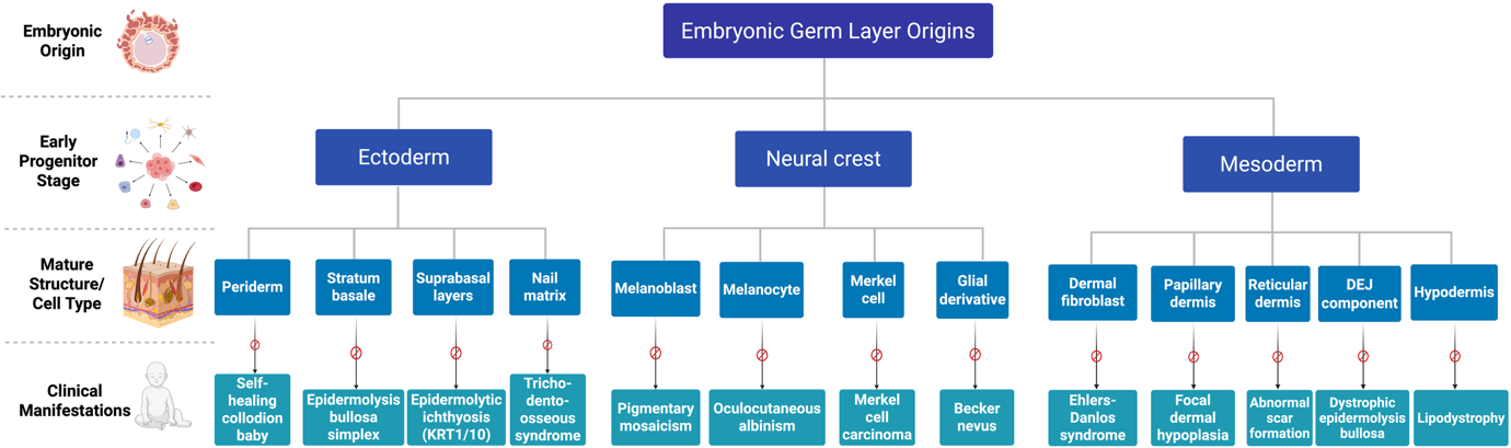
Despite its relevance, dermatoembryology remains underrepresented in dermatology training and diagnostic practice [11]. A developmentally informed, layer-specific perspective could strengthen early recognition of congenital anomalies, refine prenatal assessments, and foster integration between developmental biology and clinical dermatology [12]. Accordingly, this review delineates the embryologic origins of major skin components, analyzes how disruptions in developmental checkpoints produce genetically undetermined disorders, and situates these insights within diagnostic reasoning, medical education, and regenerative medicine. Unlike traditional Mendelian paradigms, our framework emphasizes embryologic timing and layer-specific vulnerability as central to understanding dermatologic phenotypes.
2. Overview of skin development
2.1. Embryological timeline
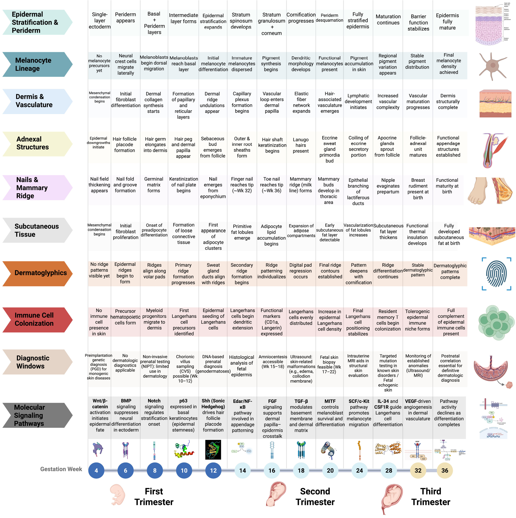
Skin development begins in the third week of embryogenesis, involving coordinated morphogenetic events derived from the ectoderm, mesoderm, and neural crest. The surface ectoderm generates the epidermis, the mesoderm forms the dermis and hypodermis, and neural crest cells contribute melanocytes and mechanosensory structures [1–3]. By week 4, the ectoderm appears as a single-cell layer that thickens between weeks 4 and 6 into a bilayer of proliferative basal cells and superficial periderm [13]. The periderm serves as a transient barrier and is normally shed into the amniotic fluid by week 21 [14]. Simultaneously, mesenchymal cells differentiate into early dermis, and neural crest–derived melanoblasts begin migrating toward the basal epidermis around embryonic day 50 (approximately week 7), continuing through weeks 8–12 under the influence of transcription factors such as SOX10, PAX3, and MITF [15].
During the second trimester, epidermal stratification accelerates, adnexal structures begin to form, and the dermoepidermal junction matures. By the third trimester, the epidermis and dermis are structurally mature, adnexal appendages such as hair follicles, glands, and nails are largely developed, and vascularization, innervation, and immune cell colonization provide barrier and sensory functions at birth [16]. Figure 2 summarizes these developmental milestones and highlights the temporal coordination of epidermal, dermal, adnexal, and immune maturation across gestation.
2.2. Epidermal and dermal layer formation
Between weeks 4 and 6, the surface ectoderm and mesoderm initiate the formation of the epidermis and dermis. The ectoderm gives rise to basal keratinocytes and the transient periderm, which expands without division and is shed by week 21 [13,14]. Stratification begins around week 11 with the appearance of an intermediate layer, and by week 24 the epidermis is composed of spinous, granular, and cornified layers [13,17]. Basal cells express keratins K5 and K14 from week 8, while suprabasal cells begin producing differentiation markers such as filaggrin, involucrin, and loricrin. Together with SPRs, envoplakin, and periplakin, these proteins are cross-linked by transglutaminases to form the cornified envelope.
The dermis, derived from mesenchymal precursors, produces collagens I, III, V, and VI between weeks 8 and 12, and by week 14 it organizes into papillary and reticular layers [18]. The dermoepidermal junction matures in parallel, with hemidesmosomes and type VII collagen fibrils providing stable adhesion between epidermis and dermis [17,18].
2.3. Melanocyte migration
Melanocytes originate from neural crest cells, which undergo epithelial-to-mesenchymal transition before migrating dorsolaterally toward the epidermis. Their migration is regulated by transcription factors including SOX10, PAX3, and MITF [19]. During this process, melanoblasts alter adhesion profiles, downregulating E-cadherin and adopting a P-cadherin phenotype to facilitate epidermal integration [19,20]. Chemotactic cues, most notably SDF-1α/CXCL12 acting through CXCR4, direct migration, while α-MSH enhances responsiveness [21]. These developmental mechanisms not only establish pigmentation during embryogenesis but also underpin postnatal processes such as wound healing.
2.4. Adnexal structure formation
The formation of adnexal structures, including hair follicles, sebaceous glands, sweat glands, and nails, occurs through reciprocal epithelial–mesenchymal interactions beginning around weeks 9–20. Hair follicles develop from epidermal placodes and interact with dermal papillae to guide follicular differentiation and shaft formation. Sebaceous glands arise from follicular epithelium and release sebum by holocrine secretion. Eccrine sweat glands develop independently from epidermal downgrowths, forming coiled secretory units within the deep dermis, whereas apocrine glands originate from follicular structures in the axillary and anogenital regions, releasing secretions via decapitation [22]. These developmental events are regulated by signaling pathways including WNT, EDA/EDAR, SHH, and BMP, and disruption of these cascades results in ectodermal dysplasias [22,23].
2.5. Molecular signaling pathways
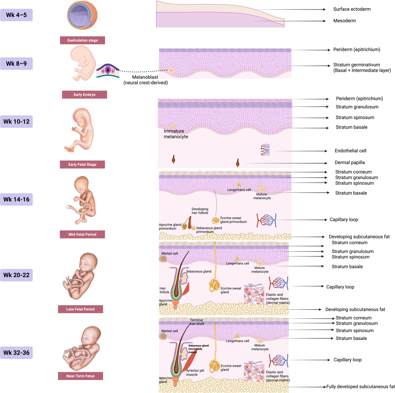
Skin morphogenesis is orchestrated by a network of evolutionarily conserved signaling pathways. Wnt signaling initiates placode formation and appendage patterning, with Dkk1 acting as a major inhibitor. Notch signaling regulates cell fate specification through lateral inhibition, whereas FGF signaling promotes epithelial–mesenchymal crosstalk critical for tissue morphogenesis. BMP signaling drives epidermal differentiation while suppressing appendage formation in the interfollicular epidermis [8,24]. MAPK/ERK signaling integrates proliferative and differentiative cues, contributing both to morphogenesis and to regenerative capacity [24]. The precise temporal coordination of these pathways ensures normal skin development, while their dysregulation produces congenital dermatologic disorders and offers potential therapeutic targets for regenerative strategies. Figure 3 illustrates the sequential progression of epidermal stratification, melanocyte differentiation, dermal remodeling, adnexal development, and hypodermal maturation throughout gestation.
3. Developmental defects and related non-genetic disorders
3.1. Periderm defects
The periderm, a transient embryonic layer, protects the developing epidermis and contributes to epithelial integrity and amniotic exchange. Failure of timely shedding produces the collodion baby phenotype, often linked to TGM1 mutations in autosomal recessive ichthyoses [25,26]. In some cases, spontaneous resolution occurs as self-healing collodion baby [27]. Persistent periderm caused by IRF6, IKKα, or SFN mutations underlies syndromes such as popliteal pterygium and Bartsocas-Papas [28]. Harlequin-like collodion due to ABCA12 variants represents a milder defect with potential for improvement under supportive care [29]. Vernix caseosa, composed of periderm remnants and sebaceous secretions, supports maturation and thermoregulation; its absence predisposes premature infants to dehydration and infection, reflecting a clinically relevant model of incomplete skin maturation [30].
3.2. Basal Layer and stratification defects
Abnormal keratinocyte proliferation and stratification in the basal layer result in distinct disorders. Mutations in KRT5 or KRT14 cause epidermolysis bullosa simplex (EBS), characterized by intraepidermal blistering of variable severity [31]. PLEC mutations lead to syndromic EBS with muscular dystrophy, while TP63 mutations disrupt stratification and adnexal development, producing AEC (Hay-Wells) syndrome with erosions, ankyloblepharon, clefting, and nail anomalies [32] (Figure 4).
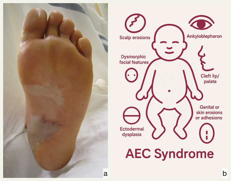
(b) Schematic of AEC (ankyloblepharon–ectodermal dysplasia–clefting) syndrome from TP63 mutations, showing scalp erosions, ankyloblepharon, clefting, nail and hair abnormalities, and impaired adnexal development.
3.3. Granular layer and cornification abnormalities
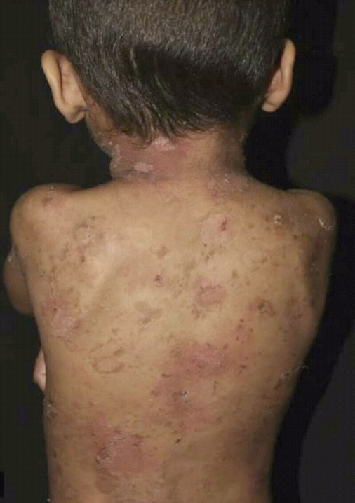
Cornification defects include epidermolytic ichthyosis, caused by dominant KRT1 or KRT10 mutations, presenting with neonatal blistering that progresses to diffuse hyperkeratosis with suprabasal cytolysis and clumped keratohyalin granules [33]. Lamellar ichthyosis, classically recessive, has also been linked to dominant NKPD1 mutations, producing milder congenital scaling [34] (Figure 5).
3.4. Embryological pigment patterning defects
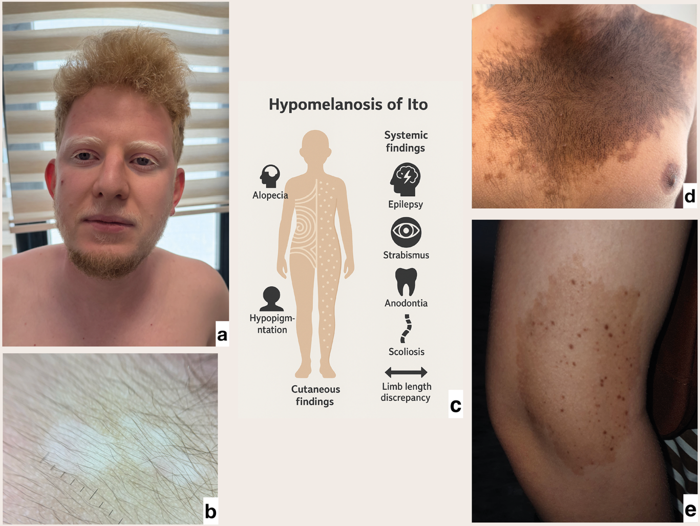
Defects in melanocyte function and patterning create pigmentary anomalies. Oculocutaneous albinism type 1 (OCA1), due to TYR mutations, manifests as generalized hypopigmentation despite normal migration [35,36]. Segmental pigmentary disorders, including hypomelanosis of Ito and nevus depigmentosus, result from postzygotic mosaicism [37,38]. Becker nevus, associated with ACTB mutations, presents with unilateral hyperpigmentation and hypertrichosis, whereas nevus spilus exhibits scattered dark macules over lighter patches [39,40] (Figure 6).
3.5. Dermal formation defects
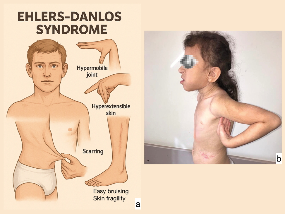
Dermal development depends on positional identity maintained by HOX gene expression [40]. Mutations in collagen genes (COL1A1, COL3A1, COL5A1) cause Ehlers-Danlos syndromes, with skin hyperextensibility, fragility, and vascular complications [41,42]. In contrast, focal dermal hypoplasia (Goltz syndrome), caused by PORCN mutations, produces linear dermal atrophy, fat herniation, and skeletal anomalies [43] (Figure 7).
3.6. Dermal–epidermal junction defects
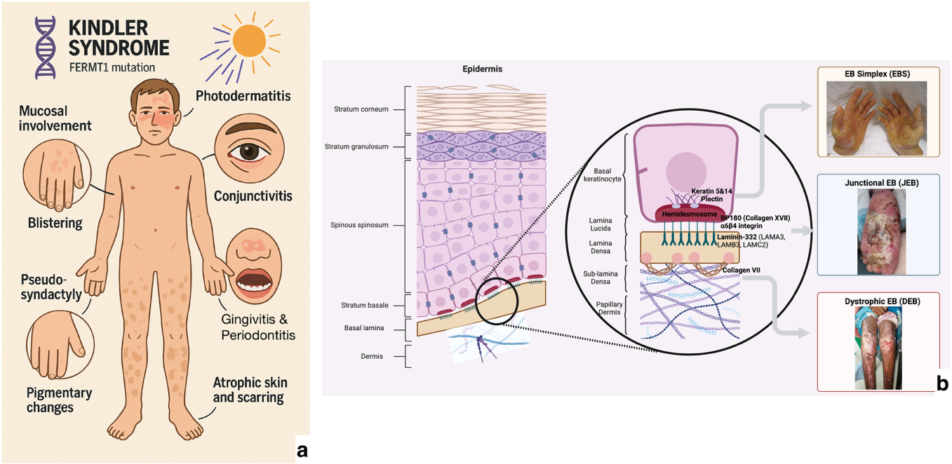
Defects at the dermoepidermal junction manifest as different subtypes of epidermolysis bullosa. Mutations in KRT5, KRT14, or PLEC produce EBS with basal keratinocyte cleavage; LAMA3, LAMB3, LAMC2, COL17A1, or ITGB4 mutations cause junctional EB (JEB) with lamina lucida blistering [44,45]; and COL7A1 mutations lead to dystrophic EB (DEB) with sublamina densa cleavage, scarring, and SCC risk [46]. Kindler syndrome (FERMT1) features mixed-level cleavage, poikiloderma, photosensitivity, and cancer predisposition [47] (Figure 8).
3.7. Adnexal development defects
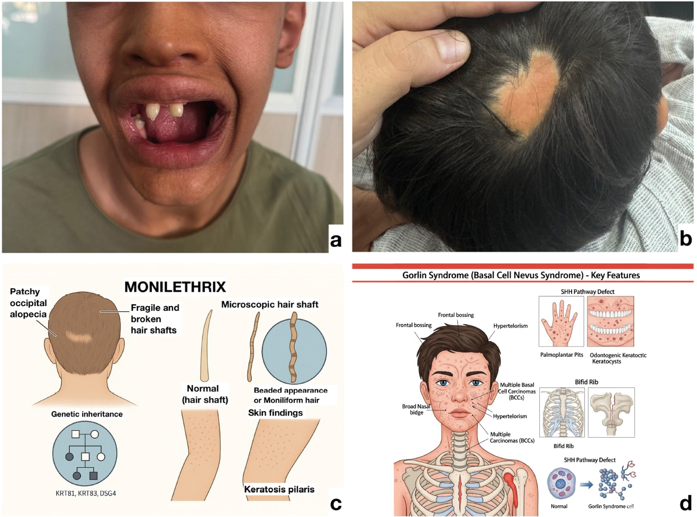
Defective appendage development produces diverse phenotypes. Hypohidrotic ectodermal dysplasia (EDA, EDAR, EDARADD) presents with hypotrichosis, hypodontia, and hypohidrosis [48]. FOXN1 mutations cause alopecia, nail dystrophy, and severe T-cell immunodeficiency [49]. Monilethrix (KRT81/83/86) is characterized by fragile, beaded hairs and keratosis pilaris [50]. Tricho-dento-osseous syndrome involves curly hair, enamel hypoplasia, and skeletal sclerosis [51]. Nevus sebaceous of Jadassohn is a congenital hamartoma with risk of secondary BCC, while Gorlin syndrome (PTCH1 mutations) links follicular morphogenesis defects to multiple BCCs and systemic anomalies [19,52,53] (Figure 9).
3.8. Embryological patterning lines
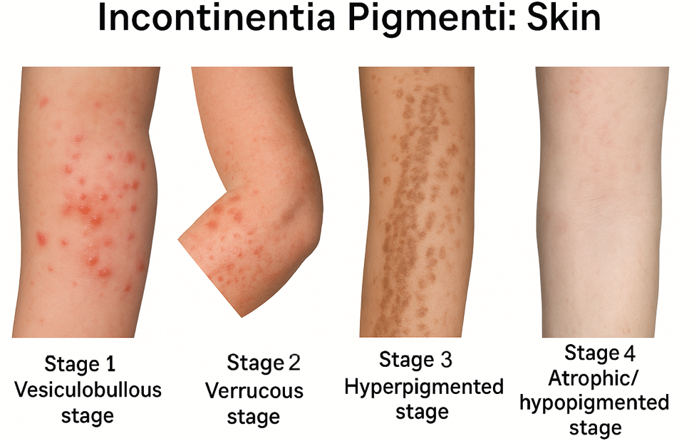
Cutaneous mosaic disorders often follow Blaschko’s lines due to postzygotic mutations [54]. Incontinentia pigmenti (IKBKG) progresses through vesiculobullous, verrucous, hyperpigmented, and atrophic stages, often with dental, ocular, and neurological abnormalities [56]. Epidermal nevi, either isolated or syndromic, similarly trace embryonic patterning lines [55,57] (Figure 10).
3.9. Externally induced developmental defects
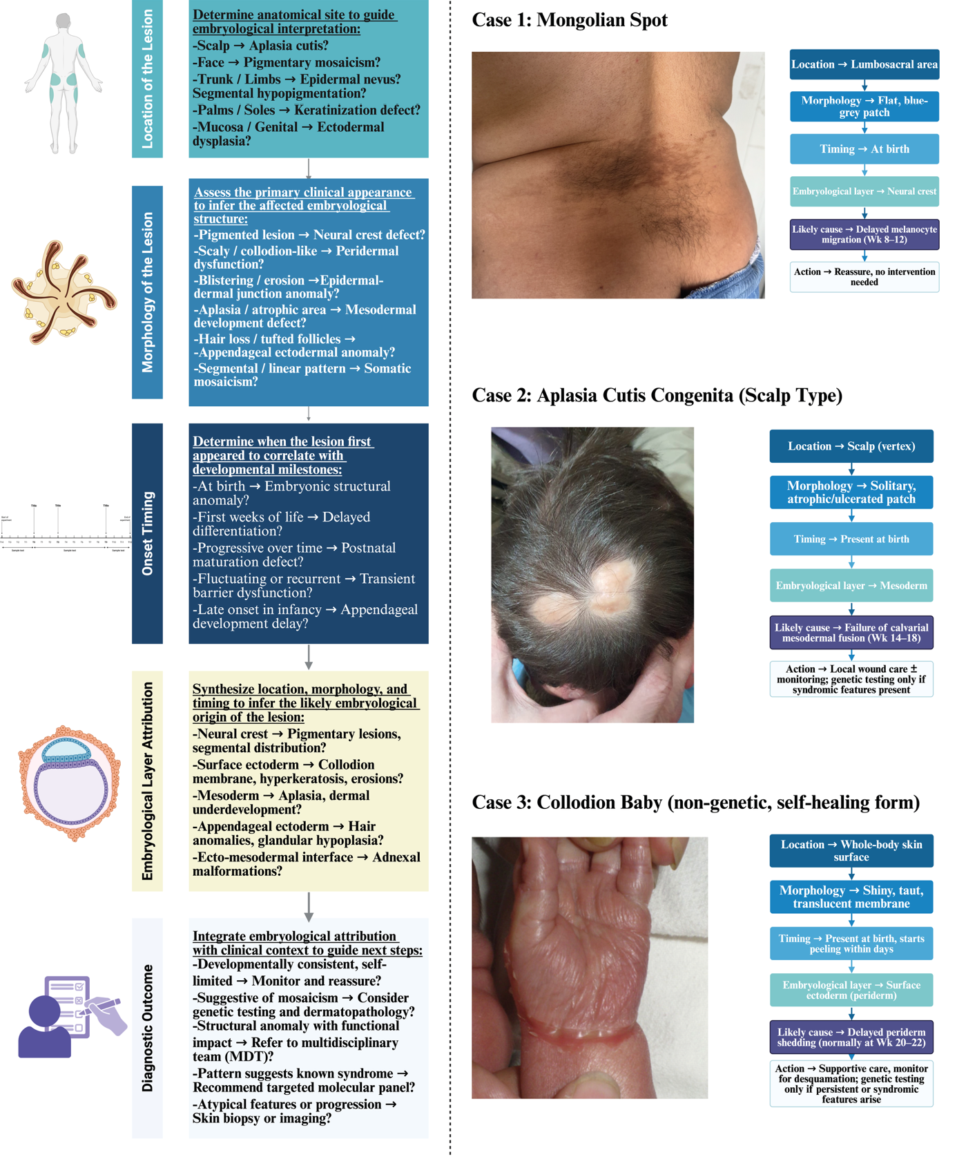
Exogenous factors may disrupt embryonic skin development. Amniotic band sequence causes constrictions and limb defects [58]. Valproic acid exposure induces neural tube and craniofacial anomalies via epigenetic and folate metabolism interference [59]. Excess retinoic acid produces craniofacial malformations through retinoid receptor activation [60]. Infections such as congenital cytomegalovirus and Zika virus impair growth and neurodevelopment, particularly with early gestational exposure [61,62]. Figure 11 presents an embryology-based diagnostic framework that integrates lesion timing, morphology, and layer attribution for congenital skin anomalies.
4. Discussion
4.1. Diagnostic utility of dermatoembryology
Dermatoembryology provides a valuable interpretive framework for prenatal dermatologic diagnosis, particularly for congenital anomalies of developmental rather than strictly genetic origin. Cutaneous structures derive from distinct embryological layers: ectoderm produces the epidermis and periderm, mesoderm forms dermal and vascular components, and neural crest cells give rise to melanocytes and some dermal elements. Each follows a defined developmental timeline: periderm forms by week 5 and regresses by week 21, while melanocyte migration occurs between weeks 8 and 12 [63]. Recognizing these sequences is critical when interpreting fetal biopsies or ultrasonography.
Persistence of the periderm beyond week 22 may indicate delayed epidermal maturation, clinically manifesting as a transient collodion membrane. Likewise, impaired melanocyte migration or differentiation may explain segmental hypopigmentation patterns that are evident at birth but not attributable to known genodermatoses. Such findings, particularly when genetic testing is inconclusive, underscore the role of developmental landmarks in prenatal diagnosis, as also supported by recent whole-exome sequencing (WES) studies [64]. Thus, dermatoembryology bridges morphology with prenatal diagnostics, offering a layer-specific perspective that enhances both invasive and non-invasive assessments. Figure 11 illustrates this integrated diagnostic approach.
4.2. Educational integration: teaching dermatologic development
Despite its relevance, dermatologic embryology remains underrepresented in medical curricula, leading to fragmented knowledge [11]. A layer-based teaching model that links ectodermal, mesodermal, and neural crest derivatives to common dermatologic conditions could strengthen both basic science education and clinical reasoning. Fakoya et al. [12] highlighted the need for an internationally standardized embryology syllabus emphasizing clinical translation, while Moraes et al. [65] showed that case-based and multimedia-enhanced teaching improves student engagement. Incorporating such approaches into dermatology education – particularly in pediatrics and prenatal dermatology – may foster early pattern recognition, diagnostic accuracy, and appreciation of developmental timing in cutaneous pathology.
4.3. Future directions: a stem cell perspective
Recent advances in stem cell biology have expanded the translational potential of dermatoembryology. Skin-derived stem cells, including epidermal stem cells (EpSCs), mesenchymal stromal cells (MSCs), and neural crest–derived melanocyte precursors, are central to both development and regeneration [66]. These cells, residing in niches such as the basal epidermis, hair follicle bulge, and dermis, are regulated by pathways including Wnt/β-catenin, Notch, and p63 [67]. Their plasticity is evident during wound healing, where follicular stem cells contribute to interfollicular repair, recapitulating developmental programs [68].
Stem cell–based therapies have shown promise in chronic wounds, autoimmune dermatoses, and hereditary blistering disorders. Adipose- and bone marrow–derived MSCs enhance re-epithelialization, angiogenesis, and immunomodulation in both experimental and clinical settings [69,70]. Induced pluripotent stem cells (iPSCs) from dermal fibroblasts offer opportunities for gene-corrected autografts in conditions such as epidermolysis bullosa [66]. Emerging human skin explant (HSE) models further demonstrate that ex vivo tissue maintains architecture, immune competence, and multipotent stem cell reservoirs (SKPs, MSCs), enabling studies on neuro-immune-cutaneous interactions relevant to both regenerative medicine and drug development [71].
5. Conclusion
Human skin develops through tightly coordinated interactions among ectodermal, mesodermal, and neural crest–derived lineages, each contributing to its structural and functional integrity. While dermatology research often emphasizes genetic causes, many congenital disorders arise instead from developmental disruptions at critical morphogenetic checkpoints. Examples include anomalies in periderm shedding, melanoblast migration, dermal extracellular matrix formation, and adnexal morphogenesis, all of which can be traced to layer-specific vulnerabilities during embryogenesis.
A dermatoembryological, layer-oriented framework improves diagnostic accuracy, particularly when genetic testing is inconclusive, and supports prenatal counseling, early biopsy decisions, and interdisciplinary management. Incorporating these principles into medical curricula also enriches dermatology education by deepening understanding of congenital anomalies and their developmental origins (Table 1).
| Embryological Layer | Stem Cell Type(s) | Differentiated Outcomes | Clinical/Research Applications |
|---|---|---|---|
| Ectoderm | Epidermal Stem Cells (EpSCs) | Keratinocytes, epidermis layers | Epidermolysis bullosa, skin grafting, wound healing |
| Neural Crest | Melanocyte SCs, EPI-NCSC | Melanocytes, neural/glial lineages | Pigment disorders, vitiligo, melanoma modeling |
| Mesoderm | Mesenchymal Stromal Cells (MSCs) | Fibroblasts, adipocytes, endothelial, myocytes | Wound healing, fibrosis modulation, immunotherapy |
| iPSC-derived (Exogenous) | iPSC-derived keratinocytes/fibroblasts | Any germ-layer lineage (pluripotent) | Gene-corrected autografts, in vitro disease modeling |
Finally, the same principles carry translational value: regenerative dermatology increasingly leverages stem cell biology and developmental pathways to restore tissue architecture. Together, these insights emphasize the diagnostic, educational, and therapeutic importance of embryology in contemporary dermatologic practice.
Abbreviations
ABCA12: ATP-binding cassette sub-family A member 12
ABS: Amniotic band sequence
ACTB: Actin beta
ADHD: Attention deficit hyperactivity disorder
AEC: Ankyloblepharon-ectodermal defects-cleft lip/palate (Hay-Wells) syndrome
ASD: Autism spectrum disorder
BMP: Bone morphogenetic protein
CXCL12: C-X-C motif chemokine ligand 12
CXCR4: C-X-C chemokine receptor type 4
DEB: Dystrophic epidermolysis bullosa
DEJ: Dermoepidermal junction
EBS: Epidermolysis bullosa simplex
E-cadherin: Epithelial cadherin
EDAR: Ectodysplasin A receptor
EDARADD: EDAR-associated death domain
EDA: Ectodysplasin A
EpSCs: Epidermal stem cells
FGF: Fibroblast growth factor
FOXN1: Forkhead box N1
HED: Hypohidrotic ectodermal dysplasia
HSE: Human skin explants
iPSCs: Induced pluripotent stem cells
IRF6: Interferon regulatory factor 6
IKKα: IκB kinase alpha
JEB: Junctional epidermolysis bullosa
KRT: Keratin
LAMA3, LAMB3, LAMC2: Laminin subunits α3, β3, γ2
MAPK/ERK: Mitogen-activated protein kinase/extracellular signal-regulated kinase
MITF: Microphthalmia-associated transcription factor
MSCs: Mesenchymal stromal cells
NKPD1: NTPase KAP family P-loop domain-containing protein 1
OCA1: Oculocutaneous albinism type 1
PAX3: Paired box gene 3
P-cadherin: Placental cadherin
PLEC: Plectin
SAM: Sterile alpha motif
SCs: Stem cells
SDF-1α: Stromal cell-derived factor 1 alpha
SFN: Stratifin
SHH: Sonic hedgehog
SKPs: Skin-derived precursors
SOX10: SRY-box transcription factor 10
TDO: Tricho-dento-osseous syndrome
TGM1: Transglutaminase 1
TP63: Tumor protein p63
TYR: Tyrosinase
VPA: Valproic acid
WES: Whole-exome sequencing
Authors’ contributions
G.K. conceived the study, conducted the literature review, drafted, and critically revised the manuscript. G.N.Y. handled the ethical approval process, critically appraised the manuscript content, and contributed to the evaluation of the study’s scope and structure. C.A. provided clinical images and case data, participated in the interpretation of clinical findings, and critically revised the manuscript for important intellectual content. Ö.S.K. contributed substantially to the conception and design of the study, supervised manuscript development, and performed critical revisions for important intellectual content. All authors read and approved the final version of the manuscript and agreed to be accountable for all aspects of the work.
Acknowledgments
Throughout the course of this study, we adhered strictly to the World Medical Association Declaration of Helsinki and the Good Clinical and Laboratory Practice standards.
The schematic illustrations included in this article were created using BioRender.com under an academic license intended for scientific research and publication purposes. These include Figures 4b, 6c, 7a, 8b, 9a–c, and 11a–b.
Clinical photographs used in Figures 4a and 5 were obtained from Wikimedia Commons and are published under the Creative Commons Attribution 4.0 International (CC BY 4.0) License. Appropriate attribution is provided, and all reuse complies with the terms of this license.
All other clinical images, including patient photographs and case examples, originate from the personal archive of the authors and are used with written informed consent from the patients and/or their legal guardians, in accordance with ethical publishing standards.
Ethics statement
The study was conducted in accordance with the principles of the Declaration of Helsinki. The study protocol was approved by the Erzincan Binali Yıldırım University Clinical Research Ethics Committee on June 13, 2025, with the decision number 456343.
References
[1] Sadler TW. Langman’s medical embryology. 12th ed. Philadelphia: Lippincott Williams & Wilkins; 2012. Available from: https://integratedsciences.lwwhealthlibrary.com/book.aspx?bookid=770
[2] Schoenwolf GC, Bleyl SB, Brauer PR, Francis-West PH. Larsen’s human embryology. 5th ed. Philadelphia: Elsevier; 2014.
[3] Fitzpatrick TB, Kang S, Goldsmith LA, Gilchrest BA, Paller AS, Leffell DJ, Wolff K, editors. Structure and function of skin. In: Fitzpatrick’s dermatology in general medicine. 10th ed. New York: McGraw-Hill; 2024. Chapter 7.
[4] Workman VL, Giblin AV, Green NH, et al. Development of a tissue-engineered skin model with epidermal, dermal and hypodermal components. In Vitro Models. 2023;2:297-306. https://doi.org/10.1007/s44164-023-00058-9.
[5] Gopee NH, Winheim E, Olabi B, et al. A prenatal skin atlas reveals immune regulation of human skin morphogenesis. Nature. 2024;635:679-689. https://doi.org/10.1038/s41586-024-08002-x.
[6] Park S. Building vs. rebuilding epidermis: Comparison embryonic development and adult wound repair. Front Cell Dev Biol. 2022;9:796080. https://doi.org/10.3389/fcell.2021.796080.
[7] Fuchs E. Scratching the surface of skin development. Nature. 2007;445(7130):834-842. https://doi.org/10.1038/nature05659.
[8] Sonnen KF, Janda CY. Signalling dynamics in embryonic development. Biochem J. 2021;478(23):4045-4070. https://doi.org/10.1042/BCJ20210043.
[9] Fu X, editor. Regenerative medicine in China. Singapore: Springer; 2021. https://doi.org/10.1007/978-981-16-1182-7.
[10] De Falco M, Pisano MM, De Luca A. Embryology and anatomy of the skin. In: Brash UT, Maitland ML, editors. Skin cancer. New York: Springer; 2014. p.3-17. https://doi.org/10.1007/978-1-4614-7357-2_1.
[11] Tumiene B, Peters H, Melegh B, et al. Rare disease education in Europe and beyond: time to act. Orphanet J Rare Dis. 2022;17:441. https://doi.org/10.1186/s13023-022-02527-y.
[12] Fakoya FA, Emmanouil-Nikoloussi E, Sharma D, Moxham BJ. A core syllabus for the teaching of embryology and teratology to medical students. Clin Anat. 2017;30(2):159-167. https://doi.org/10.1002/ca.22802.
[13] Cochard LR, Netter FH. Netter’s atlas of human embryology. Updated ed. Philadelphia: Saunders Elsevier; 2012.
[14] Chetty R. Pathology of vascular skin lesions: Clinicopathological correlations: Sangueza OP, Requena L. J Clin Pathol. 2003;56(7):559-560.
[15] Schlessinger DI, Patino SC, Belgam Syed SY, et al. Embryology, epidermis. [Updated 2022 Oct 10]. In: StatPearls [Internet]. Treasure Island (FL): StatPearls Publishing; 2025 Jan-. Available from: https://www.ncbi.nlm.nih.gov/books/NBK441867/
[16] Voiță-Mekereș F. Assessment of the embryological origin, anatomical and histological structure of the skin. Arch Pharm Pract. 2024;15(2):69-74. https://doi.org/10.51847/SWViK4kyKx.
[17] Hoeger PH, Kinsler V, Yan AC, Harper J, Oranje AP, Bodemer C, et al., editors. Harper’s textbook of pediatric dermatology. 4th ed. Oxford: Wiley-Blackwell; 2019.
[18] Singh V. Textbook of clinical embryology. New Delhi: Elsevier India; 2012.
[19] Griffiths CEM, Barker J, Bleiker TO, Hussain W, Simpson RC, editors. Rook’s textbook of dermatology. 10th ed. Oxford: Wiley-Blackwell; 2024.
[20] Mort RL, Jackson IJ, Patton EE. The melanocyte lineage in development and disease. Development. 2015;142(4):620-632. https://doi.org/10.1242/dev.106567.
[21] Keswell D, Kidson SH, Davids LM. Melanocyte migration is influenced by E-cadherin-dependent adhesion of keratinocytes in both two- and three-dimensional in vitro wound models. Cell Biol Int. 2015;39(2):169-176. https://doi.org/10.1002/cbin.10350.
[22] Yamauchi A, Hadjur C, Takahashi T, Suzuki I, Hirose K, Mahe YF. Human skin melanocyte migration towards stromal cell-derived factor-1α demonstrated by optical real-time cell mobility assay: modulation of their chemotactic ability by α-melanocyte-stimulating hormone. Exp Dermatol. 2013;22(10):664-667. https://doi.org/10.1111/exd.12232.
[23] Alsaad KO, Obaidat NA, Ghazarian D. Skin adnexal neoplasms--part 1: an approach to tumours of the pilosebaceous unit. J Clin Pathol. 2007;60(2):129-144. https://doi.org/10.1136/jcp.2006.040337.
[24] Sanz-Ezquerro JJ, Münsterberg AE, Stricker S. Signaling pathways in embryonic development. Front Cell Dev Biol. 2017;5:76. https://doi.org/10.3389/fcell.2017.00076.
[25] Tüzün Y, İşçimen A, Pehlivan Ö. Collodion baby. J Turk Acad Dermatol. 2008;2(2):Article 82201r. Available from: http://www.jtad.org/2008/2/jtad82201r.pdf
[26] Simalti AK, Sethi H. Collodion baby. Med J Armed Forces India. 2017;73(2):197-199. https://doi.org/10.1016/j.mjafi.2015.10.007.
[27] Nguyen MA, Gelman A, Norton SA. Practical events in the management of a collodion baby. JAMA Dermatol. 2015;151(9):1031-1032. https://doi.org/10.1001/jamadermatol.2015.0694.
[28] Richardson RJ, Hammond NL, Coulombe PA, et al. Periderm prevents pathological epithelial adhesions during embryogenesis. J Clin Invest. 2014;124(9):3891-3900. https://doi.org/10.1172/JCI71946.
[29] Chen H. Collodion baby. In: Puri RD, Agarwal S, editors. Atlas of genetic diagnosis and counseling. New York: Springer; 2016. p. 1-10. https://doi.org/10.1007/978-1-4614-6430-3_47-2.
[30] Bamalan OA, Moore MJ, Menezes RG. Vernix caseosa. [Updated 2023 Jul 4]. In: StatPearls [Internet]. Treasure Island (FL): StatPearls Publishing; 2025 Jan-. Available from: https://www.ncbi.nlm.nih.gov/books/NBK559238/
[31] So JY, Teng J. Epidermolysis bullosa simplex. 1998 Oct 7 [Updated 2022 Aug 4]. In: Adam MP, Feldman J, Mirzaa GM, et al., editors. GeneReviews® [Internet]. Seattle (WA): University of Washington, Seattle; 1993-2025. Available from: https://www.ncbi.nlm.nih.gov/books/NBK1369/
[32] Smith LD, Masood M, Bajaj GS, Couser NL. Genetic abnormalities of the anterior segment, eyelids, and external ocular adnexa. In: Singh AD, Damato BE, Pe’er J, Murphree AL, Perry JD, editors. Ophthalmic genetic diseases: A quick reference guide to the eye and external ocular adnexa abnormalities. Amsterdam: Elsevier; 2019. p. 15-39. https://doi.org/10.1016/B978-0-323-65414-2.00002-7.
[33] Rice AS, Crane JS. Epidermolytic hyperkeratosis. [Updated 2023 Jul 31]. In: StatPearls [Internet]. Treasure Island (FL): StatPearls Publishing; 2025 Jan-. Available from: https://www.ncbi.nlm.nih.gov/books/NBK544323/
[34] Komlosi K, Glocker C, Hsu-Rehder HH, Alter S, Kopp J, Hotz A, et al. Autosomal dominant lamellar ichthyosis due to a missense variant in the gene NKPD1. J Invest Dermatol. 2024;144(12):2754-2763.e6. https://doi.org/10.1016/j.jid.2024.03.041.
[35] Summers CG. Albinism: classification, clinical characteristics, and recent findings. Optom Vis Sci. 2009;86(6):659-662. https://doi.org/10.1097/OPX.0b013e3181a5254c.
[36] Federico JR, Krishnamurthy K. Albinism. In: StatPearls [Internet]. Treasure Island (FL): StatPearls Publishing; 2025 Jan-.
[37] Kromann AB, et al. Pigmentary mosaicism. Orphanet J Rare Dis. 2018;13:39. https://doi.org/10.1186/s13023-018-0778-6.
[38] Schaffer JV. Pigmentary mosaicism. Clin Dermatol. 2022;40(4):322-338. https://doi.org/10.1016/j.clindermatol.2022.02.005.
[39] Zhou YJ, et al. Becker’s nevus. Dermatol Ther. 2022;35(7):e15548. https://doi.org/10.1111/dth.15548.
[40] Brownell I, Loomis CA, Koss T. Skin development and maintenance. In: Bolognia JL, Schaffer JV, Cerroni L, editors. Dermatology. 5th ed. Amsterdam: Elsevier; 2024.
[41] Malfait F, Castori M, Francomano CA, et al. The Ehlers–Danlos syndromes. Nat Rev Dis Primers. 2020;6:64. https://doi.org/10.1038/s41572-020-0194-9.
[42] Miklovic T, Sieg VC. Ehlers-Danlos syndrome. In: StatPearls [Internet]. Treasure Island (FL): StatPearls Publishing; 2025 Jan-. PMID: 31747221.
[43] Ghosh SK, Dutta A, Sarkar S, Nag SS, Biswas SK, Mandal P. Focal dermal hypoplasia (Goltz syndrome): A cross-sectional study from eastern India. Indian J Dermatol. 2017;62(5):498-504. https://doi.org/10.4103/ijd.IJD_317_17.
[44] Pfendner EG, Lucky AW. Junctional epidermolysis bullosa. 2008 Feb 22 [Updated 2018 Dec 20]. In: Adam MP, Feldman J, Mirzaa GM, et al., editors. GeneReviews® [Internet]. Seattle (WA): University of Washington, Seattle; 1993–2025. Available from: https://www.ncbi.nlm.nih.gov/books/NBK1125/
[45] Khanna D, Bardhan A. Epidermolysis bullosa. [Updated 2024 Jan 11]. In: StatPearls [Internet]. Treasure Island (FL): StatPearls Publishing; 2025 Jan–. Available from: https://www.ncbi.nlm.nih.gov/books/NBK599531/
[46] Lucky AW, Pope E, Crawford S. Dystrophic epidermolysis bullosa. 2006 Aug 21 [Updated 2025 May 8]. In: Adam MP, Feldman J, Mirzaa GM, et al., editors. GeneReviews® [Internet]. Seattle (WA): University of Washington, Seattle; 1993–2025. Available from: https://www.ncbi.nlm.nih.gov/books/NBK1304/
[47] Youssefian L, Vahidnezhad H, Uitto J. Kindler syndrome. 2016 Mar 3 [Updated 2022 Jan 6]. In: Adam MP, Feldman J, Mirzaa GM, et al., editors. GeneReviews® [Internet]. Seattle (WA): University of Washington, Seattle; 1993–2025. Available from: https://www.ncbi.nlm.nih.gov/books/NBK349072/
[48] Shamim H, Hanif S. Hypohidrotic ectodermal dysplasia: A case report. Cureus. 2023;15(10):e46530. https://doi.org/10.7759/cureus.46530.
[49] Pignata C, Fusco A, Amorosi S. Human clinical phenotype associated with FOXN1 mutations. In: Maiese K, editor. Forkhead transcription factors. Advances in experimental medicine and biology. Vol. 665. New York: Springer; 2009. p. 195–206. https://doi.org/10.1007/978-1-4419-1599-3_15.
[50] Chabchoub I, Souissi A. Monilethrix. [Updated 2023 Jun 12]. In: StatPearls [Internet]. Treasure Island (FL): StatPearls Publishing; 2025 Jan–. Available from: https://www.ncbi.nlm.nih.gov/books/NBK539813/
[51] Al-Batayneh OB. Tricho-dento-osseous syndrome: Diagnosis and dental management. Int J Dent. 2012;2012:514692. https://doi.org/10.1155/2012/514692.
[52] Moody MN, Landau JM, Goldberg LH. Nevus sebaceous revisited. Pediatr Dermatol. 2012;29(1):15–23. https://doi.org/10.1111/j.1525-1470.2011.01562.x.
[53] Idriss MH, Elston DM. Secondary neoplasms associated with nevus sebaceus of Jadassohn: A study of 707 cases. J Am Acad Dermatol. 2014;70(2):332–337. https://doi.org/10.1016/j.jaad.2013.10.004.
[54] Moss C. Cytogenetic and molecular evidence for cutaneous mosaicism: The ectodermal origin of Blaschko lines. Am J Med Genet. 1999;85(4):330–333. https://doi.org/10.1002/(sici)1096-8628(19990806)85:4<330::aid-ajmg3>3.0.co;2-m.
[55] Brar BK, Mahajan BB, Puri N. Linear and whorled nevoid hypermelanosis. Indian J Dermatol Venereol Leprol. 2008;74(5):512–513. https://doi.org/10.4103/0378-6323.44325.
[56] Yadlapati S, Tripathy K. Incontinentia pigmenti (Bloch-Sulzberger syndrome). In: StatPearls [Internet]. Treasure Island (FL): StatPearls Publishing; 2025 Jan–. PMID: 35201722.
[57] Nicholson CL, Daveluy S. Epidermal nevus syndromes. [Updated 2023 Jun 12]. In: StatPearls [Internet]. Treasure Island (FL): StatPearls Publishing; 2025 Jan–. Available from: https://www.ncbi.nlm.nih.gov/books/NBK559003/.
[58] Singh AP, Gorla SR. Amniotic band syndrome. [Updated 2022 Dec 11]. In: StatPearls [Internet]. Treasure Island (FL): StatPearls Publishing; 2025 Jan–. Available from: https://www.ncbi.nlm.nih.gov/books/NBK545283/
[59] Ornoy A, Echefu B, Becker M. Valproic acid in pregnancy revisited: Neurobehavioral, biochemical and molecular changes affecting the embryo and fetus in humans and in animals: A narrative review. Int J Mol Sci. 2023;25(1):390. https://doi.org/10.3390/ijms25010390.
[60] Matt N, Ghyselinck NB, Wendling O, Chambon P, Mark M. Retinoic acid-induced developmental defects are mediated by RARbeta/RXR heterodimers in the pharyngeal endoderm. Development. 2003;130(10):2083–2093. https://doi.org/10.1242/dev.00428.
[61] Akpan US, Pillarisetty LS. Congenital cytomegalovirus infection. [Updated 2023 Aug 8]. In: StatPearls [Internet]. Treasure Island (FL): StatPearls Publishing; 2025 Jan–. Available from: https://www.ncbi.nlm.nih.gov/books/NBK541003/.
[62] Dos Santos ALS, Rosolen BB, Ferreira FC, Chiancone IS, Pereira SS, Pontes KFM, et al. Intrauterine Zika virus infection: An overview of the current findings. J Pers Med. 2025;15(3):98. https://doi.org/10.3390/jpm15030098.
[63] Dermitzakis I, Chatzi D, Kyriakoudi SA, Evangelidis N, Vakirlis E, Meditskou S, et al. Skin development and disease: A molecular perspective. Curr Issues Mol Biol. 2024;46(8):8239–8267. https://doi.org/10.3390/cimb46080487.
[64] Zhu X, Petrovski S, Xie P, Ruzzo EK, Lu YF, McSweeney KM, et al. Whole-exome sequencing in undiagnosed genetic diseases: Interpreting 119 trios. Genet Med. 2015;17(10):774–781. https://doi.org/10.1038/gim.2014.191.
[65] Moraes SG, Pereira LA. A multimedia approach for teaching human embryology: Development and evaluation of a methodology. Ann Anat. 2010;192(6):388–395. https://doi.org/10.1016/j.aanat.2010.05.005.
[66] Prodinger CM, Reichelt J, Bauer JW, Laimer M. Current and future perspectives of stem cell therapy in dermatology. Ann Dermatol. 2017;29(6):667–687. https://doi.org/10.5021/ad.2017.29.6.667.
[67] Taub AF, Pham K. Stem cells in dermatology and anti-aging care of the skin. Facial Plast Surg Clin North Am. 2018;26(4):425–437. https://doi.org/10.1016/j.fsc.2018.06.004.
[68] Farabi B, Roster K, Hirani R, Tepper K, Atak MF, Safai B. The efficacy of stem cells in wound healing: A systematic review. Int J Mol Sci. 2024;25(5):3006. https://doi.org/10.3390/ijms25053006.
[69] Nakamuta A, Yoshido K, Naoki H. Stem cell homeostasis regulated by hierarchy and neutral competition. Commun Biol. 2022;5:1268. https://doi.org/10.1038/s42003-022-04218-7.
[70] Ogliari KS, Marinowic D, Brum DE, Loth F. Stem cells in dermatology. An Bras Dermatol. 2014;89(2):286–291. https://doi.org/10.1590/abd1806-4841.20142530.
[71] Cousin I, Misery L, de Vries P, Lebonvallet N. Emergence of new concepts in skin physiopathology through the use of in vitro human skin explants models. Dermatology. 2023;239(6):849–859. https://doi.org/10.1159/000533261.