https://doi.org/10.36253/ijae-16609
A population study of variations in the brachial artery and its terminal branches: Clinical correlates
Department of Anatomy, School of Medicine, Sefako Makgatho Health Sciences University, Ga-Rankuwa, Pretoria, South Africa
*Corresponding author. Email: patriacoetzee@gmail.com
Abstract. Reports in the literature suggest that the variations in branching patterns of the upper limb arteries and their prevalence may be influenced by sex and population affinity. A comprehensive investigation of branching patterns of the brachial artery and its branches is lacking in South Africans, with many reports focusing on the axillary artery. Therefore the current study aimed to record the incidence of the variations of the brachial, radial and ulnar arteries in a sample of the South African population. One hundred and eighty (180) upper limbs of 90 South African cadavers from the Department of Anatomy at Sefako Makgatho Health Sciences University were dissected. Normal anatomy according to standard anatomy textbooks and variant branching patterns of the brachial, radial and ulnar arteries were identified and recorded. SPSS software was used to establish the differences in variant branching patterns between side and sex. Upper limb arterial variations appeared in all the dissected cadavers. The recorded variations ranged from abnormal origin of the deep brachial artery, high division of the brachial artery into the ulnar and radial arteries, tortuous brachial and radial arteries, and superficial course of radial and ulnar arteries. The prevalence of brachial, ulnar and radial artery variations in the current study is comparable to that of previous studies. However, the superficial radial artery is more prevalent in males than in females in South Africans. The results of the current study provide crucial information for planning surgical procedures and interpretation of angiograms.
Keywords: brachial artery, ulnar artery, radial artery, median artery.
Contents
Variations of the brachial artery
Variations of the radial artery
Variations of the ulnar artery
Tortuosity of the upper limb arteries and their clinical implications
Variations of the brachial artery and its terminal branches
Superficial course of the radial artery
Tortuosity of the brachial artery and its branches
Variations of the brachial artery and its terminal branches
Tortuosity of the main upper limb arteries
Introduction
The branching pattern of the upper limb arteries are highly variable, with an average incidence of 25% [1]. Literature reports describing the prevalence of the variant branching patterns of upper limb vessels usually focus on the axillary artery due to frequent incident reports on its variations [2–6]. Consequently, there is a lack of population studies on variations of brachial, radial and ulnar arteries as much focus on arterial variations is put on incident reports encountered during routine dissections in the university setting [7–10]. A study performed on a sample of African Americans and non-African-Americans showed a higher incidence of a high brachial artery bifurcation in African-Americans than in non-African-Americans suggesting that population affinity may influence this branching pattern [11]. The available reports on upper limb arterial variations are mostly based on European and Asian population groups with not many reports on the African populations [4,5,7,12,13]. Studies conducted in African populations specifically focused on the axillary artery, whereby some unique branching patterns were recorded in contrast to standard anatomy textbook descriptions [6,14,15].
Arterial variations have significant clinical implications during surgical procedures [6]. The most common variation noted in the brachial artery, is a superficial brachial artery, which could be easily mistaken for a vein during drug administration [16]. The high origin of the radial and ulnar arteries from axillary or brachial artery has also been reported [17–20]. Knowledge of variant origins of these vessels can be clinically relevant as the radial artery is commonly harvested for coronary artery bypass procedures, in which case, the ulnar artery becomes the principal arterial supply to the hand [21]. The high origin of the ulnar artery is often associated with nerve entrapment as well as trauma during intravenous cannulation [22]. Another upper limb arterial variation reported in the literature is a tortuous radial artery [23–25]. Change in the direction of the flow of blood in a tortuous vessel is often noted and could affect the histological structure of the vessel and cause atherosclerotic plaques [24]. In addition, the tortuous course of the radial artery can cause difficulty to access the radial artery during trans-radial catheterisation, as the tortuosity could hinder the passage of the catheter causing complications or influence the success of the procedure [25].
Another upper limb arterial variation in the forearm arteries is a persistent median artery which has been reported to have a prevalence of 2,8% [26]. When present, the median artery can cause symptoms consistent with median nerve disturbances, either by compression when accompanying the median nerve through the carpal tunnel, or by iatrogenic ischemia [27].
Variations of the brachial artery
Common variations of the brachial artery include the superficial brachial artery, high origin of radial and ulnar arteries, accessory brachial artery, and trifurcation of the brachial artery to common interosseous, radial and ulnar arteries [28]. The superficial brachial artery is defined as a brachial artery coursing anterior to the median nerve, instead of posterior to it [29]. A superficial brachial artery is often more susceptible to trauma and can easily be confused with superficial veins [16]. Additionally, the superficial course and variations in the brachial artery or its derivatives are clinically relevant as the brachial artery is often used as the passage during coronary artery procedures making it prone to injuries leading to bleeding or ischemia [30]. The bifurcation of the brachial artery proximal to the cubital fossa is considered a high division of the brachial artery [31]. The high origin of the radial artery has a prevalence of 3,0-18,0% [13,19,29,32]. Knowledge of the above variations is also crucial for the evaluation of angiographic images, and for vascular and reconstructive surgeries of the arm and forearm [30].
Variations of the radial artery
The radial artery usually originates from the brachial artery in the cubital fossa at the level of the neck of the radius [19]. The radial artery runs along the lateral aspect of the anterior compartment of the forearm deep to the brachioradialis muscle, lateral to the flexor carpi radialis tendon. It winds around the lateral aspect of the radius and passes through the floor of the anatomical snuffbox to pierce the first dorsal interosseous muscle [28].
The high origin of the radial artery is described as the origin from the axillary artery or from the brachial artery proximal to the cubital fossa [29]. The radial artery is commonly used in vascular, plastic and reconstructive surgery and is routinely used for arterial puncture and cannulation [19]. Radial artery anomalies may prolong and diminish the success of coronary angiography and percutaneous coronary intervention [20]. In addition, trans-radial access may be hindered by the presence of an unusual origin or course of the radial artery [33]. The superficial course of the radial artery may be more vulnerable to trauma, and thus haemorrhage [34]. It may also lead to puncture of the radial artery during venipuncture, misinterpretation of angiographic images, or severe disturbance of hand irrigation during surgical procedures on the arm [35]. Furthermore, it has been recorded that the presence of a high origin of the radial artery contributes to the development of tortuosity, which can increase the risk of failure of trans-radial catheterisation [19].
Variations of the ulnar artery
The ulnar artery passes medially, deep to the pronator teres muscle and intermediate to the flexor digitorum superficialis muscle, to reach the medial aspect of the forearm after its origin from the brachial artery in the cubital fossa. It passes superficial to the flexor retinaculum at the wrist in Guyon’s canal to enter the hand [28, 36]. The ulnar artery courses through the forearm with the ulnar nerve [37].
The superficial artery is described as an ulnar artery with a high origin in the arm that descends over the superficial muscles of the forearm [8]. Knowledge of the superficial ulnar artery has clinical significance as this variant vessel can be easily mistaken for a vein during intravenous drug administration [38]. The tortuosity of the ulnar artery can lead to ulnar nerve compression if tortuosity coincides with the passage of these two structures through the pisohammate hiatus, which may result in paresthesia and numbness of the skin in the dorsum of the hand where the ulnar nerve is distributed [39].
The median artery
During early embryonic life, the arterial axis of the forearm is represented by a transitory vessel known as the median artery. It normally regresses after the second embryonic month [27]. However, if the persistent median artery does not regress, it forms a median artery that accompanies the median nerve, which in some cases may perforate the median nerve [32]. The persistent median artery has also been associated with compressive pathology of the median nerve, which is secondary to arterial calcification, thrombosis and atherosclerosis [12]. In addition, if there is no anastomosis between the persistent median artery and the ulnar artery, it becomes of great importance to plastic surgeons and neurosurgeons as inadvertent damage to the persistent median artery may cause hand ischemia [40]. The prevalence of the variations of the median artery has shown populations difference ranging from 0,5-59,7% in Europeans, and Africans respectively, with the Europeans recording the lowest incidence [13,14,20,41].
Tortuosity of the upper limb arteries and their clinical implications
Tortuous arteries and veins are increasingly becoming a common observation throughout the human body [25]. Changes in the arterial walls due to mechanical injury, hypertension, diabetes, ageing and atherosclerosis may lead to thrombic events, embolic events, restriction of blood flow, occlusion of blood flow and ultimately change in the gross anatomical structure of the artery leading to tortuosity [24].
The mechanical stability of arteries depends on the wall stiffness [24]. Elastin is an important extracellular matrix component for arterial elasticity and stiffness and elastin degradation weakens the arterial wall [42]. Degradation of elastin in the arterial walls is said to cause weakening of the arteries leading to tortuosity, due to increased blood flow, but the underlying mechanisms are unclear [24]. The fragmentation of elastin has been reported in the walls of tortuous arteries and has been considered the cause of vessel lengthening leading to tortuosity [43].
Clinical observations have linked ageing, atherosclerosis, hypertension, genetic defects, diabetes mellitus and reduced axial tension or elongation of the arteries to some of the factors thought to cause tortuosity [24]. Certain levels of axial tension are essential in maintaining the stability of arteries and preventing tortuosity, decreasing with age as the artery lengthens [44].
Tortuosity of arteries increases the resistance in blood flow and in severe cases, obstruction or even occlusion of blood flow [45]. Han (2012) [24] classified tortuosity into four different types: curving/curling, kinking, looping and spiral twisting vessels. A previous study described three types of tortuous radial arteries [46]. The first type was the tortuous radial artery without stenosis. In this type, trans-radial catheterization was recorded to have a 98,8% success rate. The second type was the stenotic tortuous radial artery where the tortuous segment of the artery was calcified. In this type, difficulty in inserting the guide wire was reported. The third type was the hypoplastic tortuous radial artery, an additional brachioradial artery, which may influence the flow of blood and may require a different approach to gain access to the artery [46]. The afore-mentioned types of the tortuous radial artery suggest that tortuosity complicates radial catheterisation. Chronic pressure on the vessel wall is considered one of the important predisposing factors of tortuosity, resulting in the vessel wall thickening and also an increase in the length of the artery [24]. In case of an anomaly in the radial artery, like tortuosity, the ulnar artery is used as an alternative in coronary and cerebral angiography and ulnar-cephalic arteriovenous fistula when access to the radial artery fails [8].
Materials and methods
Study design
The current study is a descriptive quantitative study where the types and frequency distribution of the upper limb arterial variations were analysed and recorded in a select sample of the South African population (black and white) (Ethics Reference Number: SMUREC/M/193/2022: PG). The sampling method used in the current study was convenient sampling. Gross anatomical dissection was used to expose the upper limb arteries by using the methods described by Tank and Grant (2013)[47].
Sample size
One-hundred and eighty (180) upper limb arteries of 90 cadavers were dissected in the Department of Human Anatomy and Histology at Sefako Makgatho Health Sciences University (Table 1). Of the 90 cadavers, 14 were neonatal cadavers, 3 were fresh unembalmed adult cadavers and the remaining 73 cadavers were adult embalmed cadavers. The axillary, brachial, ulnar and radial arteries were exposed via gross anatomical dissection using a method described by Tank and Grant (2013)[47]. The dissection was completed by making use of a standard dissection kit.
| Sex | Race distribution | Sample size | |
|---|---|---|---|
| Black | White | ||
| Female | 18/90 = 20,00% | 9/90=10,00% | 27 |
| Male | 50/90 = 55,56% | 13/90 = 14,44% | 63 |
| Total | 68 | 22 | 90 |
Data analysis
The data was analysed using SPSS v.24. Frequency distribution of the variant branching patterns of the tortuosity of the radial, high origin of the radial artery, common trunk for circumflex humeral arteries and the superficial subscapular artery were recorded in the studied population from the left and right upper limbs, Black and White population as well as female and male were compared using the Chi-square statistical test. A p-value ≤ 0,05 was considered statistically significant.
Results
Variations of the brachial artery and its terminal branches
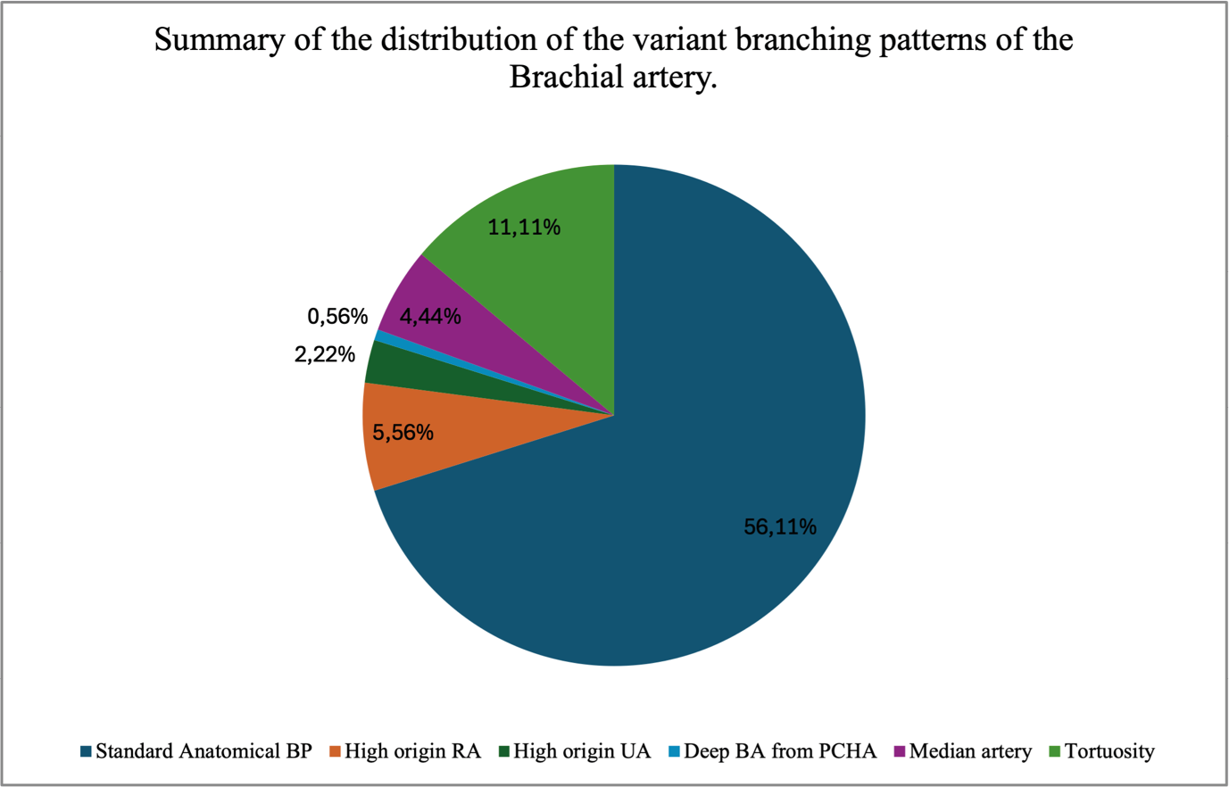
The standard anatomical branching pattern of the brachial artery was recorded in 56,11% cases (Figure 1). In the above-mentioned cases, 73 cases were recorded in males, while 28 cases were recorded in females. Additionally, 15 of the above cases were recorded in the White cadavers while 86 cases were recorded in the Black cadavers. This pattern was observed bilaterally in 96 cadavers and unilaterally in five cadavers.
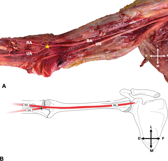
The radial artery had a high origin in 5,56% cases (Figure 2A and B). The high origin of the radial artery was found bilaterally in 4,44% and unilaterally in 1,11%. In addition, 5,00% were observed in males and 0,56% were observed in females. Considering the race distribution, 1,67% cases of high origins of the radial artery were recorded in the White cadavers and 3,89% were recorded in the Black cadavers.
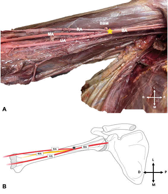
The Chi-square test comparing the frequency of the high origin of the radial artery in all 180 dissected upper limbs (136 Black, 44 White, 126 males and 54 females) showed no significant differences in the frequency of the high origin of the radial artery in the sides (p = 0,861), sex (p = 0,094) and race (p = 0,732).
In one of the above 10 cases of high origin of radial artery, the radial artery took a superficial course over the forearm muscles.
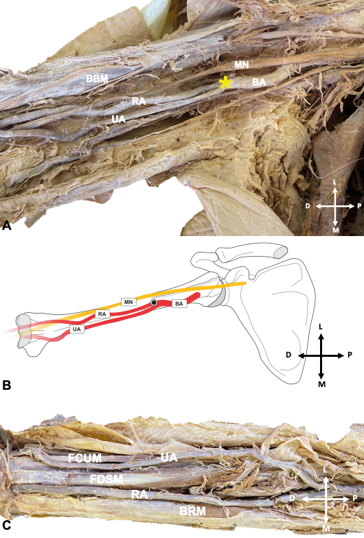
The ulnar artery had a high origin in 2,22% cases (Figure 3A and B). In one of the above four cases, the ulnar artery took a superficial course (Figure 3 C). The above cases were recorded bilaterally in two White male cadavers.
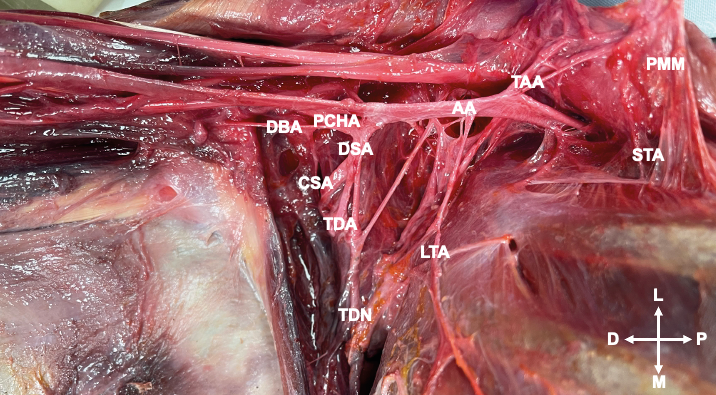
In 0,56% cases the deep brachial artery originated from the posterior circumflex humeral artery on the right of a male cadaver (Figure 4).
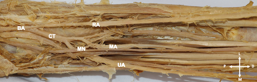
The median artery was recorded in 4,44% cases (Figure 5). It was unilateral in 2,22% cases and bilateral in 2,22% cases. In the above cases, 3,33% were observed in males, while 1,11% cases were observed in females. Considering the race distribution, 1,67% cases of the median artery were recorded in the White cadavers and 2,78% cases were recorded in the Black cadavers.
Superficial course of the radial artery
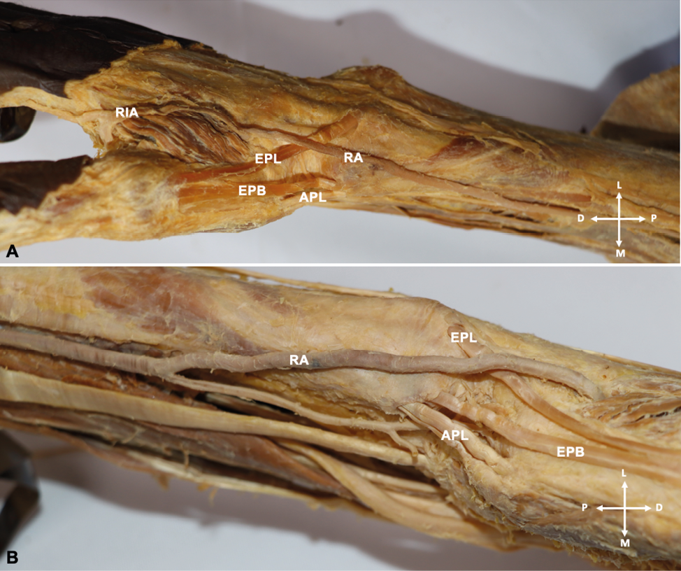
In one cadaver the radial artery ran superficial to the extensor pollicis longus and extensor pollicis brevis tendons instead of passing through the anatomical snuffbox on both sides (Figure 6 A-B). On the right side, the radial artery continued on the dorsum of the first web lying superficial to the first dorsal interosseous muscle and joining the superficial palmar arch (Figure 6 A). On the left side, before passing superficial to the extensor pollicis longus and extensor pollicis brevis tendons, the radial artery gave rise to a branch that passed through the anatomical snuff box and makes no entry into the deep palmar aspect. The radial artery then continues to loop around the first web to anastomose with the junction of the princeps pollicis artery and the radialis indicis artery (Figure 6 B). Even though the course of this branch was not followed further, it is believed to have continued to the deep palmar arch. After giving off the above branch, unlike on the right side, the radial artery did not continue superficial to the first dorsal interosseous muscle but rather penetrated and went deep to this muscle. Our literature search could not find this variation in the previous studies.
Tortuosity of the brachial artery and its branches
The brachial artery was tortuous in 11,11% cases (Figure 7A). Of these 20 cases, the tortuous brachial artery presented bilaterally in 10,00% cases and unilaterally in 1,11% cases. It was observed in males 8,33% cases and females in 2,78% cases.
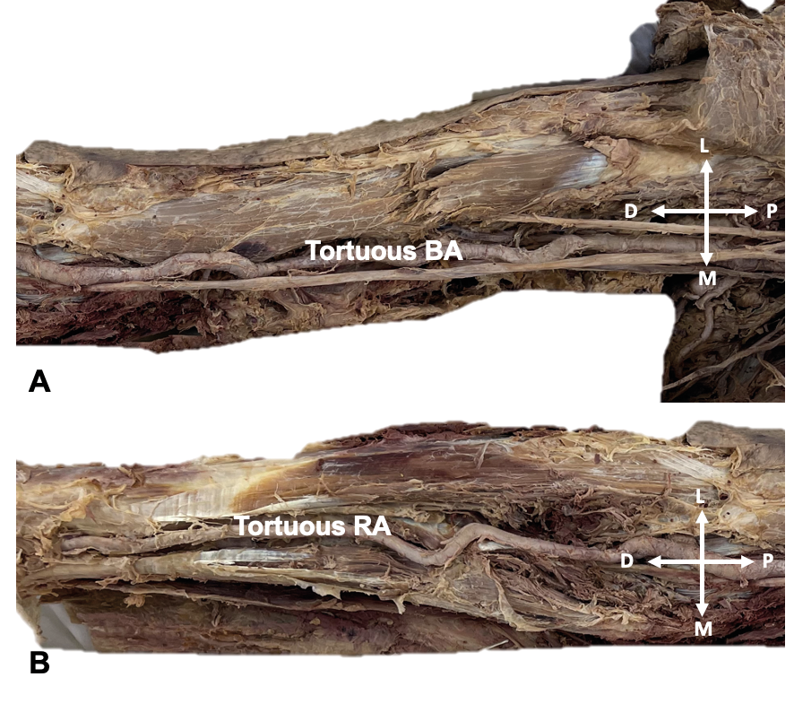
The radial artery was tortuous in 17,22% cases (Figure 7 B). The tortuosity of the radial artery was unilateral in 3,89% cases and bilateral in 13,33% cases. In addition, 9,44% were observed in male cadavers, while 7,78% were observed in female cadavers. Considering the race distribution, the tortuous radial artery was observed in 12,78% cases of White cadavers, while 4,44% cases were recorded in Black cadavers.
The Chi-square test comparing the frequency of tortuous arteries in all 180 dissected upper limbs (136 Black, 44 White, of which 126 were males and 54 were of females) showed a significantly high frequency of tortuosity of arteries in the White cadavers compared to Black cadavers (p<0,001), while side (p=0,230) and sex (p=0,230) comparisons were not statistically significant.
The frequency of the upper limb arterial variations recorded in the sections above are summarised in Figure 8 and Table 2.
| Variations | Prevalence of variation (%) (n/180) | Male (n/126) |
Female (n/54) |
||
|---|---|---|---|---|---|
| Bilateral | Unilateral | Bilateral | Unilateral | ||
| Brachial artery | |||||
| Median artery | 4.44% | 4 | 2 | 0 | 2 |
| Tortuous brachial artery | 11.11% | 14 | 1 | 4 | 1 |
| Tortuous radial artery | 17.22% | 14 | 3 | 10 | 4 |
| High origin of radial artery | 5.56% | 4 | 6 | 0 | 0 |
| High origin of ulnar artery | 2.22% | 4 | 0 | 0 | 0 |
| Superficial RA | 1.11% | 2 | 0 | 0 | 0 |
| Normal brachial artery | 56.11% | 70 | 3 | 26 | 2 |
Discussion
Introduction
The current study evaluated the variations in the brachial artery and its terminal branches to record the incidence of these variations in the studied population and the possible clinical implications that these variations may have during surgical and treatment interventions involving the upper limb vessels studied. The study records the information with limited availability in the current literature, more specifically pertaining to the African population, as much focus has been given to the axillary artery which is known to be a highly variable artery of the upper limb. Even though the above is true regarding the axillary artery, the knowledge of the variations of the brachial artery and its branches is of crucial importance as these vessels are sometimes used to perform arterial bypass and are also vulnerable to venipuncture during drug administration.
While the current study has done extensive comparisons of the variations in different sex and race groups of the studied population, care should be taken when using the results of the current study to project the possible variations of the brachial artery and its branches for surgical planning and drug administration. This is due to the fact that the limited availability of human cadaveric material did not allow a balance between sex and race groups, and therefore some statistical analysis shown in comparing the groupings might have lost statistical power. Authors nevertheless believe that the data presented in this article carriers significant scientific value that may inform the decisions for surgical procedures and treatment interventions to prevent iatrogenic injuries.
Variations of the brachial artery and its terminal branches
The current study reported 5,56% of cases of the high origin of the radial artery, in agreement with previous studies which recorded 2,84% in the European Population [13] and 14,27% in the Spanish population [48]. The presence of a high origin of the radial artery may lead to failure during trans-radial catheterisation as the catheter may be hindered, partly due to the fact that the presence of a high origin of the radial artery is associated with its tortuosity [19]. In some cases, the radial artery with high origin takes a superficial course, making it vulnerable to be mistaken as a vein during drug administration, a situation that could lead to gangrene of the forearm and hand [49] (Table 3).
| Brachial artery | Henneberg & George, 1991 & 1992 | Rodríguez-Niedenführ et al., 2001 | Li et al., 2013 | Konarik et al., 2020 | Shetty et al., 2022 | Current study |
|---|---|---|---|---|---|---|
| SA | European | Chinese | European | Indian | SA | |
| Median artery | 54% | 0% | - | 0% | - | 4,44% |
| Tortuous brachial artery | - | - | 0,4% | - | 10,00% | 11,11% |
| Tortuous radial artery | - | - | 3,6% | - | - | 17,22% |
| High origin of radial artery | - | 14,2% | - | 2,84% | 15,00% | 5,56% |
| Superficial RA | - | - | - | - | - | 1,11% |
The high origin of the ulnar artery was recorded in 2,22% of cases in the current study. Its reported frequency ranges from 0,17% to 2,00% in previous studies [27,29,50]. The presence of an ulnar artery of high origin is considered a rare anatomical variation with clinical significance [8]. The high origin of the ulnar artery is often related to ulnar nerve compression and entrapment [51]. Its clinical importance should not be underestimated as several cases of intra-arterial injection of drugs and subsequent amputations have been reported in patients with this type of variation [51]. Additionally, the ulnar artery of high origin is extremely vulnerable to iatrogenic injury during surgery [52].
A rare case of a common trunk of PCHA and deep brachial artery was recorded in 0,56% of cases in the current study. Our literature search found one study that recorded a similar variation in 2% of cases [53]. This variation is important for arteriovenous bypass, especially where the axillary artery is used [54]. It should also be considered for shoulder and axillary tissue flaps used for the reconstruction of cervical and axillary scar contractures and reconstruction [55].
The rare superficial course of the radial artery that ran on the roof of the anatomical snuff box was found in 1,11% of cases in the current study. According to our literature review, this variation has only been reported recently in a case study reported on a Korean male cadaver [56]. Clinical classifications of variants in the radial artery are important as they are the main reasons for technical failures during trans-radial catheterisations [56]. The looping of the radial artery around the first web in the current study resulted in it taking a superficial unexpected course where it could be vulnerable to injuries with potential profuse bleeding.
Median artery
The median artery was present in 4,44% of cases in the current study, in agreement with a previous study conducted in a sample of Indian population which recorded 6,6% of cases of the median artery [12]. The presence of the median artery is often coupled with idiopathic median nerve pain due to nerve entrapment [26]. A few cases of median nerve neuropathies encountered in the clinical setting are reported to be idiopathic probably due to compression by arteries that take the abnormal course or the presence of the median artery [10]. This could have been a possibility in the case observed in the current study as the median artery pierced through the median nerve.
Tortuosity of the main upper limb arteries
The tortuous brachial artery was reported in 11,11% of cases in the current study. Tortuosity of the brachial artery is a rare occurrence and is mostly reported in case reports [23,25,39]. The presence of tortuous arteries can lead to difficulty during catheterization and may result in iatrogenic injury [57]. The tortuosity of the upper limb arteries was high (28,33%) in the current study compared to previous studies (1,25%) [23,25].
The tortuous radial artery was reported in 17,22% of cases in the current study. Tortuosity of the radial artery has been reported to range from 3,6% to 23% of cases [23,25,39] which is in agreement with the results of the current study. Considering the race distribution, the tortuous radial artery was observed in 12,78% of the White South African cadavers and 4,44% recorded in the Black South African cadavers. These results suggest a higher prevalence of tortuosity of the radial artery in the White South Africans compared to the Black South Africans. It has previously been suggested that tortuosity of the arteries may be related to diabetes and hypertension [24]. Medical records were not available to relate the tortuosity to the disease in the current study.
Conclusion
All dissected cadavers showed variations in either brachial, ulnar or radial artery, confirming the high incidence of upper limb arterial variations. The incidence of variations in branching patterns of the brachial artery and its terminal branches in the South African population compares to that of the previous studies, suggesting that these variations are not race or population dependent. This study also noted that the high origin of the radial artery is more prevalent in males than in females, a point noted for the first time in the current study. Tortuosity of the brachial and radial arteries are much higher in the South African population compared to other populations.
The results of the current study provides crucial information for planning surgical procedures and interpretation of angiograms more specifically in the South African population. Such data is currently not available except the current study.
Limitations
Due to the limited availability of cadaveric material, a proper balance between race and sex was not possible and hence the current study may not sufficiently elucidate the distinct racial disparities and variations with regards to sex. In addition the variations in smaller branches might have been missed due to poor visibility after the embalming process.
Recommendations
We recommend an increase in sample size to accurately determine statistical variations between sexes and race groups to enhance the knowledge of the upper limb arterial variations. The effects of age and BMI should also be investigated. Future studies should consider including radiographic imaging to increase the sample size.
Acknowledgements
We acknowledge Professor Anna Oettle, a full Professor of the Department of Anatomy and histology of Sefako Makgatho health Sciences University for her advise in analysisng the statistics, more especially with regards possibilities of regression analyses as she specialisies in Athropology research.
References
[1] Tsoucalas G, Eleftheriou A, Panagouli E. High Bifurcation of the Brachial Artery: An Embryological Overview. Cureus. 2020 Feb 25.
[2] De Garis CF, Swartley WB. The axillary artery in white and negro stocks. Am J Anat. 1928 May;41(2):353–97.
[3] Huelke DF. Variation in the origins of the branches of the axillary artery. Anat Rec. 1959 Sep;135(1):33–41.
[4] Astik R, Dave U. Variations in branching pattern of the axillary artery: a study in 40 human cadavers. J Vasc Bras. 2012 Mar;11(1):12–7.
[5] Majumdar S, Bhattacharya S, Chatterjee A, Dasgupta H. A Study on Axillary Artery and its Branching Pattern among the Population of West Bengal, India. 2013;
[6] Xhakaza NK, Satyapal KS. Origin of the subscapular artery in the South African Black population. Folia Morphol. 2014 Nov 28;73(4):486–91.
[7] Bhat KM, Gowda S, Potu BK, Rao MS. A unique branching pattern of the axillary artery in a South Indian male cadaver. 2008;
[8] Casal D, Pais D, Toscano T, Bilhim T, Rodrigues L, Figueiredo I, et al. A rare variant of the ulnar artery with important clinical implications: a case report. BMC Res Notes. 2012 Dec;5(1):660.
[9] Piagkou M, Totlis T, Panagiotopoulos NA, Natsis K. An arterial island pattern of the axillary and brachial arteries: a case report with clinical implications. Surg Radiol Anat. 2016 Oct;38(8):975–8.
[10] Nkomozepi P, Xhakaza N, Swanepoel E. Superficial brachial artery: a possible cause for idiopathic median nerve entrapment neuropathy. Folia Morphol. 2017 Sep 7;76(3):527–31.
[11] Pham XBD, Kim JJ, Parrish AB, Tom C, Ihenachor EJ, Mina D, et al. Racial and Gender Differences in Arterial Anatomy of the Arm. Am Surg. 2016 Oct ;82(10):973–6.
[12] Kumar Singla R. Prevalence of the Persistant Median Artery. J Clin Diagn Res. 2012;
[13] Konarik M, Musil V, Baca V, Kachlik D. Upper limb principal arteries variations: A cadaveric study with terminological implication. Bosn J Basic Med Sci. 2020 Apr 6.
[14] Henneberg M, George BJ. High incidence of the median artery of the forearm in a sample of recent Southern African cadavers. 1991;
[15] Cheruiyot I, Bundi B, Munguti J, Olabu B, Ngure B, Ogeng’o J. Prevalence and anatomical pattern of the median artery among adult black Kenyans. Anat J Afr. 2017 Nov 30;6(3):1015–23.
[16] Claassen H, Schmitt O, Wree A. Large patent median arteries and their relation to the superficial palmar arch with respect to history, size consideration and clinic consequences. Surg Radiol Anat. 2008 Feb;30(1):57–63.
[17] Aharinejad S, Nourani F, Hollensteiner H. Rare case of high origin of the ulnar artery from the brachial artery. Clin Anat. 1997;10(4):253–8. A
[18] Natsis K, Papadopoulou AL, Paraskevas G, Totlis T, Tsikaras P. High origin of a superficial ulnar artery arising from the axillary artery: anatomy, embryology, clinical significance and a review of the literature. Folia Morphol. 2006;65(4).
[19] Haładaj R, Wysiadecki G, Dudkiewicz Z, Polguj M, Topol M. The High Origin of the Radial Artery (Brachioradial Artery): Its Anatomical Variations, Clinical Significance, and Contribution to the Blood Supply of the Hand. BioMed Res Int. 2018 Jun 11;2018:1–11.
[20] Lufukuja G. High Bifurcation of Brachial Artery and its Clinical Significance. 2018;
[21] Habib J, Baetz L, Satiani B. Assessment of collateral circulation to the hand prior to radial artery harvest. Vasc Med. 2012 Oct;17(5):352–61.
[22] Bozer C, Ulucam E, Kutoglu T. A case of originated high superficial ulnar artery. Trakia J Sci. 2004;2(3).
[23] Carter Y, Bennett DJ, Molla V, Wink AE, Collins AJ, Giannaris EL. A case of distal limb arterial tortuosity and dilation: observations and potential clinical significance. Folia Morphol. 2022 Aug 24;81(3):791–7.
[24] Han HC. Twisted Blood Vessels: Symptoms, Etiology and Biomechanical Mechanisms. J Vasc Res. 2012 ; 49(3):185–97.
[25] Mishra AK, Mishra A. Tortuous forearm arteries and its clinical implications: A Case Report. 2017;
[26] Osiak K, Elnazir P, Mazurek A, Pasternak A. Prevalence of the persistent median artery in patients undergoing surgical open carpal tunnel release: A case series. Transl Res Anat. 2021 Jun;23:100113.
[27] Natsis K, Iordache G, Gigis I, Kyriazidou A, Lazaridis N, Noussios G, et al. Persistent median artery in the carpal tunnel: anatomy, embryology, clinical significance, and review of the literature. Folia Morphol. 2009;68(4).
[28] Standring S, editor. Gray’s anatomy: the anatomical basis of clinical practice. Forty-first edition. New York: Elsevier Limited; 2016. 1562 p.
[29] Rodríguez-Niedenführ M, Vázquez T, Nearn L, Ferreira B, Parkin I, Sañudo JR. Variations of the arterial pattern in the upper limb revisited: a morphological and statistical study, with a review of the literature. J Anat. 2001 Nov; 199(5):547–66.
[30] Chakravarthi KK. Anatomical Variations of Brachial Artery - Its Morphology, Embryogenesis and Clinical Implications. J Clin Diagn Res. 2014.
[31] Shetty H, Patil V, Mobin N, Gowda MHN, Puttamallappa VS, Vamadevaiah RM, et al. Study of course and termination of brachial artery by dissection and computed tomography angiography methods with clinical importance. Anat Cell Biol. 2022 Sep 30;55(3):284–93.
[32] Keen JA. A study of the arterial variations in the limbs, with special reference to symmetry of vascular patterns. Am J Anat . 1961 May;108(3):245–61.
[33] Zheng Y, Shao L, Mao J yuan. Bilaterally symmetrical congenital absence of radial artery: a case report. BMC Surg. 2014 Dec;14(1):15.
[34] Singh R. Abnormal origin of posterior circumflex humeral artery and subscapular artery: case report and review of the literature. J Vasc Bras. 2017 Aug 21;16(3):248–51.
[35] Deligonul U, Gabliani G, Kern MJ, Vandormael M. Percutaneous brachial catheterization: The hidden hazard of high brachial artery bifurcation. Cathet Cardiovasc Diagn. 1988; 14(1):44–5.
[36] Moore KL, Dalley IAF, Agur AMR. Clinically Oriented Anatomy. 2018;
[37] Singh D, Malhotra M, Agarwal S. Variations in the Axillary Artery Branching Pattern. J Clin Diagn Res. 2020.
[38] Yazar F, Kirici Y, Ozan H, Aldur MM. An unusual variation of the superficial ulnar artery. Surg Radiol Anat. 1999 May;21(2):155–7.
[39] Mahajan R, Raheja S, Tuli A, Singh S, Agarwal S. Tortuous ulnar artery and Gantzer’s muscle: A rare presentation with clinical implications. 2015;
[40] Eid N, Ito Y, Shibata MA, Otsuki Y. Persistent median artery: Cadaveric study and review of the literature. Clin Anat. 2011 Jul;24(5):627–33.
[41] Henneberg M, George BJ. A further study of the high incidence of the median artery of the forearm in Southern Africa. 1992;
[42] Dobrin PB, Canfield TR. Elastase, collagenase, and the biaxial elastic properties of dog carotid artery. Am J Physiol-Heart Circ Physiol. 1984 Jul 1;247(1):H124–31. A
[43] Leeson CR, Leeson TS. Histology. 4th ed. Philadelphia: W.B. Saunders; 1981. 600 p.
[44] Nichols WW, O’Rourke M, Vlachopoulos C, Edelman ER. McDonald’s Blood Flow in Arteries: Theoretical, Experimental and Clinical Principles. 7th ed. Boca Raton: CRC Press; 2022.
[45] Hutchins GM, Miner MM, Bulkley BH. Tortuosity as an index of the age and diameter increase of coronary collateral vessels in patients after acute myocardial infarction. Am J Cardiol. 1978 Feb;41(2):210–5.
[46] Wysiadecki G, Polguj M, Haładaj R, Topol M. Low origin of the radial artery: a case study including a review of literature and proposal of an embryological explanation. Anat Sci Int. 2017 Mar;92(2):293–8. A
[47] Tank PW, Grant JCB. Grant’s dissector: Patrick W. Tank. 15th ed. Philadelphia: Lippincott Williams & Wilkins; 2013. 285 p.
[48] Gonzalez-Compta X. Origin of the radial artery from the axillary artery and associated hand vascular anomalies. J Hand Surg. 1991 Mar;16(2):293–6.
[49] Singh R, Malhotra R, Wadhawan M. Anomalies of radial and ulnar arteries. J Vasc Bras. 2017 Mar;16(1):56–9.
[50] Jacquemin G, Lemaire V, Medot M, Fissette J. Bilateral case of superficial ulnar artery originating from axillary artery. Surg Radiol Anat. 2001 Jun;23(2):139–43.
[51] Vollala VR, Jetti R, Soni S. High Origin of An Ulnar Artery – Development and Surgical Significance. 2011;34(6).
[52] Senanayake KJ, Salgado S, Rathnayake MJ, Fernando R, Somarathne K. A rare variant of the superficial ulnar artery, and its clinical implications: a case report. J Med Case Reports. 2007 Dec;1(1):128. A
[53] Paula RCD, Erthal R, Fernandes RMP, Babinski MA, Silva JG, Chagas CAA. Anomalous origin of the deep brachial artery (profunda brachii) observed in bilateral arms: case report. J Vasc Bras. 2013 Mar;12(1):53–6.
[54] Morsy MA, Khan A, Chemla ES. Prosthetic axillary-axillary arteriovenous straight access (necklace graft) for difficult hemodialysis patients: A prospective single-center experience. J Vasc Surg. 2008 Nov;48(5):1251-1254.e1.
[55] Olinger A, Benninger B. Branching patterns of the lateral thoracic, subscapular, and posterior circumflex humeral arteries and their relationship to the posterior cord of the brachial plexus. Clin Anat. 2010 May;23(4):407–12.
[56] Park HJ, Kim S, Kim S, Song WC, Kim HJ. The radial artery running over the anatomical snuffbox: a case report. Surg Radiol Anat. 2022 May;44(5):659–63.
[57] Ariyo O. A High Origin Subscapular Trunk and its Clinical Implications. Anat Physiol. 2018;08(02).