https://doi.org/10.36253/ijae-16577
Overview of the gut-liver-brain axis with particular emphasis on ferroptosis
1 Department of Anatomical, Histological, Forensic Medicine and Orthopaedics Sciences, Sapienza University of Rome, Italy
2 Division of Health Sciences, Department of Movement, Human and Health Sciences, University of Rome Foro Italico, Italy
3 Integrated Research Center (PRAAB), Campus Biomedico University of Rome, Italy
4 Department of Life Science, Health and Health Professions, Link Campus University, Rome, Italy
5 Interdisciplinary Department of Well-Being, Health and Environment Sustainability (BESSA), Sapienza University, Rieti, Italy
*Corresponding author. E-mail: g.vivacqua@unicampus.it
Abstract. Ferroptosis is a form of cellular death involved in the origin, progression, but also regulation of several human diseases. Its regulatory role in the gut-liver-brain axis (GLBA) has not been clarified. Therefore, we sought to summarize the possible correlations between ferroptosis and the GLBA. In this review, we first introduce the phenotype and the main mechanisms of this relatively newly described form of regulated cell death. Then, we analyse the anatomy of the GLBA, describing the connections between the gut and the liver, followed by the anatomical pathways from the gut to the brain and from the liver to the brain. After the morphological aspects, we summarize the main biological modulators of the GLBA, highlighting their physiological and pathological roles. In the end, we discuss in detail the regulatory role of ferroptosis on neuroinflammation and oxidative stress along GLBA, highlighting the key aspects that could be of significant clinical importance as future diagnostic and therapeutic targets.
Keywords: ferroptosis, gut-brain axis, neuroinflammation.
Contents
1. Ferroptosis: a new form of non-apoptotic cell death
1.1. Molecular and morphological phenotypes
2. The anatomy of the gut-liver-brain axis
2.4. The gut - liver - brain axis (GLBA)
3. Principal biochemical modulators along the gut-brain axis
4. Involvement of ferroptosis in the gut-liver-brain axis
1. Ferroptosis: a new form of non-apoptotic cell death
Ferroptosis is an iron-dependent, lipid peroxidation-driven form of regulated cell death (RCD). First discovered by Brent R. Stockwell et al. (2012), it was described as erastin-induced cell death characterized by a peculiar phenotype [1]. Morphological (such as loss of membrane integrity, swollen of the cytoplasm and smaller mitochondria), biochemical and genetic features distinguish ferroptosis from the other types of RCD described to date [2]. The regulation of this unique process of cell death is closely linked to the interplay between iron, lipid and amino acid metabolism resulting in oxidative perturbations of the intracellular microenvironment and loss of redox homeostasis. Recent advances in the discovery of ferroptosis markers revealed how ferroptosis is the main feature of a series of physiological processes, including tumor suppression pathways and adequate immunological response [3]. The relevance of ferroptosis is further highlighted by its role in the development of pathological conditions, spanning from kidney injuries and endocrine imbalances to neurodegenerative disorders [4], including Alzheimer’s Disease (AD) and Parkinson’s Disease (PD) along with synucleinopathy [5].
1.1. Molecular and morphological phenotypes
The buildup of peroxidized lipids, resulting in the unrepaired damage of cell membranes, is the ultimate driver of ferroptosis [6,7]. The contribution of different organelles in the biochemical evolution of ferroptosis explains the main morphological changes affecting these subcellular compartments. Mitochondria can add up to cysteine-starvation mediated GSH-depletion-induced damage by means of electrons leakage from the cellular respiration machinery, which would facilitate the initiation of a Fenton reaction by producing hydrogen peroxide. The endoplasmic reticulum, being the critical site of lipid peroxidation, is fundamental for clustering of oxidized Poly-Unsaturated Fatty Acids (PUFA) and phospholipids (PLs) leading to progression of ferroptosis [7]. Overall, the modifications at the cellular and subcellular levels include: mitochondria shrinkage and cristae reduction or disappearance, increased endoplasmic reticulum viscosity and ER membrane stiffness, loss of plasma membrane structural and functional integrity, and possible formation of pores that can promote membrane rupture [8-11].
1.2. Mechanism
The molecular mechanism of ferroptosis is driven by the increase of intracellular ferrous iron (Fe[2]+), capable of subsequently promoting both non-enzymatic and enzymatic lipid peroxidation by initiating a Fenton reaction or by exploiting its role as a critical cofactor of lipoxygenases [12]. The detrimental effects of lipid peroxidation fall into two, possibly overlapping, categories [13]: direct dampening of cellular membranes stability and formation of lipid pores, with consequent increase the permeability of the membranes, or the activation of downstream signaling pathways that result in membrane perforation [14,15].
Iron homeostasis, primarily controlled by the hepatic secretion of hepcidin [16], is met by a tightly balanced exchange of iron between extracellular and intracellular compartments, in which the transition metal is found as ferric (Fe[3]+) and ferrous (Fe[2]+) iron, respectively [17]. The expansion of the cellular labile iron pool (LIP), reaching more than 5% of total cell iron [18], promotes reactive oxygen species (ROS) formation through the reaction of ferrous iron with hydrogen peroxide, according to Fenton chemistry [19]. The rise in intracellular Fe[2]+ could be secondary to a variety of conditions, which include: (i) an increase of transferrin receptor (TFR1) expression, (ii) RAS mutations or NCOA4-mediated ferritinophagy, which induces a decrease in ferritin levels [20,21], and (iii) excessive degradation of heme by heme oxygenase 1 (HO-1) [22] (Figure 1). Lipid peroxidation can occur through both non-enzymatic and enzymatic pathways.
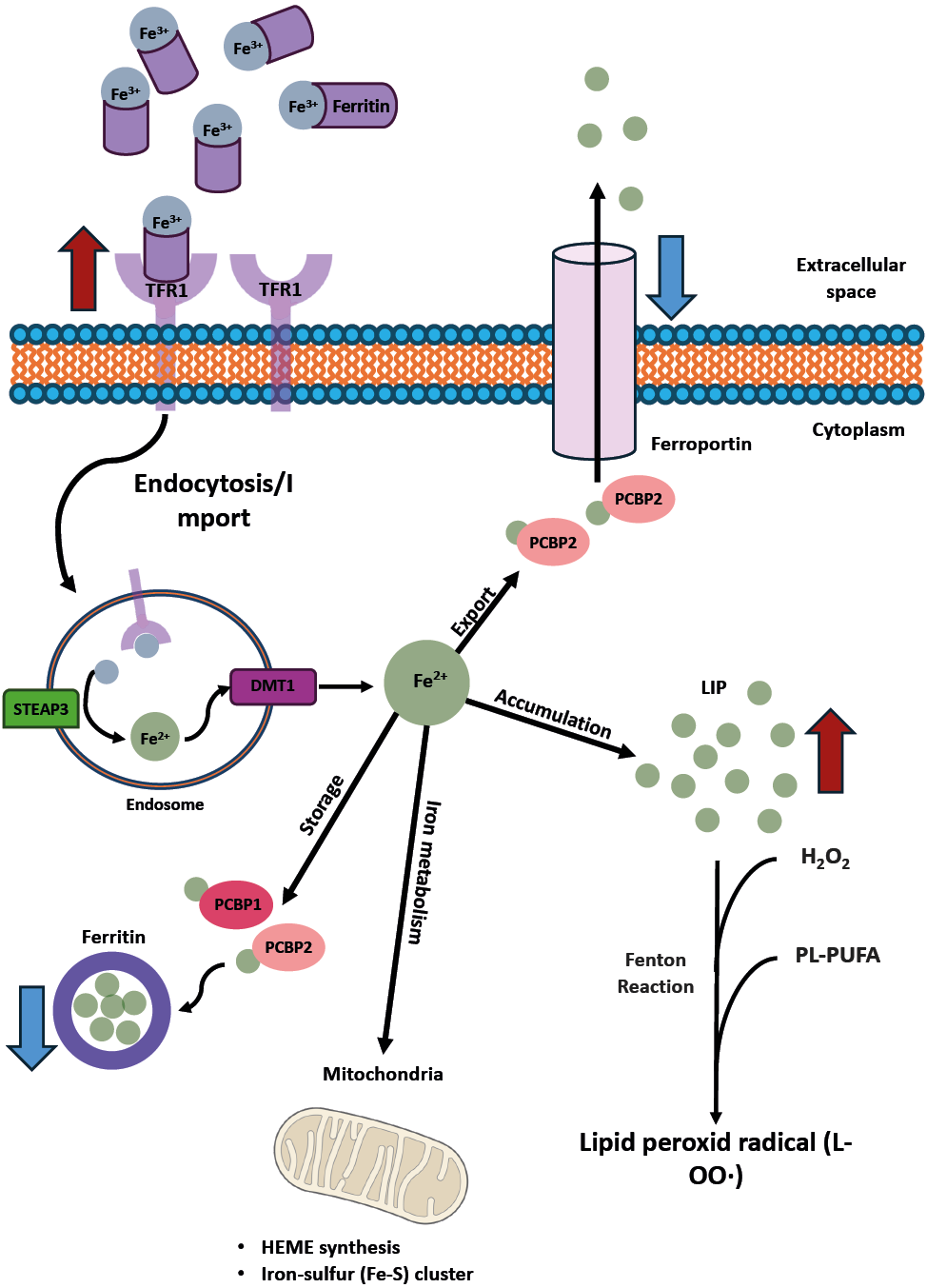
Non-enzymatic lipid peroxidation
Non-enzymatic lipid peroxidation consists of free radical driven reactions. Initiation requires a Fenton reaction to take place: the abundant ferrous iron moieties react with hydrogen peroxide derived from oxidoreductase activity, such as that of cytochromes P450 oxidoreductase (POR) and P450 reductase (CPR) localized on the smooth endoplasmic reticulum, producing ROS [23]. The hydroxyl radicals then extract hydrogen from the bis-allylic position of poly-unsaturated fatty acids (PUFAs) to form lipid radicals, which further react with molecular oxygen (O2) to produce lipid peroxy-radicals. More hydrogen moieties are removed from PUFAs to form lipid hydroperoxides and new lipid peroxy-radicals, the latter providing a way of amplification of the oxidation reaction [24,25] (Figure 2).
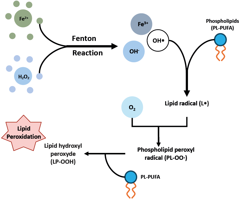
Enzymatic peroxidation
Enzymatic lipid peroxidation, as the name implies, consists of a series of enzymatically driven reactions. In the endoplasmic-reticulum-associated subcellular compartments, selective oxidation of phosphatidylethanolamines (PEs) followed by lipids oxygenation takes place [26]. Firstly acyl-CoA synthetases, such as ACSL4 (especially acting on arachidonic acid), activate free fatty acids (FFAs) by adding to them a coenzyme A moiety. Then, lysophospholipid acyltransferases, such as LPCAT3, catalyze the transfer of the fatty acyl chain from fatty acyl-CoA and incorporate into PEs to form various classes of phospholipids. Finally, lipoxygenases familiy (LOXs) generate doubly and triply oxygenated (15-hydroperoxy)-diacylated PE compounds [26,27] (Figure 3).
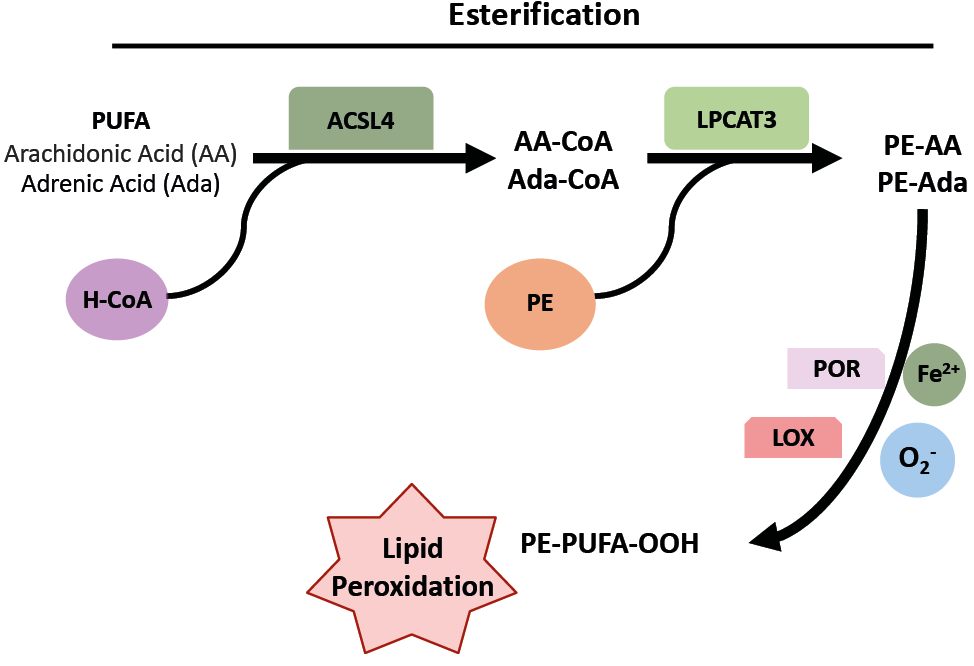
2. The anatomy of the gut-liver-brain axis
2.1. The gut-liver axis
The gut-liver axis (GLA) concept was first introduced by Marshall in 1998 and refers to the connection between the gastrointestinal (GI) tract and the liver. This axis is characterized by a complex network of bidirectional anatomical and functional interactions between the GI tract and the liver. Over the years, the GLA has gained increasing attention due to its implications in various pathologies. Key components of this axis include portal circulation, gut microbiota, intestinal tight junctions, bile acids, and mucosal hormones [28-30]
Portal circulation
Most of the venous blood from the GI tract, primarily from the small and large intestines, drains into the portal vein. This vein transports nutrient-rich blood, along with signalling molecules produced by the gut microbiota, neuroendocrine and immune cells of the gut, directly to the liver. Moreover, the portal circulation carries to the liver the products of hemocatheresis occurring in the spleen, including globins, heme and iron produced by the catabolism of haemoglobin.
Gut microbiota
The gut hosts diverse microbial communities that play essential roles in maintaining intestinal integrity and producing signalling molecules. A balanced microbiota is critical to produce various metabolites, particularly short-chain fatty acids (SCFAs), such as acetate, propionate, and butyrate [31]. Acetate, absorbed in the proximal colon, is rapidly transported to the liver, where it contributes to cholesterol biosynthesis [32]. Propionate, a substrate for lipogenesis, gluconeogenesis and protein synthesis in the liver [33], was observed to inhibit hepatic cholesterol synthesis [34]. Butyrate was demonstrated to be central for the health of colon enterocytes by providing energy to colonic epithelial cells [35]. However, recent studies suggest that excessive accumulation of butyrate induces cholestasis, hepatocyte death, and neutrophil-driven inflammation in the liver, potentially leading to icteric hepatocellular carcinoma [36]. Additionally, SCFAs, especially butyrate, stimulate the release of fasting-induced adipocyte factor (FIAF) from L cells in the gut [37]; FIAF subsequently inhibits lipoprotein lipase (LPL) activity, preventing triglyceride accumulation in both adipose tissue and liver. [7,38], Therefore, the microbiota plays a central role in the GLA and dysbiosis can lead to an imbalance of signalling molecules, contributing to liver damage. Not surprisingly, an imbalanced gut microbiota can result in increased intestinal ethanol production, contributing to the development of non-alcoholic steatohepatitis (NASH) and non-alcoholic fatty liver disease (NAFLD), today included in metabolic dysfunction-associated steatotic liver disease (MASLD) [39,40].
Tight Junctions (TJs)
Tight junctions (TJs) are components of the gut mucosal barrier, a multifaceted system of physical, chemical, microbial, and immunological defences that limit the spread of intestinal antigens [41]. TJs are composed of proteins such as claudins, TJ-associated marvel proteins (TAMPs), junctional adhesion molecules (JAMs), and zona occludens-1 (ZO-1) [42-44]. In the context of the GLA, the loss of barrier integrity, for example due to inflammation, can increase portal leakage of lipopolysaccharide (LPS), with the activation of a well-characterized pathogen-associated molecular pattern (PAMPs). LPS activates Kupffer cells, the liver’s resident macrophages, are highly sensitive to LPS, that activate it [45]. LPS-activated Kupffer cells promote liver inflammation and fibrosis by driving the transcription of pro-inflammatory cytokines and the activation of the stellate cells into the Disse’s space [31,46]. Under healthy conditions, the portal vein carries small amounts of pathogens and bacteria, which are effectively managed by the liver’s immune system. However, pathological states can result in an excessive influx of microorganisms into the portal vein, contributing to liver disease [47,48].
In neurodegenerative conditions, α-syn aggregates can travel via portal circulation from the gut to the liver. Moreover, aggregated α-syn can be either a consequence as a driver of bowel inflammation with alteration of the intestinal barrier and increased passage of LPS to the liver [49,50].
Bile
Bile is a digestive fluid secreted by the hepatocytes and stored in the gallbladder to reach the duodenum. Within the context of the GLA, bile acids play a crucial signalling role. The gut microbiota converts primary bile acids, such as cholic acid (CA) and chenodeoxycholic acid (CDCA), into secondary bile acids, including lithocholic acid (LCA) and deoxycholic acid (DCA) [51]. Both primary and secondary bile acids are absorbed by enterocytes and enter the portal circulation. Bile acids act as signalling molecules by interacting with nuclear farnesoid X receptors (FXR) in both intestine and liver, regulating cholesterol, lipid, and energy metabolism [52]. Reabsorbed bile acids can also activate Takeda G protein-coupled receptor 5 (TGR5 a membrane receptor for bile acid), causing multiple effects in hepatic stellate cells, Kupffer cells, cholangiocytes, and enterocytes [53,54]. Activation and transdifferentiation of hepatic stellate cells into myofibroblasts can promote liver fibrosis [55]. Another important receptor, Shingosine-1-phosphate receptor 2 (S1PR2), is present in the liver and bile ducts and is activated by conjugated bile acids such as taurine or glycine-conjugated bile acids [53] Inhibition of S1PR2 has shown promising results as a potential treatment for cholestatic liver diseases. Knockout mice for S1PR2 showed reduced inflammation and hepatic fibrosis in liver [56]. Dysregulation of the gut-liver circulation is evident in cirrhotic patients, where depletion of bacterial populations responsible for bile acid dehydroxylation has been observed [57]. Additionally, gut microbiota can convert choline, a bile component, into trimethylamine N-oxide (TMAO), a toxic metabolite associated with dysbiosis and hepatic steatosis [53,58]. Lastly, bile contains IgA, bicarbonate, and antibacterial molecules, which have bacteriostatic properties and contribute to the regulation of gut microbiota composition [47].
Mucosal hormones
The gut mucosa contains cells capable of releasing hormones, such as substance P, vasoactive intestinal peptide (VIP) and serotonin as well as fibroblast growth factor 15 (FGF15), which regulates bile-acids synthesis, hepatic glucose, and lipid metabolism [59]. FGF15 (which is equivalent in humans to FGF19) [60] also plays a crucial role in regulating postprandial glucose and energy metabolism by modulating gluconeogenesis [61]. The glucagon-like peptide 1 (GLP-1) is an incretin hormone secreted by L cells of ileum and colon. GLP-1 is involved in the regulation of glucose metabolism in the liver, stomach emptying and feeding [62]. Secretin, another crucial hormone, secreted by S cells in the duodenum, has been shown: (i) to regulate biliary secretion and proliferation [63] and, (ii) to modulate the brown adipose tissue (BAT)-brain metabolic crosstalk [64]. Moreover, one of the endocrine mechanisms responsible for weight regain includes the brain-gut axis, which supports food intake via the production of several gastrointestinal hormones, such as ghrelin, leptin and cholecystokinin (CKK) [65].
2.2. The gut-brain axis
The concept of the gut-brain axis (GBA) was first proposed by Michael D. Gershon in the late 20th century. The idea suggested a bidirectional interaction between the GI tract and brain. The importance of this axis is now emerging. Changes in GI biochemical and physiological pathways have been observed in a growing range of CNS disorders, such as PD, in which GI dysfunction often precedes of decades the onset of neurological symptoms [66]. Generally, the gut-brain axis is mediated by the enteric nervous system (ENS), autonomic nervous system (ANS), hypothalamus-pituitary-adrenal (HPA) axis, the enteroendocrine systems, and the immune system [53].
ENS anatomy
The GI tract is innervated by number of neurons (estimated at 200-600 million) that exceeds the total number of neurons in the spinal cord. This neuronal system, specific of the gut, is classified as the ENS, also known as the meta sympathetic nervous system. Studies suggested that these neurons are evolutionarily older than those of the central nervous system, and the ENS is capable of functioning independently through various reflexes without input from the CNS and the ANS. Consequently, the ENS is often referred to as the body’s “second brain” [67-69] Anatomically, the ENS can be considered a complex network of neuronal connections and ganglia embedded in the layers of the gut wall. These groups of neurons can be subdivided into two plexuses: the Auerbach’s (myenteric) plexus, located between the circular and longitudinal layers of the muscularis externa, and the Meissner’s (submucosal) plexus, situated within the submucosa [70]. The ENS comprises a complex network of neurons, some of which have been classified, such as: intrinsic primary afferent neurons, excitatory and inhibitory motor neurons, ascending and descending interneurons, secretomotor/vasodilator neurons, and intestinofugal neurons. [71,72]
ENS - ANS cross talk
Although the ENS can function independently, it is still strongly influenced by the ANS (both sympathetic and parasympathetic). The sympathetic system reaches mostly the myenteric plexus, whereas the post ganglionic neurons of the parasympathetic nervous system (mostly constituted by hepatic and celiac branches of the vagus nerve) are found both within the myenteric and submucosal plexuses (Figure 4). The importance of the ANS and ENS in the context of the gut can be understood by considering the numerous functions in which they are involved: gut motility, gut permeability, epithelial fluid homeostasis, luminal osmolarity, bile secretion, carbohydrate concentration, mechanical distortion of the mucosa, bicarbonate production, mucus production and secretion, as well as mucosal immune responses and handling of intestinal fluids [73,74].
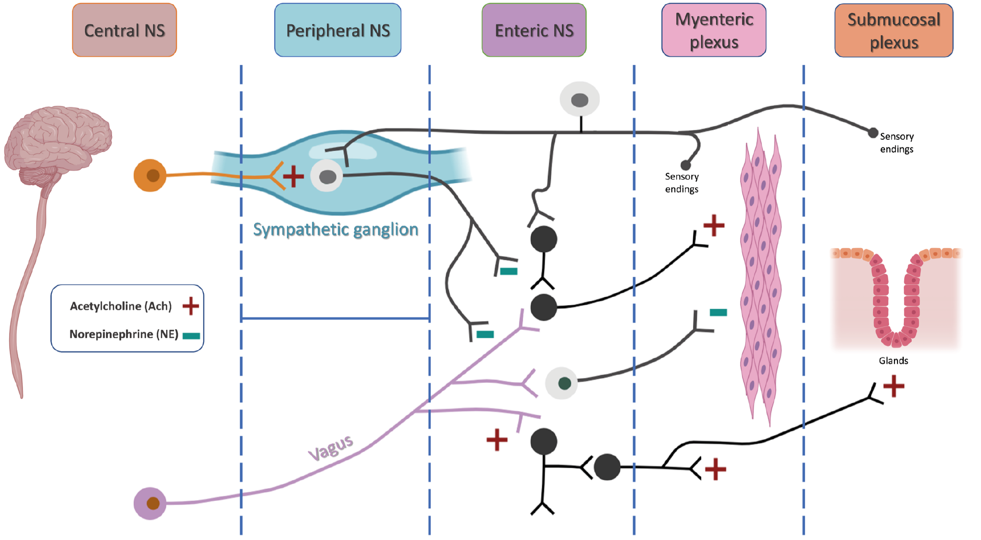
The neuroactive components of the GBA
A diverse array of neuroactive substances exists within the GBA, encompassing gut-derived hormones, neuroactive molecules, metabolites produced by gut microbiota, and various microbial products [75]. Notable examples include bacteria-derived metabolites such as short-chain-fatty-acids (SCFAs), gamma-aminobutyric acid (GABA), serotonin (5-HT), glutamate, acetylcholine, dopamine, norepinephrine, and gut neuropeptides, including peptide YY (PYY), GLP-1, CCK, and ghrelin [53]. A central role in detecting these substances is played by vagal terminal afferent fibres, which have been described in three locations: in the terminals ending of the intestinal muscular layers, in the GI mucosa, and in a subset of enteroendocrine cells (now referred as neuropods) that form synapses with vagal neurons [76]. Due to their wide range of receptor expression, vagal afferents are considered polymodal, meaning that they can detect multiple types of stimuli, including mechanical, chemical, and hormonal signals [77]. Signals from these afferent nerves ascend towards the nucleus tractus solitarius (NTS) in the CNS [78] where they are relayed to other brainstem nuclei and forebrain structures [79]. Nervous communication is bilateral, thus substances targeting the CNS are also released by the ENS and ANS, including GABA, glutamate, acetylcholine, dopamine, norepinephrine, and other bioactive amines [80].
The microbiota in the gut brain axis
As previously mentioned, the role of gut microbiota is becoming increasingly central in biomedical research, evolving the concept of the GBA into the microbiota-gut-brain axis [81,82]. In addition to interacting with neuronal terminals in the gut, microbial metabolites can enter the circulation and directly affect the CNS. Therefore, the composition of the gut microbiota and its related products, are crucial for the interactions between gut and brain, as well as for the determination of different neurological and psychiatric disorders. Several examples emphasize the molecular pathways involved in this interaction: SCFAs can cross the blood-brain-barrier (BBB) and reduce LPS-induced neurological inflammation in primary microglia and the hippocampus, as well as decrease circulating pro-inflammatory cytokines [83-85] When imbalances in gut microbiota occur, like in patients affected by irritable bowel syndrome (IBS), the production of secondary bile acids decreases, leading to increased inflammation in the brain [53]. Lastly, it has been observed that trimethylamine N-oxide (TMAO, a metabolite of the gut microbiota) accelerates brain aging and causes age-associated cognitive impairments [86].
The HPA system in the gut brain axis
The HPA axis is one of the body’s primary systems for regulating the release of cortisol and other stress hormones. This response is also mediated by several components, including gut microbiota, the vagus nerve, and the immune system. Studies on germ-free (GF) mice have demonstrated that these mice exhibit an exaggerated HPA response when exposed to stress, releasing more adrenocorticotropic hormone (ACTH) and cortisol [87,88]. In humans, patients with IBS show higher levels of ACTH and cortisol in response to stress, along with altered microbiota composition [89-91]. The HPA axis interacts with other gut-brain communication routes, including the vagus nerve. Studies in rodents indicate that stimulating the vagus nerve increases corticotropin-releasing factor (CRF) production in the brain and raises ACTH and corticosterone levels [92]. The HPA axis also connects to the immune system, where stress and inflammation can influence gut-brain signalling. Stress increases gut permeability, allowing bacteria to cross into the body, triggering an immune response that activates in turn the HPA axis [93].
Enteroendocrine system
Enteroendocrine cells (EECs), though comprising only 1% of all gut epithelial cells, are vital for maintaining gut balance due to the various functions of the molecules they release [94]. There are multiple types of EECs, all of which act as sensors that help in regulating the processes such as insulin secretion and food intake based on gut contents [94]. Two well-studied types of EECs in the context of the gut-brain axis are enteroendocrine L cells and enterochromaffin cells.
L cells release hormones like GLP-1 and PYY, which reduce hunger and regulate food intake [95]. These hormones act on receptors in both gut and brain, either directly or indirectly through the vagus nerve, to signal feelings of satiety also involving the circuits of the parabrachial nucleus [96,97]. L cells also communicate with the ENS through neuropods, allowing for fast and precise signalling between the gut and brain [97-99]. The activation of L cells in different parts of the gut depends on what is contained in the lumen: in the upper gut, they are triggered by nutrients like carbohydrates and fats, while in the lower gut they are driven by bacterial metabolites like SCFAs, which can stimulate GLP-1 and PYY secretion [100-102].
Enterochromaffin cells produce most of the serotonin (5-HT) out of the CNS, which is important for intestinal motility, pain perception, and inflammatory response [103]. Although serotonin from the gut doesn’t directly affect the brain due to the BBB, it may influence gut-brain communication through vagal signals [103] and inflammation [104-106].
2.3. The liver-brain axis
Increasing attention is being given to the liver’s connection with the brain, which occurs primarily through two main routes: neuronal connections and vascular pathways [107]. Understanding these complex neural circuits is essential to appreciate the liver’s involvement in conditions that affect both the liver and brain, including neurodegenerative disorders [108,109].
Liver brain nervous anatomical connection
The liver, a vital organ for metabolism, detoxification, and nutrient storage, is regulated by the ANS, comprising the sympathetic and parasympathetic branches. Autonomic nerve fibers enter the liver through the hilum, forming plexuses around the liver’s primary blood vessels – an anterior plexus encircling the hepatic artery and a posterior plexus surrounding the portal vein [108].
Sympathetic fibers to the liver originate from preganglionic neurons in the thoracic spinal cord (T7–T12), traveling through the splanchnic nerves and synapsing in the celiac and superior mesenteric ganglia. From there, postganglionic sympathetic fibers innervate various structures, including the hepatic artery, portal vein, and bile ducts [110,111]. Parasympathetic innervation occurs via the hepatic branches of the left vagus nerve, which also innervates the bile ducts, portal vein, duodenum, and portions of the pancreas [112-114].
In animal species, parasympathetic innervation is generally limited to the portal triad, while sympathetic innervation varies. In humans they reach the hepatocytes [115].
The sympathetic nervous system predominantly regulates catabolic processes, while the parasympathetic system supports anabolic functions [116]. For example, vagal nerve pathways modulate hepatic lipid metabolism, influencing very low-density lipoprotein (VLDL) triglyceride secretion and reducing lipid accumulation in the liver [117].
Additionally, to adrenaline, noradrenaline and Ach, hepatic neurons synthesize and release various neurotransmitters and neuropeptides, including neuropeptide Y, substance P, VIP, glucagon-like peptide, somatostatin, neurotensin, and serotonin, all of which further influence liver and systemic homeostasis [118,119].
Hepatic afferent nerves, particularly from the vagus nerve, sense the liver’s microenvironment and relay signals to the NTS, where feedback is sent to the liver via parasympathetic and sympathetic nerves [120,121]. These signals are also transmitted to higher brain regions, such as the hypothalamus and limbic system, influencing metabolic regulation and organ function [122].
Liver brain vascular communication
In addition to neural circuits, liver and brain communicate through several vascular mechanisms, including BBB permeability, immune modulation, epigenetic alterations and amyloid β metabolism.
BBB permeability: Pro-inflammatory cytokines, including TNF-α and IL-1β, increase BBB permeability, allowing substances like ammonia, xenobiotics, and inflammatory cytokines to penetrate the brain, exacerbating neuroinflammation [123]. Excessive activation of microglia due to these cytokines attracts monocytes to the brain parenchyma, creating chronic inflammation in conditions as liver failure [124,125]. This is evident in chronic liver diseases, where neuroinflammatory processes contribute to neurological pathologies, such as hepatic encephalopathy, which occurs in approximately 30–45% of patients with cirrhosis [126]. Cholestatic mouse models, such as BDL, show debilitating CNS symptoms defined as sickness behaviors. This behavior appears to be related to elevated levels of circulating and liver-synthesized IL-6. Regulatory T cells (T-regs) appear to be responsible for modulating IL-6 synthesis and inhibiting the activity of circulating monocytes [127].
Epigenetic alterations: An example of how a metabolite produced by the liver can act centrally is β-hydroxybutyrate, a ketone body capable of crossing the BBB via specific monocarboxylate transporters and able to inhibit histone deacetylases (HDACs) the most important epigenetic control enzymes [128]. Elevated β-hydroxybutyrate levels in the brain increase brain-derived neurotrophic factor (BDNF), which plays a therapeutic role in neurodegenerative diseases and mental health disorders like depression [129,130].
Amyloid β: The liver is central to the peripheral metabolism of Aβ, and dysregulation of Aβ clearance is a critical factor in the pathogenesis of AD. The connection between the liver and brain is thus closely linked to the progression of AD [131,132].
α-Synuclein: Accumulation of α-synuclein in the liver may start liver inflammation and fibrosis [133] or represents a mechanism of clearance, which prevent a massive transmission of α-synuclein aggregates from the gut to the brain [134,135]. For this reason, liver could be directly involved in progression of PD either by cytokines release as by regulation of the transmission and clearance of α-synuclein aggregates.
2.4. The gut - liver - brain axis (GLBA)
The GLBA represents a network of bidirectional communication among the GI tract, the liver, and the brain, highlighting the interdependence of these organs in maintaining homeostasis and in contributing also to cognitive process. Understanding the GLBA is primary to elucidate the pathophysiology and the mechanisms of certain diseases, and to develop innovative therapeutic strategies that target this multifaceted network.
Studies suggested that different pathological conditions inducing dysbiosis could have acute or chronic systemic consequences, such as a weakened of the immune system, inflammation or central disorders, as well as neuropsychiatric disorders, including anxiety, depression or neurodegenerative conditions [136,137]. Furthermore, events, such as maternal stress to lack of breastfeeding, mode of delivery (vaginal VS cesarean) and antibiotic exposure, are major players in the development of acute infections and long-term dysbiosis that are associated to chronic pathological conditions, including asthma, diabetes, neurodevelopmental disorders and IBS [138,139].
In addition, dysbiosis can also impact the function of the HPA, a fundamental part of the limbic system, involved in both emotional behavior and memory consolidation [140,141]. At last, the role of dysbiosis in both a cause and consequence of the “leaky gut”, a condition associated to many autoimmune disorders where intestinal permeability is increased and pathogens as well as toxins can enter more freely into the human body, is well documented [142].
3. Principal biochemical modulators along the gut-brain axis
3.1. Serotonin
Serotonin, also known as 5-hydroxytryptamine (5-HT), is a monoamine neurotransmitter [143]. Historically, its roles have been mostly associated with the regulation of basic functions of the CNS, including sleep, mood and body temperature. Its deficits are present in numerous mental disorders [144]. Despite this common knowledge, it has been demonstrated that less than 5% of the total body’s serotonin is in the CNS [145], while most of it is hosted by the intestine [146]. 5-HT is produced mostly by enterochromaffin cells in the intestinal lining, which regulates many functions of the GI tract, including motility and permeability, as well as local secretions and ENS development [147].
Alterations of serotonin levels have been found in several diseases, including IBS [148], hepatic conditions [149] and psychiatric disorders [150], underlying the importance of the GBA and how serotonin pathways affect it. Recent studies have shown that commensal bacteria participate in the production and modulation of intestinal serotonin [151]. Consequently, prebiotics and probiotics can alter both synthesis and release of serotonin, actively impacting both GI and nervous system [152]. Moreover, although enteric 5-HT can both act locally and enter the bloodstream, divergent data on the capacity of 5-HT to cross the BBB are available [153,154]. Despite this, 5-HT has been shown to alter the permeability of the BBB [155]. Study demonstrated that the endothelium of the choroid plexus, which in healthy conditions is permeable to large (70 kDa) circulating molecules, upon intestinal inflammation acts as a vascular barrier (PVB), by closing its accessibility to inflammatory and bacterial molecules [156]. Recent study hypothesizes the capacity of neurotransmitters produced in the gut to cross the choroid plexus [157]. Therefore, it can be said that alterations in serotonin pathways can be both the cause and the consequence of GI or CNS diseases, as GI tract and brain are deeply linked, regulate each other, and are in charge for the control of emotional and stress responses.
As previously mentioned, numerous therapeutic options for several diseases affect serotonin metabolism, from its secretion to its release or reuptake. Due to the high prevalence of serotonin in the GI tract, such therapies can significantly impact GI functions as the use of serotonin reuptake inhinitors [158]. People affected by IBS exhibit altered levels of post-prandial plasma serotonin, both in children [159] and adults [160], and it has been hypothesized that such individuals may have a genetic predisposition for decreased serotonin reuptake transporter (SERT) expression [160]. Potential therapeutic options are being studied via serotonin-related signaling pathways and the gut-liver axis, in the regulation of obesity [161], MASLD, and hepatic steatosis. [162] Studies involving serotonin agonists have been shown to benefit patients affected by IBS suffering from constipation, by increasing intestinal mobility and secretion [103]. Moreover, tricyclic antidepressants that were used in numerous psychiatric conditions play a role in both mood and GI symptoms in patients affected by depression as well as IBD [163]. Furthermore, alterations in serotonin signaling can exacerbate gastrointestinal symptoms in stressed individuals, emphasizing the connection between psychological mechanisms and GI function [164]. The serotonin produced by guts significantly influences the brain through immune function, vagal nerve stimulation, neuroendocrine feedback, and the HPA axis, but the relationship between serotonin levels in the brain and depression remains unclear [165].
3.2. GLP-1 and GIP
GLP-1 and glucose dependent insulinotropic polypeptide (GIP) are incretin hormones secreted by their EECs [166]. These cells constitute a small portion of the total intestinal cell population (about 1%). Despite this, they represent the largest endocrine organ of the human body, and they are distributed along the entire GI tract (Figure 5). These are divided into three different subgroups, each specialized in the production of a different gut hormone: (i) L cells, which secrete (secreting GLP-1), (ii) I cell (secreting cholecystokinin [CCK]), and (iii) K cells, which secrete (secreting GIP).
Incretin GLP-1 is present at low levels in a fasting state, and it increases within the first few minutes after food ingestion, reaching a peak of about 15 pmol/l at around 60-90 minutes after a meal [166]. The GLP-1 incretin is characterized by a very short half-life, corresponding to about 1 to 2 minutes. Therefore, only about 12% of gut-derived GLP-1 enters the systemic circulation [167]. This could relate to the possibility that GLP-1 could activate vagal terminals innervating, the gut and the hepato-portal region, mediating also a signal from the brain to the pancreatic β-cells through vasovagal reflexes that induce insulin secretion [168]. In fact, the common hepatic, celiac and gastric branches have been shown to contribute to the glucoregulatory effects of gut GLP-1 [169].
GIP is involved in the regulation of glucose in the circulation by actin on islet of Langerhans to release insulin and glucagone [170]. However, different studies demostrated contradictory data on the role of GIP in human energy homeostasis [171]. GIP receptors are expressed on the adipocytes to promote lipid storage, on neurons in the arcuate nucleus (ARC), dorsomedial nucleus, and paraventricular nucleus of the hypothalamus to control food intake [172]. An array of studies has shown the possible correlation between central GIP signaling, body weight gain and adiposity, through a possible induction of neural leptin resistance. GIP has also been shown to have a direct relation to obesity [173].
3.3. CCK and lipids
CCK is released by I cells, which are a subgroup of the enteroendocrine cell family. It is secreted in the proximal small intestine because of protein and lipid ingestion. CCK is released following lipid ingestion through a G-protein-coupled receptor 40 (GPR40) mediated mechanism, while it is released because of protein ingestion through calcium-sensing receptor (CaSR).
After being released by ECCs, CCK enters the bloodstream and reaches a peak in concentration of between 6 and 15 pmol/L, about 90 to 120 minutes after eating, timing being influenced by fat and protein content of the chymus [174]. CCK is released as a pro-hormone, or pro-CCK, which is then cleaved and activated by pro-hormone that converts pro-CCK into various forms giving rise to different types of CCK [175]. For instance, CCK8 is the most abundantly utilized form, followed by other types such as CCK58, CCK33 and CCK58, each acting at a different site along the gastrointestinal tract [176]. Different studies have demonstrated the ability of CCK to activate vagal afferent pathways, showing for instance that CCK receptor 1 activation increased cAMP/PKA pathway, which is crucial for CCK activation of central afferent terminals. Furthermore, the activation of CCK receptor1/cAMP/PKA signaling in the small intestine eventually results into vagal afferent firing [177].
Gut microbiota can affect lipid metabolism acting on bile acids, cholesterol and lipoprotein production. Some bacteria can produce enzymes to deconjugate bile acids, modifying their enterohepatic circulation and altering cholesterol levels [178]. Lipids can increase CCK release and CCK receptor antagonists are used as therapeutic targets to reduce the satiety senses after the intestinal passage of lipid in both humans and experimental models [179,180]. In detail, lipids promote the secretion of CCK and activate VANs to decrease food intake maybe be due to chylomicron formation and activation of a sensory mechanism on the basolateral region of EECs [181,182]. In addition, Pluronic L-81, that can block chylomicron formation, significantly reduces the anorexigenic effects of lipid administration decreasing CCK release, celiac and cervical vagal afferent activation, which are typical effects after lipid infusion [183,184].
In summary, EECs in the GI tract can secrete different gut peptides affecting energy and glucose homeostasis. Overall, there is strong evidence that suggests how CCK can impact on energy and glucose homeostasis through the activation of a gut-brain vagal pathway, and more studies are required to analyze the mechanism of the process [185].
4. Involvement of ferroptosis in the gut-liver-brain axis
Iron dysregulation and ferroptosis have been shown to be active players in the progression of different neurodegenerative disorders, including Amyotrophic Lateral Sclerosis (ALS), multiple sclerosis (MS) and PD [186,188,189]. In neurodegeneration, multiple features of ferroptosis are reported, such as accumulation of lipid peroxidation products, depletion of glutathione, excess of extracellular glutamate and increased lipoxygenase (LOX) activity [190,191]. Microglia are sensitive to iron overload-induced ferroptosis and clinical investigations suggest how ferroptosis inhibitors may represent a therapeutic approach in these types of diseases. In fact, in a human induced pluripotent stem cell-derived tri-culture system containing neurons, astrocytes and microglia, the latter showed the highest transcriptional response to iron accumulation identifying a subgroup of microglia with a distinct ferroptosis-associated transcriptomic signature (FAS). The removal of microglia in the culture system decreased neuronal lipid peroxidation and reduced the death of neurons [192]. Microglia may induce ferroptosis in neurons through the production of pro-inflammatory cytokines, such as IL-1β, IL-6 and IL-8 [193]. Microglial uptake of iron may be protective at the beginning of the disease, while a neurotoxic state is later established and leads to the cellular damage. In addition, several data strongly support the concept that the dying microglia may release factors that escalate neuronal death. Moreover, it has been demonstrated that increased endogenous levels of α-syn oligomers are correlated with high cytosolic calcium influx by changes in plasmalemmal membrane potential or activation of glutamate receptor [194]. In the absence of lipid peroxidation, the α-syn-induced calcium dysregulation is also abolished, and physiological calcium signalling is restored [195].
On these bases, ferroptosis can induce and promote diseases by affecting the GLA and the interaction among these organs, microbiota and environmental factors. For example, the imbalance of intestinal microbiota can enhance microbial metabolites, such as SCFAs, and gut permeability, thereby inducing inflammatory response and NAFLD [196]. The involvement of ferroptosis in NAFLD is mainly linked to the increase of lipid peroxidation. Moreover, the most important signaling pathways may incorporate Nrf2- GPX4 and AKT-GSK3β-Nrf2 pathways [197]. These imbalances of gut microbiota can be modulated by Pleurotus geesteranus polysaccharides through the reduction of oxidative stress acting on Nrf2/HO-1 and TLR4/NF-kB signaling pathways with the result to protect the intestinal barrier [198]. Furthermore, ferroptosis can mediate liver-brain axis affecting the hepatic metabolism level of amino acids and causing neuroinflammation through AKT/Nrf2/GPX4 and Nrf2-ARE pathways leading to ROS production [199]. Nrf2 can block the process of ferroptosis by its nuclear translocation and production of proteins related to iron metabolism, such as ferritin, ferroportin and SLC7A11 [200].
In summary, ferroptosis may modulate the gut-brain axis influencing the progression of the neurodegenerative disorders, acting on two main targets: Nrf2 and HO-1 [201,202]. Nrf2 play a crucial role in the preservation of epithelial tight junction and gut barrier [203,204], whereas, HO-1 represents an important factor regulating the antioxidant and neuro-protective responses [205]. The deposition of iron, GPX4 inactivation and lipid peroxidation accumulation cause damage of cell membrane, cellular junctions and subcellular organelles that contribute to: (i) the pathogenesis of IBD, (ii) initiate and continue liver injury, and (iii) to induce neurodegenerative disease onset mainly reaching the brainstem through the hepatic afferent vagus nerve [206,207]. For all these reasons, iron chelators represent an important approach for the process of ferroptosis, but many of them may have difficulties in the progression along the GLBA and may disrupt homeostatic redox functions [208]. However, some preclinical studies have used lipid peroxidation inhibitors targeting lipid peroxidation and oxidative stress, such as vitamin E derivative vatiquinone acid and activators of the antioxidant NRF2 pathway [209].
4.1. Specific neuronal territories as main targets of ferroptosis and neurodegeneration along the gut-liver-brain axis
Ferroptosis has been demonstrated to be an important factor in the development of pathological processes in the brain. This is arguably secondary to the relatively higher susceptibility of the CNS to iron-induced lipid peroxidation, because of an intrinsic increased energy consumption and lipid content, as well as lower tolerance to ROS [210].
Specifically, hippocampal neurons show increased levels of mitochondrial ferroptosis following general anesthesia, partially explaining the mechanism of anesthesia-induced ischemia-reperfusion injury [211]. As isoflurane is often used in general anesthesia, ferroptosis inhibitors and mitochondrial activators (such as ferrostatin-1 and dimethyl fumarate, respectively) could be used to decrease hippocampal susceptibility to ferroptosis [212]. Moreover, ferroptosis significantly induces memory decline. Very interestingly, this seems to be slowed down by hormone-replacement therapy in post-menopausal women [213], possibly via anti-neuroinflammation and anti-oxidative stress mechanisms [214].
The importance of ferroptosis in neurological diseases is underlined by the potential treatment strategies that can be utilized in future. For instance, in the hippocampus of animal model experiments mimicking diabetes-related cognitive dysfunction, sinomenine showed a neuroprotective potential against erastin-induced ferroptosis via the GBA [215] and by increasing EGF expression (known to reduce oxidative stress via the Nrf2/HO-1 signaling pathway) [216]. Moreover, another study showed the potential to slow down diabetes-induced cognitive impairment by inhibition of ferroptosis via activation of AMPK [217]. The hippocampus is also highly susceptible to sepsis-associated encephalopathy (SAE), secondarily to ROS formation and altered ferroptosis-related protein expressions [218]. Similarly, ferroptosis has been shown to have a causative effect in traumatic brain injury (TBI) [219] as well as hemorrhagic and ischemic strokes [220,221]. Reduction of ferroptosis and improved mitochondrial dysfunction were achieved by administration of irisin [218], a molecule proven to be a potential therapeutic target for cerebral ischemia [222], neurodegenerative diseases [223] and TBI [224], through its anti-oxidative, anti-inflammatory and anti-ferroptotic activities. Interestingly, also low-dose acetaminophen prevents hippocampal ferroptosis in septic mice via the GPX4 and FSP1 pathways [225]. In the hippocampus, ferroptosis is significantly involved in AD [226,227], opening new potential diagnostic and therapeutic possibilities against the most prevalent neurodegenerative disease in the general population [228].
Moreover, putamen and globus pallidus are particularly susceptible to iron accumulation in PD [229] and in other diseases that involve iron accumulation in the brain [230]. Progressive loss of spontaneous activity in the putamen also contributes to impaired task performance in patients with PD [231]. In a metanalysis of post-mortem measurements of iron levels in the brain of PD patients, significantly high levels of iron deposition were observed in the whole Basal Ganglia system, including the substantia nigra, the caudate nucleus and the globus pallidus, suggesting a crucial role of ferroptosis in the pathogenesis of PD [232]. Ferroptosis plays a role also in the development of Huntington’s disease (HD), an inherited, neurodegenerative disorder. This neuro-disorder involves striatum, and it is characterized by the gradual development of choreic movements. Studies suggested high levels of iron in HD patients [233,234]. Unsurprisingly, ferrostatin-1 showed the potential to prevent iron-dependent oxidative stress and the capacity to reverse decrease neuronal cell death in cellular models of HD [235]. Moreover, laduviglusib, a highly selective inhibitor of Glycogen synthase kinase 3, was demonstrated to target ferroptosis-related genes in striatal neurons in the setting of HD [236].
Doublecortin deficiency and the presence of ferroptosis were observed in a post-mortem analysis of the caudate nucleus of AD patients [237]. Also, the nigrostriatal system can be specifically pathologically affected by ferroptosis. This can be perhaps exemplified by the effect of methamphetamine (the second most used recreational drug, that works through an amphetamine-type neuronal stimulation) [238], whose use was associated to increased iron deposits in both the substantia nigra and caudate nucleus. Very interestingly, the administration of iron chelators in the setting of methamphetamines use proved to decrease the pathological nigrostriatal changes via attenuated iron deposition and ROS formation, as well as dopaminergic cell death [239].
Several studies have demonstrated that neuroinflammation, or rather the activation of the neuroimmune cells into proinflammatory states, is implicated in neurodegenerative diseases, such as AD, Synucleinopathies, ALS and HD not only because of neurodegeneration but also as a pivotal player in this process [240]. In this context, CNS inflammatory response is orchestrated by an interaction of microglial cells, infiltrating myeloid cells, astrocytes, the BBB cells (endothelial cells, pericytes, astrocytes) and the action of signaling molecules (cytokines, chemokines, and growth factors), which produce both central and peripheral reaction [241].
Interestingly, the process of neuroinflammation could be triggered and highlighted by both gut and liver produced molecules as well as by ferroptosis [242-244]. Therefore, ferroptosis contributes to the connections of the gut-liver-brain axis, as well as to the interplay between neuroinflammation and neurodegeneration in the context of different brain areas.
Concluding remarks
Ferroptosis, an iron-dependent form of programmed cell death, is emerging as a critical factor in neurodegeneration, intricately linked with neuroinflammation and the GLBA. The process of ferroptosis is characterized by the accumulation of lethal lipid peroxides, leading to cell death and dysfunction. In neurodegenerative diseases like AD and PD [245-247], this pathway is significantly activated, contributing to the degeneration of vulnerable neuronal populations.
Neuroinflammation, characterized by the activation of the brain’s immune cells and the release of pro-inflammatory cytokines, exacerbates ferroptotic mechanisms. The inflammatory milieu enhances oxidative stress, fuelling iron accumulation and lipid peroxidation [248], thereby contributing to neuronal death. This implies a vicious cycle where neuroinflammation and ferroptosis amplify each other.
Furthermore, the GLBA plays a substantial role in modulating these interactions. The gut microbiota influences systemic inflammation and oxidative stress through the production of metabolites and the modulation of immune responses. Dysbiosis can lead to increased gut permeability, allowing inflammatory mediators to enter circulation and reach the brain [249,250], thereby directly affecting neuroinflammatory states and potentially triggering ferroptotic pathways [250,251].
The liver also contributes to this axis, as it regulates systemic iron metabolism and inflammatory responses. Liver dysfunction can lead to altered iron homeostasis and increased production of inflammatory mediators, both of which can influence ferroptotic activity in the brain [252].
This interplay suggests that targeting the GLBA could potentially offer therapeutic strategies to mitigate ferroptosis and neuroinflammation. By maintaining gut microbiota balance and ensuring proper liver function, it may be possible to modulate systemic inflammation and oxidative stress, thus reducing neuronal vulnerability to ferroptosis and slowing the progression of different neurodegenerative disorders. Understanding these complex interactions underscores the need for integrated therapeutic approaches that address the multifaceted nature of GLBA.
References
[1] Yang, W., Li, X., Li, X., Li, X. & Yu, S. Neuronal hemoglobin in mitochondria is reduced by forming a complex with α-synuclein in aging monkey brains. Oncotarget 7, 7441–7454 (2016).
[2] Galluzzi, L. et al. Molecular mechanisms of cell death: recommendations of the Nomenclature Committee on Cell Death 2018. Cell Death Differ 25, 486–541 (2018).
[3] Stockwell, B. R. Ferroptosis turns 10: Emerging mechanisms, physiological functions, and therapeutic applications. Cell 185, 2401–2421 (2022).
[4] Yang, W. S. & Stockwell, B. R. Ferroptosis: Death by Lipid Peroxidation. Trends Cell Biol 26, 165–176 (2016).
[5] Ward, R. J., Zucca, F. A., Duyn, J. H., Crichton, R. R. & Zecca, L. The role of iron in brain ageing and neurodegenerative disorders. Lancet Neurol 13, 1045–60 (2014).
[6] Stockwell, B. R. et al. Ferroptosis: A Regulated Cell Death Nexus Linking Metabolism, Redox Biology, and Disease. Cell 171, 273–285 (2017).
[7] Stockwell, B. R. Ferroptosis turns 10: Emerging mechanisms, physiological functions, and therapeutic applications. Cell 185, 2401–2421 (2022).
[8] Linkermann, A. et al. Synchronized renal tubular cell death involves ferroptosis. Proc Natl Acad Sci U S A 111, 16836–41 (2014).
[9] Chng, C.-P., Sadovsky, Y., Hsia, K. J. & Huang, C. Site-Specific Peroxidation Modulates Lipid Bilayer Mechanics. Extreme Mech Lett 42, (2021).
[10] Hao, L., Zhong, Y.-M., Tan, C.-P. & Mao, Z.-W. Quantitative tracking of endoplasmic reticulum viscosity during ferroptosis by an iridium complex via TPPLM. Chem Commun (Camb) 57, 5040–5042 (2021).
[11] Nie, Q., Hu, Y., Yu, X., Li, X. & Fang, X. Induction and application of ferroptosis in cancer therapy. Cancer Cell Int 22, 12 (2022).
[12] Chen, X., Yu, C., Kang, R. & Tang, D. Iron Metabolism in Ferroptosis. Front Cell Dev Biol 8, 590226 (2020).
[13] Koppula, P., Zhuang, L. & Gan, B. Cytochrome P450 reductase (POR) as a ferroptosis fuel. Protein Cell 12, 675–679 (2021).
[14] Cobler, L., Zhang, H., Suri, P., Park, C. & Timmerman, L. A. xCT inhibition sensitizes tumors to γ-radiation via glutathione reduction. Oncotarget 9, 32280–32297 (2018).
[15] Feng, H. & Stockwell, B. R. Unsolved mysteries: How does lipid peroxidation cause ferroptosis? PLoS Biol 16, e2006203 (2018).
[16] Ganz, T. Hepcidin, a key regulator of iron metabolism and mediator of anemia of inflammation. Blood 102, 783–8 (2003).
[17] Vogt, A.-C. S. et al. On Iron Metabolism and Its Regulation. Int J Mol Sci 22, (2021).
[18] Epsztejn, S. et al. H-ferritin subunit overexpression in erythroid cells reduces the oxidative stress response and induces multidrug resistance properties. Blood 94, 3593–603 (1999).
[19] Nie, Q., Hu, Y., Yu, X., Li, X. & Fang, X. Induction and application of ferroptosis in cancer therapy. Cancer Cell Int 22, 12 (2022).
[20] Quiles Del Rey, M. & Mancias, J. D. NCOA4-Mediated Ferritinophagy: A Potential Link to Neurodegeneration. Front Neurosci 13, 238 (2019).
[21] Yang, W. S. & Stockwell, B. R. Synthetic lethal screening identifies compounds activating iron-dependent, nonapoptotic cell death in oncogenic-RAS-harboring cancer cells. Chem Biol 15, 234–45 (2008).
[22] Kwon, M.-Y., Park, E., Lee, S.-J. & Chung, S. W. Heme oxygenase-1 accelerates erastin-induced ferroptotic cell death. Oncotarget 6, 24393–403 (2015).
[23] Koppula, P., Zhuang, L. & Gan, B. Cytochrome P450 reductase (POR) as a ferroptosis fuel. Protein Cell 12, 675–679 (2021).
[24] Lin, L.-S. et al. Simultaneous Fenton-like Ion Delivery and Glutathione Depletion by MnO2 -Based Nanoagent to Enhance Chemodynamic Therapy. Angew Chem Int Ed Engl 57, 4902–4906 (2018).
[25] Reis, A. & Spickett, C. M. Chemistry of phospholipid oxidation. Biochim Biophys Acta 1818, 2374–87 (2012).
[26] Kagan, V. E. et al. Oxidized arachidonic and adrenic PEs navigate cells to ferroptosis. Nat Chem Biol 13, 81–90 (2017).
[27] Gijón, M. A., Riekhof, W. R., Zarini, S., Murphy, R. C. & Voelker, D. R. Lysophospholipid acyltransferases and arachidonate recycling in human neutrophils. J Biol Chem 283, 30235–45 (2008).
[28] Pabst, O. et al. Gut-liver axis: barriers and functional circuits. Nat Rev Gastroenterol Hepatol 20, 447–461 (2023).
[29] Tilg, H., Adolph, T. E. & Trauner, M. Gut-liver axis: Pathophysiological concepts and clinical implications. Cell Metab 34, 1700–1718 (2022).
[30] Milosevic, I. et al. Gut-Liver Axis, Gut Microbiota, and Its Modulation in the Management of Liver Diseases: A Review of the Literature. Int J Mol Sci 20, (2019).
[31] He, J. et al. Short-Chain Fatty Acids and Their Association with Signalling Pathways in Inflammation, Glucose and Lipid Metabolism. Int J Mol Sci 21, (2020).
[32] Coppola, S., Avagliano, C., Calignano, A. & Berni Canani, R. The Protective Role of Butyrate against Obesity and Obesity-Related Diseases. Molecules 26, (2021).
[33] Kawasoe, J. et al. Propionic Acid, Induced in Gut by an Inulin Diet, Suppresses Inflammation and Ameliorates Liver Ischemia and Reperfusion Injury in Mice. Front Immunol 13, 862503 (2022).
[34] Wright, R. S., Anderson, J. W. & Bridges, S. R. Propionate inhibits hepatocyte lipid synthesis. Proc Soc Exp Biol Med 195, 26–9 (1990).
[35] McNabney, S. M. & Henagan, T. M. Short Chain Fatty Acids in the Colon and Peripheral Tissues: A Focus on Butyrate, Colon Cancer, Obesity and Insulin Resistance. Nutrients 9, (2017).
[36] Singh, V. et al. Dysregulated Microbial Fermentation of Soluble Fiber Induces Cholestatic Liver Cancer. Cell 175, 679-694.e22 (2018).
[37] Bäckhed, F. et al. The gut microbiota as an environmental factor that regulates fat storage. Proc Natl Acad Sci U S A 101, 15718–23 (2004).
[38] van Eenige, R. et al. Angiopoietin-like 4 governs diurnal lipoprotein lipase activity in brown adipose tissue. Mol Metab 60, 101497 (2022).
[39] Chen, J. & Vitetta, L. Gut Microbiota Metabolites in NAFLD Pathogenesis and Therapeutic Implications. Int J Mol Sci 21, (2020).
[40] Israelsen M, Francque S, Tsochatzis EA, Krag A. Steatotic liver disease. Lancet 404(10464), 1761-1778 (2024).
[41] Pabst, O. et al. Gut-liver axis: barriers and functional circuits. Nat Rev Gastroenterol Hepatol 20, 447–461 (2023).
[42] Kuo, W.-T. et al. The Tight Junction Protein ZO-1 Is Dispensable for Barrier Function but Critical for Effective Mucosal Repair. Gastroenterology 161, 1924–1939 (2021).
[43] Günzel, D. & Fromm, M. Claudins and other tight junction proteins. Compr Physiol 2, 1819–52 (2012).
[44] Tsukita, S., Tanaka, H. & Tamura, A. The Claudins: From Tight Junctions to Biological Systems. Trends Biochem Sci 44, 141–152 (2019).
[45] Yiu, J. H. C., Dorweiler, B. & Woo, C. W. Interaction between gut microbiota and toll-like receptor: from immunity to metabolism. J Mol Med (Berl) 95, 13–20 (2017).
[46] Ciesielska, A., Matyjek, M. & Kwiatkowska, K. TLR4 and CD14 trafficking and its influence on LPS-induced pro-inflammatory signaling. Cell Mol Life Sci 78, 1233–1261 (2021).
[47] Yu, X. et al. Ferroptosis: An important mechanism of disease mediated by the gut-liver-brain axis. Life Sci 347, 122650 (2024).
[48] Albillos, A., de Gottardi, A. & Rescigno, M. The gut-liver axis in liver disease: Pathophysiological basis for therapy. J Hepatol 72, 558–577 (2020).
[49] Espinosa-Oliva, A. M. et al. Inflammatory bowel disease induces pathological α-synuclein aggregation in the human gut and brain. Neuropathol Appl Neurobiol 50, e12962 (2024).
[50] Casini, A. et al. TNBS colitis induces architectural changes and alpha-synuclein overexpression in mouse distal colon: A morphological study. Cell Tissue Res 399, 247–265 (2025).
[51] Simbrunner, B., Trauner, M. & Reiberger, T. Review article: therapeutic aspects of bile acid signalling in the gut-liver axis. Aliment Pharmacol Ther 54, 1243–1262 (2021).
[52] Schoeler, M. & Caesar, R. Dietary lipids, gut microbiota and lipid metabolism. Rev Endocr Metab Disord 20, 461–472 (2019).
[53] Yan, M. et al. Gut liver brain axis in diseases: the implications for therapeutic interventions. Signal Transduct Target Ther 8, 443 (2023).
[54] Guo, C., Chen, W.-D. & Wang, Y.-D. TGR5, Not Only a Metabolic Regulator. Front Physiol 7, 646 (2016).
[55] Xie, G. et al. Conjugated secondary 12α-hydroxylated bile acids promote liver fibrogenesis. EBioMedicine 66, 103290 (2021).
[56] Wang, Y. et al. The role of sphingosine 1-phosphate receptor 2 in bile-acid-induced cholangiocyte proliferation and cholestasis-induced liver injury in mice. Hepatology 65, 2005–2018 (2017).
[57] Kakiyama, G. et al. Modulation of the fecal bile acid profile by gut microbiota in cirrhosis. J Hepatol 58, 949–55 (2013).
[58] Arias, N. et al. The Relationship between Choline Bioavailability from Diet, Intestinal Microbiota Composition, and Its Modulation of Human Diseases. Nutrients 12, (2020).
[59] Gadaleta, R. M. & Moschetta, A. Metabolic Messengers: fibroblast growth factor 15/19. Nat Metab 1, 588–594 (2019).
[60] Somm, E. & Jornayvaz, F. R. Fibroblast Growth Factor 15/19: From Basic Functions to Therapeutic Perspectives. Endocr Rev 39, 960–989 (2018).
[61] Potthoff, M. J. et al. FGF15/19 regulates hepatic glucose metabolism by inhibiting the CREB-PGC-1α pathway. Cell Metab 13, 729–38 (2011).
[62] Andersen, A., Lund, A., Knop, F. K. & Vilsbøll, T. Glucagon-like peptide 1 in health and disease. Nat Rev Endocrinol 14, 390–403 (2018).
[63] Munshi, M. K. et al. Regulation of biliary proliferation by neuroendocrine factors: implications for the pathogenesis of cholestatic liver diseases. Am J Pathol 178, 472–84 (2011).
[64] Sun, L. et al. Secretin modulates appetite via brown adipose tissue-brain axis. Eur J Nucl Med Mol Imaging 50, 1597–1606 (2023).
[65] Barrea, L. et al. The challenge of weight loss maintenance in obesity: a review of the evidence on the best strategies available. Int J Food Sci Nutr 73, 1030–1046 (2022).
[66] Bove, C. & Travagli, R. A. Neurophysiology of the brain stem in Parkinson’s disease. J Neurophysiol 121, 1856–1864 (2019).
[67] Fleming, M. A., Ehsan, L., Moore, S. R. & Levin, D. E. The Enteric Nervous System and Its Emerging Role as a Therapeutic Target. Gastroenterol Res Pract 2020, 8024171 (2020).
[68] Furness, J. B. & Stebbing, M. J. The first brain: Species comparisons and evolutionary implications for the enteric and central nervous systems. Neurogastroenterology and motility 30, (2018).
[69] Rao M, Gershon MD. The bowel and beyond: the enteric nervous system in neurological disorders. Nat Rev Gastroenterol Hepatol. 13(9),517–28 (2016).
[70] Santucci, N. R. & Velez, A. Physiology of lower gastrointestinal tract. Aliment Pharmacol Ther 60 Suppl 1, S1–S19 (2024).
[71] Wallrapp, A. & Chiu, I. M. Neuroimmune Interactions in the Intestine. Annu Rev Immunol 42, 489–519 (2024).
[72] Spencer, N. J. & Hu, H. Enteric nervous system: sensory transduction, neural circuits and gastrointestinal motility. Nat Rev Gastroenterol Hepatol 17, 338–351 (2020).
[73] Wehrwein, E. A., Orer, H. S. & Barman, S. M. Overview of the Anatomy, Physiology, and Pharmacology of the Autonomic Nervous System. Compr Physiol 6, 1239–78 (2016).
[74] Santucci, N. R. & Velez, A. Physiology of lower gastrointestinal tract. Aliment Pharmacol Ther 60 Suppl 1, S1–S19 (2024).
[75] Chakrabarti, A. et al. The microbiota-gut-brain axis: pathways to better brain health. Perspectives on what we know, what we need to investigate and how to put knowledge into practice. Cell Mol Life Sci 79, 80 (2022).
[76] Kaelberer, M. M. et al. A gut-brain neural circuit for nutrient sensory transduction. Science 361, (2018).
[77] Berthoud, H. R., Blackshaw, L. A., Brookes, S. J. H. & Grundy, D. Neuroanatomy of extrinsic afferents supplying the gastrointestinal tract. Neurogastroenterology and motility 16 Suppl 1, 28–33 (2004).
[78] Bonaz, B., Sinniger, V. & Pellissier, S. Vagus Nerve Stimulation at the Interface of Brain-Gut Interactions. Cold Spring Harb Perspect Med 9, (2019).
[79] Rinaman, L. Ascending projections from the caudal visceral nucleus of the solitary tract to brain regions involved in food intake and energy expenditure. Brain Res 1350, 18–34 (2010).
[80] Doifode, T. et al. The impact of the microbiota-gut-brain axis on Alzheimer’s disease pathophysiology. Pharmacol Res 164, 105314 (2021).
[81] Cryan, J. F. & Dinan, T. G. Mind-altering microorganisms: the impact of the gut microbiota on brain and behaviour. Nat Rev Neurosci 13, 701–12 (2012).
[82] Rhee, S. H., Pothoulakis, C. & Mayer, E. A. Principles and clinical implications of the brain-gut-enteric microbiota axis. Nat Rev Gastroenterol Hepatol 6, 306–14 (2009).
[83] Kumar, A. et al. Gut Microbiota in Anxiety and Depression: Unveiling the Relationships and Management Options. Pharmaceuticals (Basel) 16, (2023).
[84] Su, Y.-L. et al. Phlorizin alleviates cholinergic memory impairment and regulates gut microbiota in d-galactose induced mice. Exp Gerontol 165, 111863 (2022).
[85] Wenzel, T. J., Gates, E. J., Ranger, A. L. & Klegeris, A. Short-chain fatty acids (SCFAs) alone or in combination regulate select immune functions of microglia-like cells. Mol Cell Neurosci 105, 103493 (2020).
[86] Li, D. et al. Trimethylamine-N-oxide promotes brain aging and cognitive impairment in mice. Aging Cell 17, e12768 (2018).
[87] Clarke, G. et al. The microbiome-gut-brain axis during early life regulates the hippocampal serotonergic system in a sex-dependent manner. Mol Psychiatry 18, 666–73 (2013).
[88] Sudo, N. et al. Postnatal microbial colonization programs the hypothalamic-pituitary-adrenal system for stress response in mice. J Physiol 558, 263–75 (2004).
[89] Grenham, S., Clarke, G., Cryan, J. F. & Dinan, T. G. Brain-gut-microbe communication in health and disease. Front Physiol 2, 94 (2011).
[90] Eisenstein, M. Microbiome: Bacterial broadband. Nature 533, S104-6 (2016).
[91] Dinan, T. G. et al. Hypothalamic-pituitary-gut axis dysregulation in irritable bowel syndrome: plasma cytokines as a potential biomarker? Gastroenterology 130, 304–11 (2006).
[92] Hosoi, T., Okuma, Y. & Nomura, Y. Electrical stimulation of afferent vagus nerve induces IL-1β expression in the brain and activates HPA axis. American Journal of Physiology-Regulatory, Integrative and Comparative Physiology 279, R141–R147 (2000).
[93] Demaude, J. Phenotypic changes in colonocytes following acute stress or activation of mast cells in mice: implications for delayed epithelial barrier dysfunction. Gut 55, 655–661 (2006).
[94] Gribble, F. M. & Reimann, F. Enteroendocrine Cells: Chemosensors in the Intestinal Epithelium. Annu Rev Physiol 78, 277–99 (2016).
[95] De Silva, A. & Bloom, S. R. Gut Hormones and Appetite Control: A Focus on PYY and GLP-1 as Therapeutic Targets in Obesity. Gut Liver 6, 10–20 (2012).
[96] Latorre, R., Sternini, C., De Giorgio, R. & Greenwood-Van Meerveld, B. Enteroendocrine cells: a review of their role in brain-gut communication. Neurogastroenterology and motility 28, 620–30 (2016).
[97] Steinert, R. E. et al. Ghrelin, CCK, GLP-1, and PYY(3-36): Secretory Controls and Physiological Roles in Eating and Glycemia in Health, Obesity, and After RYGB. Physiol Rev 97, 411–463 (2017).
[98] Kaelberer, M. M. et al. A gut-brain neural circuit for nutrient sensory transduction. Science 361, (2018).
[99] Bohórquez, D. V et al. Neuroepithelial circuit formed by innervation of sensory enteroendocrine cells. J Clin Invest 125, 782–6 (2015).
[100] Tolhurst, G. et al. Short-chain fatty acids stimulate glucagon-like peptide-1 secretion via the G-protein-coupled receptor FFAR2. Diabetes 61, 364–71 (2012).
[101] Psichas, A. et al. The short chain fatty acid propionate stimulates GLP-1 and PYY secretion via free fatty acid receptor 2 in rodents. Int J Obes 39, 424–429 (2015).
[102] Lin, H. V. et al. Butyrate and Propionate Protect against Diet-Induced Obesity and Regulate Gut Hormones via Free Fatty Acid Receptor 3-Independent Mechanisms. PLoS One 7, e35240 (2012).
[103] Mawe, G. M. & Hoffman, J. M. Serotonin signalling in the gut--functions, dysfunctions and therapeutic targets. Nat Rev Gastroenterol Hepatol 10, 473–86 (2013).
[104] Golubeva, A. V et al. Microbiota-related Changes in Bile Acid & Tryptophan Metabolism are Associated with Gastrointestinal Dysfunction in a Mouse Model of Autism. EBioMedicine 24, 166–178 (2017).
[105] Mawe, G. M., Coates, M. D. & Moses, P. L. Review article: intestinal serotonin signalling in irritable bowel syndrome. Aliment Pharmacol Ther 23, 1067–76 (2006).
[106] Shajib, M. S., Baranov, A. & Khan, W. I. Diverse Effects of Gut-Derived Serotonin in Intestinal Inflammation. ACS Chem Neurosci 8, 920–931 (2017).
[107] Liu K, Yang L, Wang G, Liu J, Zhao X, Wang Y, Li J, Yang. Metabolic stress drives sympathetic neuropathy within the liver. J. Cell Metab. 33(3), 666-675 (2021).
[108] Miller, B. M., Oderberg, I. M. & Goessling, W. Hepatic Nervous System in Development, Regeneration, and Disease. Hepatology 74, 3513–3522 (2021).
[109] Matsubara, Y., Kiyohara, H., Teratani, T., Mikami, Y. & Kanai, T. Organ and brain crosstalk: The liver-brain axis in gastrointestinal, liver, and pancreatic diseases. Neuropharmacology 205, 108915 (2022).
[110] Berthoud, H. R. & Neuhuber, W. L. Functional and chemical anatomy of the afferent vagal system. Auton Neurosci 85, 1–17 (2000).
[111] Magni, F. & Carobi, C. The afferent and preganglionic parasympathetic innervation of the rat liver, demonstrated by the retrograde transport of horseradish peroxidase. J Auton Nerv Syst 8, 237–60 (1983).
[112] Waise, T. M. Z., Dranse, H. J. & Lam, T. K. T. The metabolic role of vagal afferent innervation. Nat Rev Gastroenterol Hepatol 15, 625–636 (2018).
[113] Berthoud, H.-R. & Neuhuber, W. L. Vagal mechanisms as neuromodulatory targets for the treatment of metabolic disease. Ann N Y Acad Sci 1454, 42–55 (2019).
[114] Berthoud, H. R., Carlson, N. R. & Powley, T. L. Topography of efferent vagal innervation of the rat gastrointestinal tract. Am J Physiol 260, R200-7 (1991).
[115] Fukuda, Y., Imoto, M., Koyama, Y., Miyazawa, Y. & Hayakawa, T. Demonstration of Noradrenaline-Immunoreactive Nerve Fibres in the Liver. Journal of International Medical Research 24, 466–472 (1996).
[116] Imai, J. & Katagiri, H. Regulation of systemic metabolism by the autonomic nervous system consisting of afferent and efferent innervation. Int Immunol 34, 67–79 (2022).
[117] Metz, M. et al. Leptin increases hepatic triglyceride export via a vagal mechanism in humans. Cell Metab 34, 1719-1731.e5 (2022).
[118] Stoyanova, I. I. & Gulubova, M. V. Peptidergic nerve fibres in the human liver. Acta Histochem 100, 245–56 (1998).
[119] Akiyoshi, H., Gonda, T. & Terada, T. A comparative histochemical and immunohistochemical study of aminergic, cholinergic and peptidergic innervation in rat, hamster, guinea pig, dog and human livers. Liver 18, 352–9 (1998).
[120] Patterson, T. T., Nicholson, S., Wallace, D., Hawryluk, G. W. J. & Grandhi, R. Complex Feed-Forward and Feedback Mechanisms Underlie the Relationship Between Traumatic Brain Injury and the Gut-Microbiota-Brain Axis. Shock 52, 318–325 (2019).
[121] Coulter, A. A., Rebello, C. J. & Greenway, F. L. Centrally Acting Agents for Obesity: Past, Present, and Future. Drugs 78, 1113–1132 (2018).
[122] Sawchenko, P. E. Central connections of the sensory and motor nuclei of the vagus nerve. J Auton Nerv Syst 9, 13–26 (1983).
[123] Cheon, S. Y. & Song, J. The Association between Hepatic Encephalopathy and Diabetic Encephalopathy: The Brain-Liver Axis. Int J Mol Sci 22, (2021).
[124] Kerfoot, S. M. et al. TNF-alpha-secreting monocytes are recruited into the brain of cholestatic mice. Hepatology 43, 154–62 (2006).
[125] D’Mello, C., Le, T. & Swain, M. G. Cerebral microglia recruit monocytes into the brain in response to tumor necrosis factoralpha signaling during peripheral organ inflammation. J Neurosci 29, 2089–102 (2009).
[126] Cheon, S. Y. & Song, J. The Association between Hepatic Encephalopathy and Diabetic Encephalopathy: The Brain-Liver Axis. Int J Mol Sci 22, (2021).
[127] Nguyen, K., D’Mello, C., Le, T., Urbanski, S. & Swain, M. G. Regulatory T cells suppress sickness behaviour development without altering liver injury in cholestatic mice. J Hepatol 56, 626–31 (2012).
[128] Sleiman, S. F. et al. Exercise promotes the expression of brain derived neurotrophic factor (BDNF) through the action of the ketone body β-hydroxybutyrate. Elife 5, (2016).
[129] Ieraci, A., Mallei, A., Musazzi, L. & Popoli, M. Physical exercise and acute restraint stress differentially modulate hippocampal brain-derived neurotrophic factor transcripts and epigenetic mechanisms in mice. Hippocampus 25, 1380–92 (2015).
[130] Sleiman, S. F. et al. Exercise promotes the expression of brain derived neurotrophic factor (BDNF) through the action of the ketone body β-hydroxybutyrate. Elife 5, (2016).
[131] Ray, W. J. & Buggia-Prevot, V. Novel Targets for Alzheimer’s Disease: A View Beyond Amyloid. Annu Rev Med 72, 15–28 (2021).
[132] Cheng, Y. et al. Physiological β-amyloid clearance by the liver and its therapeutic potential for Alzheimer’s disease. Acta Neuropathol 145, 717–731 (2023).
[133] Mao, Z., Zhang, Y., Liang, Y., Xia, C. & Tang, L. Liver X receptor α contribution to neuroinflammation and glial cells activation induced by MPTP: Implications for Parkinson’s disease. Neuroscience 560, 109–119 (2024).
[134] Reyes, J. F. et al. Accumulation of alpha-synuclein within the liver, potential role in the clearance of brain pathology associated with Parkinson’s disease. Acta Neuropathol Commun 9, 46 (2021).
[135] Hallbeck, M., Ekmark-Lewén, S., Kahle, P. J., Ingelsson, M. & Reyes, J. F. Accumulation of alpha-synuclein pathology in the liver exhibits post-translational modifications associated with Parkinson’s disease. iScience 27, 111448 (2024).
[136] Góralczyk-Bińkowska, A., Szmajda-Krygier, D. & Kozłowska, E. The Microbiota-Gut-Brain Axis in Psychiatric Disorders. Int J Mol Sci 23, (2022).
[137] Socała, K. et al. The role of microbiota-gut-brain axis in neuropsychiatric and neurological disorders. Pharmacol Res 172, 105840 (2021).
[138] Ancona, A. et al. The gut-brain axis in irritable bowel syndrome and inflammatory bowel disease. Dig Liver Dis 53, 298–305 (2021).
[139] Hummel, S. et al. Associations of breastfeeding with childhood autoimmunity, allergies, and overweight: The Environmental Determinants of Diabetes in the Young (TEDDY) study. Am J Clin Nutr 114, 134–142 (2021).
[140] Ilie, O.-D., Ciobica, A., McKenna, J., Doroftei, B. & Mavroudis, I. Minireview on the Relations between Gut Microflora and Parkinson’s Disease: Further Biochemical (Oxidative Stress), Inflammatory, and Neurological Particularities. Oxid Med Cell Longev 2020, 4518023 (2020).
[141] Christovich, A. & Luo, X. M. Gut Microbiota, Leaky Gut, and Autoimmune Diseases. Front Immunol 13, 946248 (2022).
[142] Moser, G., Fournier, C. & Peter, J. Intestinal microbiome-gut-brain axis and irritable bowel syndrome. Wien Med Wochenschr 168, 62–66 (2018).
[143] Green, A. R. Neuropharmacology of 5-hydroxytryptamine. Br J Pharmacol 147 Suppl 1, S145-52 (2006).
[144] Lin, S.-H., Lee, L.-T. & Yang, Y. K. Serotonin and mental disorders: a concise review on molecular neuroimaging evidence. Clin Psychopharmacol Neurosci 12, 196–202 (2014).
[145] Khlevner, J., Park, Y. & Margolis, K. G. Brain-Gut Axis: Clinical Implications. Gastroenterol Clin North Am 47, 727–739 (2018).
[146] Gershon, M. D. 5-Hydroxytryptamine (serotonin) in the gastrointestinal tract. Curr Opin Endocrinol Diabetes Obes 20, 14–21 (2013).
[147] Gross, E. R. et al. Neuronal serotonin regulates growth of the intestinal mucosa in mice. Gastroenterology 143, 408–17.e2 (2012).
[148] Crowell, M. D. Role of serotonin in the pathophysiology of the irritable bowel syndrome. Br J Pharmacol 141, 1285–93 (2004).
[149] Lesurtel, M., Soll, C., Humar, B. & Clavien, P.-A. Serotonin: a double-edged sword for the liver? Surgeon 10, 107–13 (2012).
[150] Marazziti, D. Understanding the role of serotonin in psychiatric diseases. F1000Res 6, 180 (2017).
[151] Reigstad, C. S. et al. Gut microbes promote colonic serotonin production through an effect of short-chain fatty acids on enterochromaffin cells. FASEB J 29, 1395–403 (2015).
[152] Sampson, T. R. et al. Gut Microbiota Regulate Motor Deficits and Neuroinflammation in a Model of Parkinson’s Disease. Cell 167, 1469-1480.e12 (2016).
[153] Xie, A. et al. Bacterial Butyrate in Parkinson’s Disease Is Linked to Epigenetic Changes and Depressive Symptoms. Mov Disord 37, 1644–1653 (2022).
[154] Desbonnet, L. et al. Effects of the probiotic Bifidobacterium infantis in the maternal separation model of depression. Neuroscience 170, 1179–88 (2010).
[155] Sittipo, P., Choi, J., Lee, S. & Lee, Y. K. The function of gut microbiota in immune-related neurological disorders: a review. J Neuroinflammation 19, 154 (2022).
[156] Carloni, S. et al. Identification of a choroid plexus vascular barrier closing during intestinal inflammation. Science 374, 439–448 (2021).
[157] Carloni, S. & Rescigno, M. The gut-brain vascular axis in neuroinflammation. Semin Immunol 69, 101802 (2023).
[158] Sjöstedt, P., Enander, J. & Isung, J. Serotonin Reuptake Inhibitors and the Gut Microbiome: Significance of the Gut Microbiome in Relation to Mechanism of Action, Treatment Response, Side Effects, and Tachyphylaxis. Front Psychiatry 12, 682868 (2021).
[159] Faure, C., Patey, N., Gauthier, C., Brooks, E. M. & Mawe, G. M. Serotonin Signaling Is Altered in Irritable Bowel Syndrome With Diarrhea but Not in Functional Dyspepsia in Pediatric Age Patients. Gastroenterology 139, 249–258 (2010).
[160] Sikander, A., Rana, S. V. & Prasad, K. K. Role of serotonin in gastrointestinal motility and irritable bowel syndrome. Clin Chim Acta 403, 47–55 (2009).
[161] Li, H.-Y. et al. Theabrownin inhibits obesity and non-alcoholic fatty liver disease in mice via serotonin-related signaling pathways and gut-liver axis. J Adv Res 52, 59–72 (2023).
[162] Ko, M. et al. Correction: Modulation of serotonin in the gut-liver neural axis ameliorates the fatty and fibrotic changes in non-alcoholic fatty liver. Dis Model Mech 16, (2023).
[163] Ford, A. C. et al. American College of Gastroenterology monograph on the management of irritable bowel syndrome and chronic idiopathic constipation. Am J Gastroenterol 109 Suppl 1, S2-26; quiz S27 (2014).
[164] Murnane, K. S. Serotonin 2A receptors are a stress response system: implications for post-traumatic stress disorder. Behavioural pharmacology 30, 151–162 (2019).
[165] Lee, S.-H., Han, C. & Shin, C. IUPHAR review: Microbiota-gut-brain axis and its role in neuropsychiatric disorders. Pharmacol Res 216, 107749 (2025).
[166] Nauck, M. A. & Meier, J. J. Incretin hormones: Their role in health and disease. Diabetes Obes Metab 20 Suppl 1, 5–21 (2018).
[167] Zhang T, Perkins MH, Chang H, Han W, de Araujo IE. An inter-organ neural circuit for appetite suppression. Cell 185(14), 2478-2494 (2022).
[168] Ikezawa Y, Yamatani K, Ohnuma H, Daimon M, Manaka H , Sasaki H. Glucagon-like peptide-1 inhibits glucagon-induced glycogenolysis in perivenous hepatocytes specifically. Regul Pept. 111(1-3), 207-10 (2003).
[169] Ionut, V. et al. Hepatic portal vein denervation impairs oral glucose tolerance but not exenatide’s effect on glycemia. Am J Physiol Endocrinol Metab 307, E644-52 (2014).
[170] Taminato, T., Seino, Y., Goto, Y., Inoue, Y. & Kadowaki, S. Synthetic gastric inhibitory polypeptide. Stimulatory effect on insulin and glucagon secretion in the rat. Diabetes 26, 480–4 (1977).
[171] Ebert, R., Nauck, M. & Creutzfeldt, W. Effect of exogenous or endogenous gastric inhibitory polypeptide (GIP) on plasma triglyceride responses in rats. Horm Metab Res 23, 517–21 (1991).
[172] Adriaenssens, A. E. et al. Glucose-Dependent Insulinotropic Polypeptide Receptor-Expressing Cells in the Hypothalamus Regulate Food Intake. Cell Metab 30, 987-996.e6 (2019).
[173] Vilsbøll, T., Krarup, T., Deacon, C. F., Madsbad, S. & Holst, J. J. Reduced postprandial concentrations of intact biologically active glucagon-like peptide 1 in type 2 diabetic patients. Diabetes 50, 609–13 (2001).
[174] Burton-Freeman, B., Davis, P. A. & Schneeman, B. O. Interaction of fat availability and sex on postprandial satiety and cholecystokinin after mixed-food meals. Am J Clin Nutr 80, 1207–14 (2004).
[175] Sayegh, A. I. The role of cholecystokinin receptors in the short-term control of food intake. Prog Mol Biol Transl Sci 114, 277–316 (2013).
[176] Sayegh, A. I., Washington, M. C., Raboin, S. J., Aglan, A. H. & Reeve, J. R. CCK-58 prolongs the intermeal interval, whereas CCK-8 reduces this interval: not all forms of cholecystokinin have equal bioactivity. Peptides (N.Y.) 55, 120–5 (2014).
[177] Rasmussen, B. A. et al. Duodenal activation of cAMP-dependent protein kinase induces vagal afferent firing and lowers glucose production in rats. Gastroenterology 142, 834-843.e3 (2012).
[178] Cani, P. D. et al. Metabolic endotoxemia initiates obesity and insulin resistance. Diabetes 56, 1761–72 (2007).
[179] Matzinger, D. The role of long chain fatty acids in regulating food intake and cholecystokinin release in humans. Gut 46, 689–694 (2000).
[180] Duca, F. A. & Lam, T. K. T. Gut microbiota, nutrient sensing and energy balance. Diabetes Obes Metab 16 Suppl 1, 68–76 (2014).
[181] Beglinger, S. et al. Role of fat hydrolysis in regulating glucagon-like Peptide-1 secretion. J Clin Endocrinol Metab 95, 879–86 (2010).
[182] Degen, L. et al. Effect of CCK-1 receptor blockade on ghrelin and PYY secretion in men. Am J Physiol Regul Integr Comp Physiol 292, R1391-9 (2007).
[183] Lu, W. J. et al. Chylomicron formation and secretion is required for lipid-stimulated release of incretins GLP-1 and GIP. Lipids 47, 571–80 (2012).
[184] Randich, A. et al. Responses of celiac and cervical vagal afferents to infusions of lipids in the jejunum or ileum of the rat. Am J Physiol Regul Integr Comp Physiol 278, R34-43 (2000).
[185] Wachsmuth, H. R., Weninger, S. N. & Duca, F. A. Role of the gut-brain axis in energy and glucose metabolism. Exp Mol Med 54, 377–392 (2022).
[186] Kwan, J. Y. et al. Iron accumulation in deep cortical layers accounts for MRI signal abnormalities in ALS: correlating 7 tesla MRI and pathology. PLoS One 7, e35241 (2012).
[187] Angelova, P. R. et al. Alpha synuclein aggregation drives ferroptosis: an interplay of iron, calcium and lipid peroxidation. Cell Death Differ 27, 2781–2796 (2020).
[188] Ryan, S. K. et al. Therapeutic inhibition of ferroptosis in neurodegenerative disease. Trends Pharmacol Sci 44, 674–688 (2023).
[189] Agmon, E., Solon, J., Bassereau, P. & Stockwell, B. R. Modeling the effects of lipid peroxidation during ferroptosis on membrane properties. Sci Rep 8, 5155 (2018).
[190] Angelova, P. R. et al. Alpha synuclein aggregation drives ferroptosis: an interplay of iron, calcium and lipid peroxidation. Cell Death Differ 27, 2781–2796 (2020).
[191] Lei, P., Bai, T. & Sun, Y. Mechanisms of Ferroptosis and Relations With Regulated Cell Death: A Review. Front Physiol 10, 139 (2019).
[192] Ryan, S. K. et al. Microglia ferroptosis is regulated by SEC24B and contributes to neurodegeneration. Nat Neurosci 26, 12–26 (2023).
[193] Li, J. et al. Ferroptosis: past, present and future. Cell Death Dis 11, 88 (2020).
[194] Galvagnion, C. et al. Chemical properties of lipids strongly affect the kinetics of the membrane-induced aggregation of α-synuclein. Proc Natl Acad Sci U S A 113, 7065–70 (2016).
[195] Angelova, P. R. et al. Alpha synuclein aggregation drives ferroptosis: an interplay of iron, calcium and lipid peroxidation. Cell Death Differ 27, 2781–2796 (2020).
[196] Ji, J., Wu, L., Wei, J., Wu, J. & Guo, C. The Gut Microbiome and Ferroptosis in MAFLD. J Clin Transl Hepatol 11, 174–187 (2023).
[197] Ye, Q. et al. Atractylodin alleviates nonalcoholic fatty liver disease by regulating Nrf2-mediated ferroptosis. Heliyon 9, e18321 (2023).
[198] Song, X. et al. Glucopyranose from Pleurotus geesteranus prevent alcoholic liver diseases by regulating Nrf2/HO-1-TLR4/NF-κB signalling pathways and gut microbiota. Food Funct 13, 2441–2455 (2022).
[199] Matsubara, Y., Kiyohara, H., Teratani, T., Mikami, Y. & Kanai, T. Organ and brain crosstalk: The liver-brain axis in gastrointestinal, liver, and pancreatic diseases. Neuropharmacology 205, 108915 (2022).
[200] Ren, B. et al. Protective effects of sesamol on systemic oxidative stress-induced cognitive impairments via regulation of Nrf2/Keap1 pathway. Food Funct 9, 5912–5924 (2018).
[201] Wei, X. et al. Astragalus polysaccharide ameliorated complex factor-induced chronic fatigue syndrome by modulating the gut microbiota and metabolites in mice. Biomed Pharmacother 163, 114862 (2023).
[202] Zhang, H. et al. Gut Microbiota Mediates the Susceptibility of Mice to Sepsis-Associated Encephalopathy by Butyric Acid. J Inflamm Res 15, 2103–2119 (2022).
[203] Singh, R. et al. Enhancement of the gut barrier integrity by a microbial metabolite through the Nrf2 pathway. Nat Commun 10, 89 (2019).
[204] Singh, R. et al. Enhancement of the gut barrier integrity by a microbial metabolite through the Nrf2 pathway. Nat Commun 10, 89 (2019).
[205] Cai, Y. et al. Vitamin D suppresses ferroptosis and protects against neonatal hypoxic-ischemic encephalopathy by activating the Nrf2/HO-1 pathway. Transl Pediatr 11, 1633–1644 (2022).
[206] Margolis, K. G., Cryan, J. F. & Mayer, E. A. The Microbiota-Gut-Brain Axis: From Motility to Mood. Gastroenterology 160, 1486–1501 (2021).
[207] Benarroch, E. What Is the Role of Ferroptosis in Neurodegeneration? Neurology 101, 312–319 (2023).
[208] Nuñez, M. T. & Chana-Cuevas, P. New Perspectives in Iron Chelation Therapy for the Treatment of Neurodegenerative Diseases. Pharmaceuticals (Basel) 11, (2018).
[209] Kahn-Kirby, A. H. et al. Targeting ferroptosis: A novel therapeutic strategy for the treatment of mitochondrial disease-related epilepsy. PLoS One 14, e0214250 (2019).
[210] Pekkurnaz, G. & Wang, X. Mitochondrial heterogeneity and homeostasis through the lens of a neuron. Nat Metab 4, 802–812 (2022).
[211] Yu, X. et al. Ferroptosis involved in sevoflurane-aggravated young rats brain injury induced by liver transplantation. Neuroreport 33, 705–713 (2022).
[212] Liu, P. et al. Ferroptosis contributes to isoflurane-induced neurotoxicity and learning and memory impairment. Cell Death Discov 7, 72 (2021).
[213] Tian, Y. et al. 17β-oestradiol inhibits ferroptosis in the hippocampus by upregulating DHODH and further improves memory decline after ovariectomy. Redox Biol 62, 102708 (2023).
[214] Kouhestani, S., Jafari, A. & Babaei, P. Kaempferol attenuates cognitive deficit via regulating oxidative stress and neuroinflammation in an ovariectomized rat model of sporadic dementia. Neural Regen Res 13, 1827–1832 (2018).
[215] Chen, J. et al. Cognitive protection of sinomenine in type 2 diabetes mellitus through regulating the EGF/Nrf2/HO-1 signaling, the microbiota-gut-brain axis, and hippocampal neuron ferroptosis. Phytother Res 37, 3323–3341 (2023).
[216] Ding, C. et al. EGF released from human placental mesenchymal stem cells improves premature ovarian insufficiency via NRF2/HO-1 activation. Aging 12, 2992–3009 (2020).
[217] Xie, Z. et al. Activated AMPK mitigates diabetes-related cognitive dysfunction by inhibiting hippocampal ferroptosis. Biochem Pharmacol 207, 115374 (2023).
[218] Wang, J. et al. Irisin protects against sepsis-associated encephalopathy by suppressing ferroptosis via activation of the Nrf2/GPX4 signal axis. Free Radic Biol Med 187, 171–184 (2022).
[219] Fang, J. et al. Overexpression of GPX4 attenuates cognitive dysfunction through inhibiting hippocampus ferroptosis and neuroinflammation after traumatic brain injury. Free Radic Biol Med 204, 68–81 (2023).
[220] Zhang, Y., Zhang, X., Wee Yong, V. & Xue, M. Vildagliptin improves neurological function by inhibiting apoptosis and ferroptosis following intracerebral hemorrhage in mice. Neurosci Lett 776, 136579 (2022).
[221] Xie, B.-S. et al. Inhibition of ferroptosis attenuates tissue damage and improves long-term outcomes after traumatic brain injury in mice. CNS Neurosci Ther 25, 465–475 (2019).
[222] Liu, Y., Zhu, C., Guo, J., Chen, Y. & Meng, C. The Neuroprotective Effect of Irisin in Ischemic Stroke. Front Aging Neurosci 12, 588958 (2020).
[223] Young, M. F., Valaris, S. & Wrann, C. D. A role for FNDC5/Irisin in the beneficial effects of exercise on the brain and in neurodegenerative diseases. Prog Cardiovasc Dis 62, 172–178 (2019).
[224] Guo, P., Jin, Z., Wang, J., Sang, A. & Wu, H. Irisin Rescues Blood-Brain Barrier Permeability following Traumatic Brain Injury and Contributes to the Neuroprotection of Exercise in Traumatic Brain Injury. Oxid Med Cell Longev 2021, 1118981 (2021).
[225] Chu, J. et al. Acetaminophen impairs ferroptosis in the hippocampus of septic mice by regulating glutathione peroxidase 4 and ferroptosis suppressor protein 1 pathways. Brain Behav 13, e3145 (2023).
[226] Hambright, W. S., Fonseca, R. S., Chen, L., Na, R. & Ran, Q. Ablation of ferroptosis regulator glutathione peroxidase 4 in forebrain neurons promotes cognitive impairment and neurodegeneration. Redox Biol 12, 8–17 (2017).
[227] Wang, B. et al. Ferroptosis-related biomarkers for Alzheimer’s disease: Identification by bioinformatic analysis in hippocampus. Front Cell Neurosci 16, 1023947 (2022).
[228] Yuan, H., Pratte, J. & Giardina, C. Ferroptosis and its potential as a therapeutic target. Biochem Pharmacol 186, 114486 (2021).
[229] Wang, J.-Y. et al. Meta-analysis of brain iron levels of Parkinson’s disease patients determined by postmortem and MRI measurements. Sci Rep 6, 36669 (2016).
[230] Hayflick, S. J., Kurian, M. A. & Hogarth, P. Neurodegeneration with brain iron accumulation. Handb Clin Neurol 147, 293–305 (2018).
[231] Liu, A. et al. Decreased subregional specificity of the putamen in Parkinson’s Disease revealed by dynamic connectivity-derived parcellation. Neuroimage Clin 20, 1163–1175 (2018).
[232] Wang, J.-Y. et al. Meta-analysis of brain iron levels of Parkinson’s disease patients determined by postmortem and MRI measurements. Sci Rep 6, 36669 (2016).
[233] Chen, J. et al. Iron accumulates in Huntington’s disease neurons: protection by deferoxamine. PLoS One 8, e77023 (2013).
[234] Rosas, H. D. et al. Alterations in brain transition metals in Huntington disease: an evolving and intricate story. Arch Neurol 69, 887–93 (2012).
[235] Skouta, R. et al. Ferrostatins inhibit oxidative lipid damage and cell death in diverse disease models. J Am Chem Soc 136, 4551–6 (2014).
[236] Liu, M., Zhao, J., Xue, C., Yang, J. & Ying, L. Uncovering the ferroptosis related mechanism of laduviglusib in the cell-type-specific targets of the striatum in Huntington’s disease. BMC Genomics 25, 633 (2024).
[237] Barrett, E. et al. Reduced GLP-1R availability in the caudate nucleus with Alzheimer’s disease. Front Aging Neurosci 16, 1350239 (2024).
[238] Deng, B. et al. MicroRNAs in Methamphetamine-Induced Neurotoxicity and Addiction. Front Pharmacol 13, 875666 (2022).
[239] Hu, S. et al. Iron chelation prevents nigrostriatal neurodegeneration in a chronic methamphetamine mice model. Neurotoxicology 99, 24–33 (2023).
[240] Schain, M. & Kreisl, W. C. Neuroinflammation in Neurodegenerative Disorders-a Review. Curr Neurol Neurosci Rep 17, 25 (2017).
[241] Millán Solano, M. V. et al. Effect of Systemic Inflammation in the CNS: A Silent History of Neuronal Damage. Int J Mol Sci 24, (2023).
[242] Guo, X., Wei, R., Yin, X. & Yang, G. Crosstalk between neuroinflammation and ferroptosis: Implications for Parkinson’s disease progression. Front Pharmacol 16, 1528538 (2025).
[243] Hu, X. et al. Emerging role of STING signalling in CNS injury: inflammation, autophagy, necroptosis, ferroptosis and pyroptosis. J Neuroinflammation 19, 242 (2022).
[244] Cui, Y. et al. Microglia and macrophage exhibit attenuated inflammatory response and ferroptosis resistance after RSL3 stimulation via increasing Nrf2 expression. J Neuroinflammation 18, 249 (2021).
[245] Ward, R. J., Dexter, D. T. & Crichton, R. R. Iron, Neuroinflammation and Neurodegeneration. Int J Mol Sci 23, (2022).
[246] Ong, W.-Y. & Farooqui, A. A. Iron, neuroinflammation, and Alzheimer’s disease. J Alzheimers Dis 8, 183–200; discussion 209-15 (2005).
[247] Zeng, T. et al. Nrf2 regulates iron-dependent hippocampal synapses and functional connectivity damage in depression. J Neuroinflammation 20, 212 (2023).
[248] Ward, R. J., Zucca, F. A., Duyn, J. H., Crichton, R. R. & Zecca, L. The role of iron in brain ageing and neurodegenerative disorders. Lancet Neurol 13, 1045–60 (2014).
[249] Pretorius, L., Kell, D. B. & Pretorius, E. Iron Dysregulation and Dormant Microbes as Causative Agents for Impaired Blood Rheology and Pathological Clotting in Alzheimer’s Type Dementia. Front Neurosci 12, 851 (2018).
[250] Tizabi, Y., Bennani, S., El Kouhen, N., Getachew, B. & Aschner, M. Heavy Metal Interactions with Neuroglia and Gut Microbiota: Implications for Huntington’s Disease. Cells 13, (2024).
[251] Kania, B., Sotelo, A., Ty, D. & Wisco, J. J. The Prevention of Inflammation and the Maintenance of Iron and Hepcidin Homeostasis in the Gut, Liver, and Brain Pathologies. J Alzheimers Dis 92, 769–789 (2023).
[252] Butterworth, R. F. Metal toxicity, liver disease and neurodegeneration. Neurotox Res 18, 100–5 (2010).