https://doi.org/10.36253/ijae-16176
Visual Thinking Strategies for medico-anatomical teaching and rheumatological diagnostics: the case of M. L. Greville Cooksey’s Maria Virgo (1915)
1 Department of Experimental and Clinical Medicine, University of Florence, Florence, Italy
2 Department of History, Archaeology, Geography, Fine and Performing Art, University of Florence, Florence, Italy
3 Department of Biomedical, Experimental and Clinical Sciences “Mario Serio”, University of Florence, Florence, Italy
4 Department of Anthropology, Faculty of Biology and Environmental Protection, University of Lodz, Łódź, Poland
5 Department of Psychology and Health Sciences, Telematic University Pegaso
6 School of Biomedicine, The University of Adelaide, Adelaide, SA, Australia
7 Archaeology, College of Humanities, Arts and Social Sciences, Flinders University, Adelaide, SA, Australia
*Corresponding author. E-mail: ferdinando.paternostro@unifi.it
Abstract. The paper exemplifies the use of Visual Thinking Strategies method in the biomedical area as implemented in the degree course in Medicine and Surgery at the University of Florence through the analysis of the Pre-Raphaelite painting Maria Virgo by May Louise Greville Cooksey. The team includes an art historian, a medical historian, two palaeopathologists, a rheumatologist, an endocrinologist and two anatomist, who have adopted their disciplines’ diagnostic methodologies. The nodose hands of the Virgin Mary in the painting remind the art historian of Botticelli’s and Filippino Lippi’s Madonne, models for the Pre-Raphaelites, whereas the rheumatologist conjectures that she suffers from knuckle pads.
Keywords: Visual Thinking Strategies, fictopathography, medical didactics, rheumatology, art and medicine, anatomy.
Contents
4. Multicentric reticulohistiocytosis
5. Knuckle pads (also known as heloderma)
Introduction
Visual Thinking Strategies (VTS) are an educational strategy that trains students, using works of art, counting through inclusive discussions, supporting critical thinking, visual literacy, communication and collaboration skills. The VTS were born in the USA, developed by Abigail Housen, Professor of Art Education and Director of the Graduate Program at Massachusetts College of Art, and Philip Yenawine, when he was Director of Education at the Museum of Modern Art, New York (Housen 1987). Their longitudinal research studies on the impact of VTS have demonstrated that, in addition to growth in aesthetic understanding, VTS support the development of creative and critical thinking skills (Yenawine 2013). In front of a work of art, in which possible signs of illness are detected, the intention is not to set up a pathographic interpretation, but to solicit the students’ attention to the details and signs, in the absence of symptoms (Hailey et al. 2015).
The interpretative ways that are offered are innumerable, thus we are not looking for the definitive answer, but rather training in observation, which will become an acquired posture in the clinical exercise of the profession. The literature on these topics has increased significantly, in parallel with illness stories, which do not belong to scientific texts, but are the expression of the patients’ voices.
In this way, alongside the biologically identifiable disease, ample space is given to illness – as the subjective experience of being unwell – and to sickness, understood as its social dimension within a network of relationships.
Therefore, VTS represent an original interpretation for students in the biomedical area, in this way they know how to develop the clinical eye, highlighting the signs, the ability to discuss a differential diagnosis and highlight the elements that escape the untrained eye (Housen 1997).
Every year, 5th year students of the degree course in Medicine and Surgery of the University of Florence (Florence, Italy) are accompanied to the Uffizi Gallery to carry out this exercise, accompanied by a medical doctor and an art historian: in this manner, the work of art is analysed, going deep into the students’ reactions and their capacity of highlighting possible signs of disease.
The work of art is silent in a certain sense: virtual patients do not report their symptoms and, for this reason, students must sharpen their eyes and highlight the possible signs.
A similar exercise is performed with reading: looking to learn to see, reading to learn to listen (Lippi 2014).
The VTS method proposes some questions to which the observer is asked to answer:
What’s going on in this picture?
What do you see that makes you say that?
What else can you find?
To these, others can be added, aimed at the medical student, who can practice suggesting hypotheses and rejecting them, hypothesizing a clinical history, listing the possible tests to formulate a diagnosis:
What is the most likely history from this patient?
What would you find on further examination?
What else could you do in the clinic to further confirm the diagnosis? (Lippi et al. 2019)
This is what was called Fictopathography, “to refer to the diagnoses which are pronounced about characters who aren’t real” and can be based on literature, cinema and television characters (Jutel & Russell 2023).
We have broadened this concept to works of art, broadening the fictional approach to works of art and therefore integrating VTS, through a series of more purely medical and clinical questions.
The examples we could propose are countless, but we have chosen a work recently showcased at the Exhibition Pre-Raphaelites-Modern Renaissance, which was held in Forlì from February to June 2024.
Materials and Methods
The title of the artwork in question is Maria Virgo and the author is May Louise Greville Cooksey (Birmingham, 1878-Freshfield, Lancashire, 1943), a British painter of ecclesiastical subjects, figures and landscapes in Pre-Raphaelite style. The work is an oil on panel from the Russell-Cotes Art Gallery and Museum in Bournemouth (GB) (Russel-Cotes Museum 2024) – Link to museum’s webpage: Photo credit: Bournemouth, Russell-Cotes Art Gallery & Museum, licensed under CC BY-NC-ND (https://artuk.org/discover/artworks/maria-virgo-58583). Cooksey, an alumna of the Leamington School of Arts, the Liverpool School of Arts and the South Kensington School of Arts (where she later taught), converted to Catholicism in 1899, then went to Italy on a scholarship in 1903. After her return from Italy, she devoted herself to religious paintings such as this, which was exhibited at the Royal Academy of Arts in 1915 (Cooksey 1999).
The team involved in this case study includes an art historian (E.Z.), a medical historian (D.L.), two palaeopathologists (F.M.G., E.V.), a rheumatologist (D.C.) two anatomists (I.B., F.P.) and the president of the degree course in medicine and surgery at the University of Florence, responsible for the training project, who also serves as an academic endocrinologist (L.V.). When retrospectively diagnosing artworks, the methodologies applied in retrospective diagnoses in palaeopathology are implemented (Traversari et al. 2017; Galassi et al. 2023; Varotto & Ballestriero 2018).
Results and Discussion
The Virgin Mary is portrayed in profile against a brocade background – a possible reminiscence of Gentile da Fabriano’s Madonna at the San Matteo National Museum in Pisa – holding a lily, a symbol of purity associated with the Madonna, moreover because the Archangel Gabriel usually brings a lily to Mary in the iconography of the Annunciation; indeed, this lily looks like the one in Leonardo da Vinci’s Annunciation (1475, Florence, Uffizi). The type of the Virgin recalls examples by Botticelli and Filippino Lippi, from her delicate features to the transparent veil on her hair, as does the sombre palette, in line with the Pre-Raphaelite tenet of looking up to 14th and 15th century Italian painters preceding Raphael, especially Botticelli and other Florentines. In fact, her nodose hands remind an art historian of those visible in works from Botticelli’s later years (e. g. Lamentation over the dead Christ, Milan, Poldi Pezzoli Museum), in works by Filippino Lippi (e. g. Annunciation, San Gimignano, City Museum), Piero di Cosimo (e. g. Madonna and Child with angels, Venice, Fondazione Giorgio Cini), all artists imitated by later Pre-Raphaelites (Acidini 2024; Catalogue entry XV.31 2024).
The hands of the Virgin Mary show nodosities at the metacarpophalangeal (MCP) and proximal interphalangeal (PIP) joints (Fig. 1). The pathology that first catches our attention is primary nodular arthrosis of the hand. Arthrosis (the osteoarthritis of the Anglo-Saxon Authors) is a degenerative joint disease characterized by erosion of articular cartilage, hypertrophy of the bones at the margins (the so-called osteophytes), sclerosis of the subchondral bone, and a series of biochemical and morphological changes in the synovial membrane and joint capsule. The sequence of pathological changes in osteoarthritis includes softening, ulceration, and focal disintegration of articular cartilage, which is followed by inflammation of the synovial membrane. Typical clinical symptoms are pain and stiffness, especially after prolonged activity (Di Cesare et al. 2017).
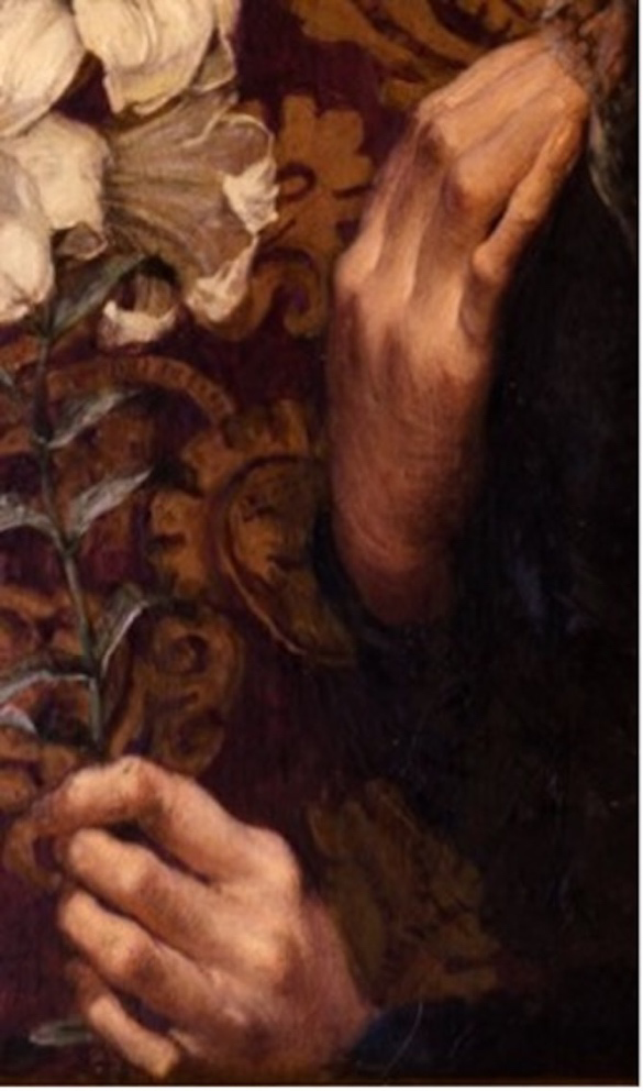
It is known that this disease is significantly more common in women than in men and that it generally occurs in the climacteric or perimenopausal period. The disease characteristically affects the PIP (Bouchard’s nodes) and distal interphalangeal (DIP) joints (Heberden’s nodes), rarely the MCPs. The female figure depicted in the picture is that of a young woman who has joint swellings in both the PIPs and MCPs. The involvement of the latter (MCPs) and the young age of the virgin make this diagnostic hypothesis unlikely, although suggestive. In fact, cases of osteoarthritis are described in the literature even at ages < 40 years (Culvenor et al. 2019).
Another disease that we need to differentially diagnose is undoubtedly rheumatoid arthritis, which is known to be a chronic inflammatory disease that characteristically affects the small joints of the hands and feet symmetrically. In our picture we note that while symmetricity is respected, another key detail is present, namely sparing of DIPs. The absence of involvement of the DIPs is a peculiar feature of RA that differentiates this disease from arthrosis on the one hand and from other inflammatory rheumatisms on the other, especially psoriatic arthritis. However, if we look closely at Virgo’s hands, the swellings of the fingers give the impression of having a nodular, solid, hard appearance, whereas in RA the joint inflammation is localized at the level of the synovial membrane, where it causes a thickening of the same, the so-called synovial cloth, giving the fingers a spindle-like, soft, doughy appearance, and not hard as in arthrosis.
If we move on to consider not the joint itself, but the skin overlying the small joints of the hands, it opens up a range of diagnostic hypotheses that we will have to examine, that is it is necessary to make a differential diagnosis of the various nodosities that may occur at the DIP, PIP and MCP.
A rheumatologist would say to consider the following pathological conditions:
1. Rheumatoid nodules
They are observed that in some forms of RA, usually those with high titers of rheumatoid factor, are predominantly located at the extensor surface of the fingers.
2. Meynet nodules
They are characteristic of rheumatic fever or acute articular rheumatism (AAR), a disease of pediatric age, but also observable in adults, characterized by migrating arthritis following streptococcal angina. In AAR there is possible involvement of the heart and nervous system. Solid, painless, roundish nodules may develop in the subcutaneous tissue, the diameter of which may vary from a few millimeters to 1 or 2 cm. The skin over the lesions is not inflamed. They are found at bony surfaces or bumps or near tendons. Subcutaneous Meynet nodules generally appear only after the first few weeks of disease, usually only in patients with carditis (Guilherme et al. 2016).
3. Xanthomatosis
Xanthomatosis is characterized by the presence of yellowish nodular formations, called xanthomas, appearing in various locations (particularly in the skin and tendons), which result from the accumulation of histiocytic cells engulfing lipid substances. Tendon xanthomatosis often accompanies familial hypercholesterolemia. The Achilles tendon is the most commonly affected tendon due to xanthomatosis. Regarding hand involvement, it is known that xanthomas can occur at the level of the flexor tendons and, more rarely, at the extensor tendons. In the literature, rare are the descriptions concerning bilateral xanthomatosis involving the extensor tendons of the hand and, at the same time, causing reduced range of motion of the fingers (Brenn et al. 2019).
4. Multicentric reticulohistiocytosis
Multicentric reticulohistiocytosis (MRH) is a rare multisystem disease of unknown etiology, occurring most frequently in women characterized by localized erythematous-nodular lesions in the skin, mucous membranes, and subcutaneous tissue. There is a predilection for the hands followed by the face, extremities, and trunk. The skin lesions are multiple, symmetrical papules and nodules, ranging from several millimeters to a few centimeters. The skin lesions are often accompanied by progressive, symmetrical erosive arthritis, which is often the onset symptom. The joints of the hand, wrist, and knee are predominantly affected, followed by the shoulder and ankles. The clinical course is characterized by remission and recurrence, which in most cases results in erosive, disfiguring, and disabling arthritis. The disease is often accompanied by generalized systemic symptoms, such as weight loss, fever, malaise, myalgia, and lymphadenopathy.
Of considerable clinical importance is the frequent association of MRH with cancerous conditions such as melanoma, breast, stomach, lung, ovarian, colon, and pancreatic carcinoma, and with noncancerous conditions such as thyroid disease, diabetes mellitus, and tuberculosis.
Histological examination showed the presence of a dermal infiltrate consisting of histiocytes and multinucleated giant cells containing abundant eosinophilic cytoplasm with the characteristic “ground glass” appearance (Brenn et al. 2019; Richetta et al. 2000).
5. Knuckle pads (also known as heloderma)
These are benign, round, plaque-like, smooth, firm, well-circumscribed subcutaneous fibrotic nodules localized mainly at the dorsal portion of the proximal interphalangeal (PIP) joints and, less frequently, the metacarpophalangeal (MCP) joints of the hands; the thumbs and toes are less frequently involved. They are sometimes associated with Dupuytren’s contracture and camptodactyly. Knuckle pads are mostly asymptomatic, painless, and have a progressive growth, reaching sizes up to 40 mm in diameter. They also do not cause any functional impact on the joints, such as decreased flexibility or tendon disruption. Histologically, epidermal abnormalities such as hyperkeratosis and acanthosis can be observed, whereas at the dermal level, a proliferative stage can be distinguished, in which fibroblasts immersed in collagen bands to form an unencapsulated dermal nodule and a less cellular fibrotic stage, with spindle-shaped fibroblasts and thickened, irregular collagen bundles are recognizable. The histologic picture is similar to that found in Dupuytren’s contracture or Ledderhose disease, with which knuckle pads are often associated (Lagier et al. 1975).
They were first described by Garrod in 1893 (Garrod 1893). Some authors have identified knuckle pads in some of Michelangelo’s works such as the David, the Moses, the statue of Victory and that of Giuliano de Medici (Allison & Allison 1966).
Knuckle pads have been associated with repetitive friction or pressure (e.g., habitual chewing or sucking of the fingers, repetitive work activities, athletic activities such as boxing and surfing, and bulimia nervosa with self-induced vomiting), fibrosing disorders (Dupuytren’s contracture, Ledderhose syndrome/plantar fascial fibromatosis, and Peyronie’s disease), and autosomal dominant syndrome (Bart-Pumphrey syndrome), but idiopathic cases have also been described (Giovannini et al. 2021).
Conclusions
In conclusion, the careful observation of the nodular lesions on the hands of Maria Virgo portrayed by May Louise Greville Cooksey led us to undertake a differential diagnostic exercise and to formulate several diagnostic hypotheses, each of them suggestive and possible, while emphasizing the differences that distinguish them from each other. Of all the diagnostic hypotheses examined, the most plausible is, in the rheumatologist’s opinion, the knuckle pads hypothesis.
References
Acidini C. (2024) L’amore e l’esilio. Firenze nello specchio preraffaellita fra Dante e Botticelli, in: Preraffaelliti. Rinascimento moderno. Dario Cimorelli editore, Milan, pp. 59-73.
Allison J.R. Jr, Allison J.R. Sr. (1966) Knuckle pads. Arch. Dermatol. 93:311-316.
Brenn T., Jason L., Hornick J.L. (2019) Cutaneous Mesenchymal Tumors. In: Practical Soft Tissue Pathology: a Diagnostic Approach, Second Edition. Elsevier, Philadelphia, pp. 403-457.
Catalogue entry XV.31. In: Preraffaelliti. Rinascimento moderno. Milan: Dario Cimorelli editore, 2024.
Cooksey M.L.G. (1999) In: SaurAllgemeines Künstlerlexikon. K.G. Saur Verlag, Munich-Leipzig: 1999, p. 21.
Culvenor A.G., Øiestad B.E., Hart H.F., Stefanik J.J., Guermazi A., Crossley K.M. (2019) Prevalence of knee osteoarthritis features on magnetic resonance imaging in asymptomatic uninjured adults: a systematic review and meta-analysis. Br. J. Sports Med. 53:1268-1278. doi: 10.1136/bjsports-2018-099257.
Di Cesare P.E., et al. (2017) Pathogenesis of Osteoarthritis. In: Kelley & Firestein’s Textbook of Rheumatology, Tenth Edition. Elsevier, Philadelphia, pp. 1685-1704.
Galassi F.M., Lippi D., Zucchini E., Bianucci R., Varotto E. (2023) Palaeodermatological exposé on the historical case of Ferdinando II de’ Medici (AD 1610-1670). J. Eur. Acad. Dermatol. Venereol. 37:2415-2418. https://doi.org/10.1111/jdv.19436.
Garrod A.E. (1893) On an unusual form of nodule upon joints of the fingers. St. Bartholomew’s Hosp. Rep. 1893;29:157-161.
Giovannini I., Zandonella Callegher S., Errichetti E., De Vita S., Zabotti A. (2021) Knuckle pads mimic early psoriatic arthritis. Reumatismo 73:67-69 https://doi.org/10.4081/reumatismo.2021.1354.
Guilherme L. et al. (2016) Rheumatic Fever and Post-streptococcal Arthritis. In: Kelley & Firestein’s Textbook of Rheumatology. Tenth Edition. Elsevier, Philadelphia, pp. 1956-1972.
Hailey D, Miller A, Yenawine P. (2015) Understanding Visual Literacy: The Visual Thinking Strategies Approach. In: Baylen D., D’Alba A. (eds). Essentials of Teaching and Integrating Visual and Media Literacy. Springer, New York, pp. 49-73.
Housen A. (1983) The eye of the Beholder: Measuring Aesthetic Development. Dissertation. Harvard Graduate School of Education.
Housen A. (1997) The Eye of the Beholder: Research, Theory and Practice. https://VTShome.org/wp-content/uploads/2016/08/Eye-of-the-Beholder.pdf (accessed April 5, 2020).
Huri G., Joachim N. (2013) An unusual case of hand xanthomatosis. Case Rep. Orthop. 2013; 183018. https://doi.org/10.1155/2013/183018.
Jutel A. Russell G. (2023) Past, present and imaginary: Pathography in all its forms. Health. 27: 886-902. https://doi.org/10.1177/13634593211060759.
Lagier R., Meineeke R. (1975) Pathology of “Knuckle Pads”. Study of Four Cases. Virchows Arch. A. Pathol. Anat. Histol. 365:185-191.
Lippi D. (2014) Reading different literature helps one to learn to listen. Intern. Emerg. Med. 2014;9:493-495. https://doi.org/10.1007/s11739-014-1078-3.
Lippi D, Bianucci R, Donell S. (2019) The visual arts and medical education. Knee Surg. Sports Traumatol. Arthrosc. 27:3397-3399. https://doi.org/10.1007/s00167-019-05744-4.
Richetta A., Faiola R., Divona L., Calabretta F., Sansolini T., Innocenzi D., Bottoni U., Calvieri S. (2000) Multicentric reticulocytosis (Description of a case). Ital. J. Dermatol. Venereol. 135: 501-504.
Russell-Cotes Museum. Maria Virgo. https://russellcotes.com/collection-piece/maria-virgo/ (accessed on 18 September 2024).
Traversari M., Ballestriero R., Galassi F.M. (2017) A likely case of goiter in the Madonna col Bambino dormiente (1465/1470) by Andrea Mantegna (1431-1506). J. Endocrinol. Invest. 40:237-238. https://doi.org/10.1007/s40618-016-0548-z.
Varotto E., Ballestriero R. (2018) 17th-century sculptural representation of leprosy in Perugia’s Cathedral. Infection. 46:893-895. https://doi.org/10.1007/s15010-018-1237-y.
Yenawine P. (2013) Visual Thinking Strategies: Using Art to Deepen Learning Across School Disciplines. Harvard Education Press, Cambridge (MA).