https://doi.org/10.36253/ijae-16050
Middle cerebral artery: a systematic review and meta-analysis
1 Faculty of Medicine of ABC, Santo André, São Paulo, Brazil
2 Department of Morphophysiology, Faculty of Medicine of ABC, Santo André, São Paulo, Brazil
*Corresponding author. E-mail: nicolepalma2002@gmail.com
Abstract. The middle cerebral artery is the largest and most intricate artery in the brain, making a thorough understanding of its anatomical variations and anomalies crucial. Despite its importance, considerable debate surrounds the classification of these variations, the existence of anomalies, and their prevalence. This study seeks to elucidate the prevalence of anatomical variations and anomalies in the middle cerebral artery and, as a secondary objective, to examine their correlation with clinically significant events, such as aneurysms. Methods: A systematic review was conducted in accordance with PRISMA guidelines, using databases such as PubMed, Embase, BVS, and Cochrane. Relevant terms from MeSH, DeCS, and Emtree were used alongside the “Open Grey” platform. A meta-analysis was carried out to determine the overall prevalence of anomalies, along with a subgroup analysis based on the methods used for the inspection and detection of arterial anomalies. Results: The overall prevalence of anomalies was found to be 1.4%. Through subgroup analysis, studies utilizing colored material injection revealed an almost-7 fold higher prevalence of anomalies than imaging techniques studies, and this was statistically significant (3.9% vs 0.5%, p <0.01). Conclusion: Imaging techniques may not adequately detect all anatomical variations of the middle cerebral artery. Longitudinal observational studies are necessary for better understanding of our findings.
Keywords: middle cerebral artery, neuroanatomy, cerebral arteries, cerebral arterial diseases, cerebrovascular disorders.
Contents
3.2. Inclusion and exclusion criteria
1. Introduction
The middle cerebral artery (MCA) is the largest and most complex artery in the brain [1]. Its vascular territory includes important areas, such as the basal ganglia, descending and corticospinal tracts, and cortical regions, essential for motor and sensory functions [2]. Considering that a great part of the brain hemisphere is nurtured by the MCA, this artery is frequently used in surgical interventions [3], making the understanding of its anatomical variations vital.
Typically, between days 32 and 40 of embryonic development, the MCA develops from the primitive internal carotid artery near the anterior cerebral artery. By days 47–48, it becomes more prominent and develops branches that supply areas of the cerebral hemispheres. Alterations in this process can result in anomalies such as duplications, fenestrations, and accessory arteries. Although the embryological origin and prevalence of MCA are poorly understood [4], these anomalies are associated with various clinical presentations, including aneurysms [5].
The MCA is the major site of a third of all cerebral aneurysms with diverse morphological traits. [6]. Anatomical modifications, such as in M1 segment length, may be correlated with this type of vascular event [7], which is estimated to be present in 3.2% of the population around 50 years of age [8].
Despite the significance of the MCA, its patterns and abnormalities remain unclear and no controversies exist about the classification criteria for different presentation forms [2]. The prevalence and characteristics of its various presentations, including bifurcation, trifurcation, and tetrafurcation, remain inadequately addressed in the literature. Approximately 50% of MCA aneurysms lead to rupture, and half of the individuals who experience subarachnoid hemorrhages due to these ruptures suffer severe long-term effects [9]. These insights underscore the necessity for different therapeutic strategies, highlighting the importance of anatomical studies of the MCA and its variations. This review aims to summarize collected data related to the MCA and its pattern identification, based on literature, aiming to impact the initial diagnosis and patient management.
2. Literature review
The MCA is a critical vascular structure that supplies a substantial portion of the cerebral cortex [10]. Despite its significance, current literature reveals that variations in branching patterns and associated abnormalities of the MCA remain inadequately understood, and a consensus on classification is yet to be achieved [2].
The branching patterns of the MCA are defined by the number of blood vessels in which the main trunk of the artery divides. Many variations exist between branching patterns, however literature determined the segment M1 bifurcates into two main trunks in approximately 69.9% of the cases [11]. The artery can also divide into three trunks (trifurcation), four trunks (tetrafurcations), or maintain the permanence of the main trunk without ramification (monofurcation). Kashtiara et al. estimates that the prevalence of tetrafurcation, monofurcation, and trifurcation to be 1%, 1.9% and 27%, being the trifurcation the most common branching after bifurcation.
It also outlined uncommon variations, classified as abnormalities. Literature estimates that the occurrence of these presentations on the MCA is lower than that of other important cerebral arteries [2]. Its most frequent abnormalities are accessory artery, duplication, and fenestration. The accessory MCA is a vessel that originates from the anterior cerebral artery and goes through the sylvian fissure along with the MCA [11], having a prevalence estimated at 0.03% [2]. Duplication of the MCA is an abnormality represented by an artery that originated from the internal carotid artery, independently from its extent [12], having a prevalence estimated at 0.17% [2]. MCA fenestration or segment duplication has a prevalence estimated at 0.28% [13].
Despite the recognition of these variations, the prevalence rates reported in literature remain inconsistent. This discrepancy underscores the necessity for further research to provide in-depth understanding and consolidate the existing body of knowledge regarding MCA anatomy and its variations.
3. Methodology
3.1. Search strategies
This review was conducted in accordance with the guidelines set forth by the Preferred Reporting Items for Systematic Reviews and Meta-Analyses (PRISMA)[14]. This review´s guiding question was defined based on the “PICOS” method, in which: P (population) represents patients without neurological previous diseases; I, anatomical aspects; C, -; O (outcome), MCA pathologies predictability; S (study), observational study. The selection was conducted in August 2024.
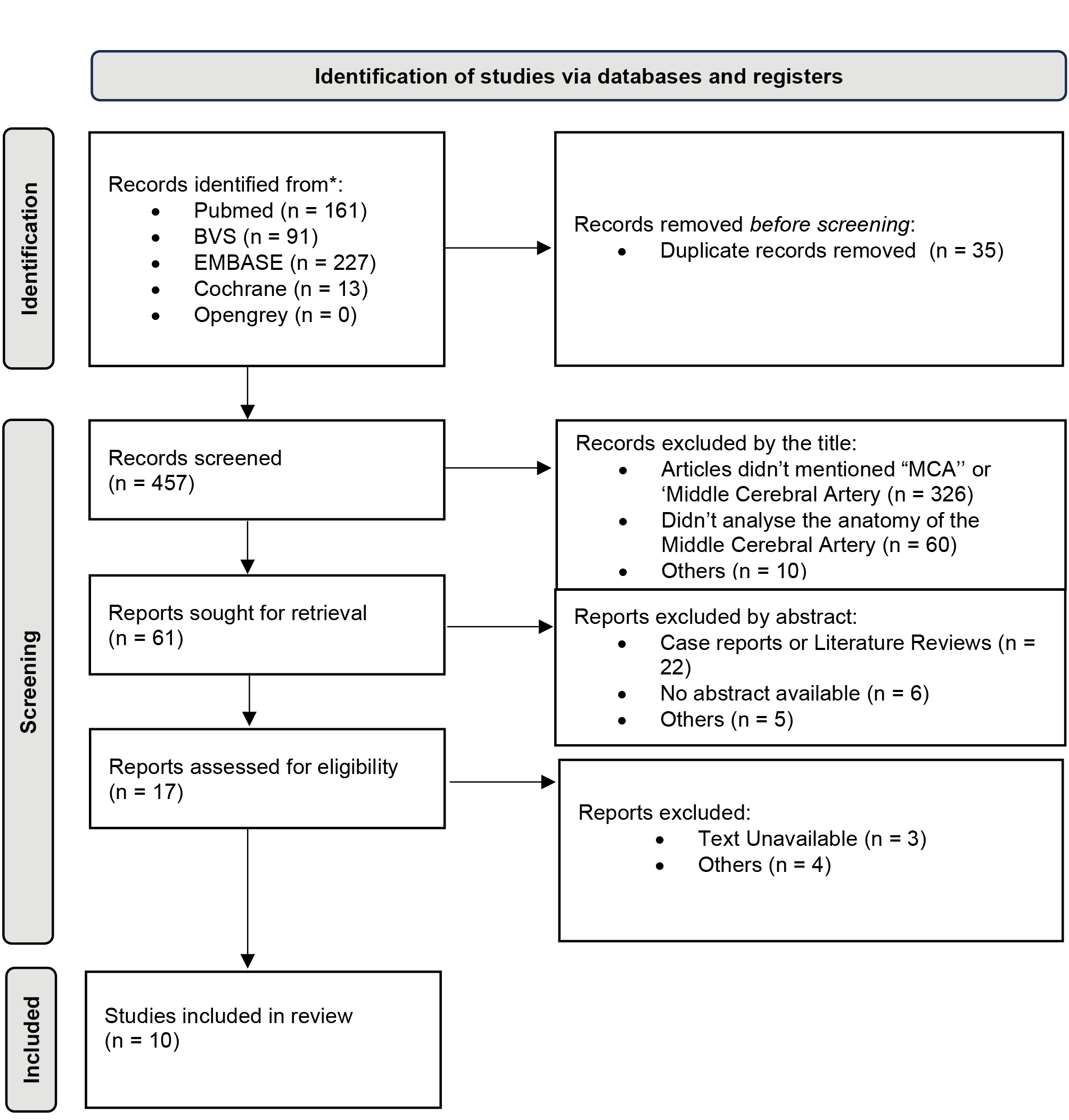
This research is based on electronic data from PubMed, Embase, BVS, and Cochrane, according to MeSH (PubMed, Cochrane), Decs (BVS), and Entree (Embase), using the most sensible and specific terms possible, along with boolean operators, shown in the Table 1. OpenGrey was used for gray literature. We used the Rayann platform to screen and organize studies [15]. A flowchart with a summary of the selected articles is presented in Figure 1.
| Data Base | Descriptors |
|---|---|
| Embase | (‘Middle Cerebral Artery’/exp OR ‘mca’) AND anatomy AND (‘variations’ OR ‘anomalies’ OR ‘patterns’) |
| Pubmed | (“Middle Cerebral Artery”[Mesh]) AND “abnormalities” [Subheading] |
| Cochrane | “middle cerebral artery” AND “anatomy” |
| BVS | (Middle Cerebral Artery) AND (anatomy ) AND (sh:(abnormalities)) |
| Open Grey | “ Middle Cerebral Artery” |
3.2. Inclusion and exclusion criteria
The articles selected for this review are those that specifically addressed MCA anatomy variations in patients without previous neurological conditions . The exclusion criteria were: articles without the pertinent issue and lack of substantial information regarding MCA variations. Additional exclusions encompassed abstracts, opinion pieces, narrative reviews, case reports, systematic reviews, and meta-analyses.
4. Data analysis
A prevalence meta-analysis was conducted based on the selected studies. The extracted variable for this meta-analysis was the overall percentage of anomalies (including duplication, accessory, and fenestration) observed in the MCA. A random effects model was used for the analysis.
In addition, a subgroup analysis was carried out based on the method used to detect artery anomalies. The results of the meta-analysis are illustrated using a forest plot graphic. All analyses were performed using the R program, version 4.3.1 (2023).
5. Risk of bias
The ROBINS-E platform was used to assess the risk of bias across studies [16]. Figure 2 illustrates the risk of bias, delineating distinct domains within the evaluated literature.
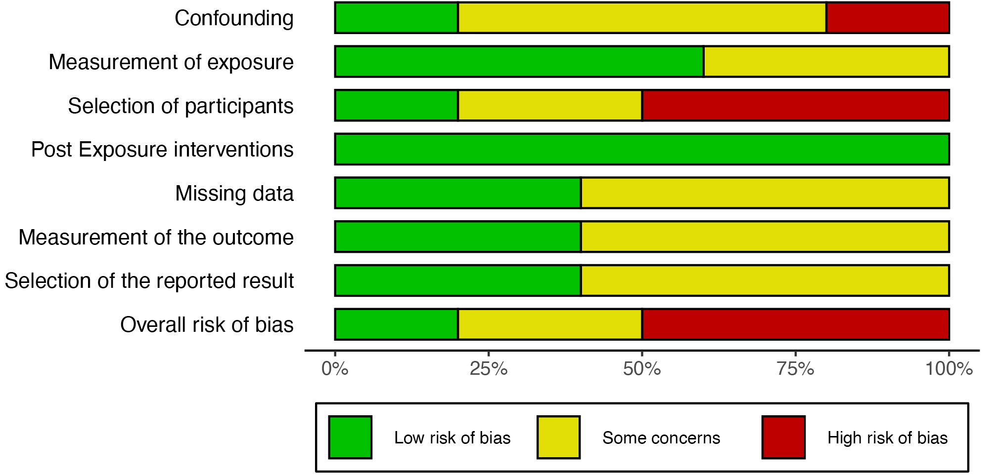
Studies differed in their risk of bias due to confounding factors. In some cases, as seen in a study by Brzegowy and Sharma, the variable control of age and sex was inadequate, raising concerns and moderate to high risk. In other studies, the measure of exposure presented a moderate risk due to lack of details of the instrument’s precision or pattern.
Participant selection bias was higher in studies that used small or representative samples, as seen on Celliers, that did not detail the choice criteria for selecting the cerebral hemispheres to be studied. No risk of post-exposure intervention was observed in studies that did not include it. The risk of bias due to the absence of data was moderate in some studies, mainly regarding treatment. The outcome measure bias varied from low to moderate. The result selection reported bias was considered low to moderate risk, with some concerns regarding the selectivity in reporting only significant findings. In general, the graphic highlights that multiple studies present bias to some extent.
6. Results
Based on the information presented, 492 articles were initially filtered, with 35 excluded because of duplication. Following an analysis of titles and abstracts, 17 articles remained. After a complete study of the articles, only 10 met the inclusion criteria.
The selected articles, presented in Table 2, were published between 1984 and 2023, and the number of arteries analyzed in these studies ranged from 20 to 6,982. Among the methods used, four articles utilized colored substances for arterial infusion, one article implemented antiresonance techniques, two articles explored nanotomography, two used fresh brain specimens, and one used transcranial ultrasonography.
| Study | Age (mean) | Nº of arteries | Method | Anomalies | Branching patterns | Length of the main trunk |
|---|---|---|---|---|---|---|
| Celliers K, 2016 | - | 20 | Arteries perfused with colored silicone and dissected. | Not evaluated. | 5% presented monofurcation; 80% bifurcation; 15% trifurcation. | - |
| Uchino A et al, 2012. | - | 6982 | Magnetic resonance angiographies. | 0.057% presented duplication; 0.043% presented fenestration. | They were not evaluated. | - |
| Umansky F et al, 1984. | adult individuals | 70 | Arteries injected with colored polyester and microsurgically dissected. | 3% presented accessory artery. | 6% presented monofurcation; 64% bifurcation; 29% trifurcation; 1% tetrafurcation. | 15.35 |
| Brzegowy P et al, 2017. | 52.1 | 500 | Angiotomography. | 1% presented duplication; 0,4% accessory artery; 0,2% fenestration. | 82,2% presented bifurcation; 13,8% trifurcation; 0,4% tetrafurcation. | 15.8 |
| Oo EM et al, 2021. | - | 100 | Fresh brains with microsurgical dissection. | Not evaluated. | 12% presented monofurcation; 72% bifurcation; 16% trifurcation. | 20.6 |
| Pai SB et al, 2005. | - | 10 | Fresh brains with microsurgical dissection. | Not evaluated. | 80% presented bifurcation; 20% trifurction. | 20 |
| Umansky F et al, 1988. | adult individuals | 104 | Arteries injected with colored polyester and microsurgically dissected. | 1% presented duplication; 2% accessory artery; 1% fenestreation. | 4% presented monofurcation; 60% bifurcation; 26% trifurcation; 4% tetrafurcation. | - |
| Tanriover N et al, 2003. | adult individuals | 50 | Arteries injected with colored latex. | 2% presented duplication; 4% accessory artery. | 88% presented bifurcation; 12% trifurcation. | 17.82 |
| Sharma U et al, 2023. | 43.9 | 578 | Angiotomography. | 0.34% presented duplication; 0.17% presented fenestration. | 0,17% presented monofurcation; 97,75% bifrucation; 1,04% trifurcation. | - |
| Rogge A et al, 2015. | 42 | 100 | Transcranial ultrasound with Doppler. | Not evaluated. | 63% presented bifurcation; 32% trifurcation. | 19.0 |
In terms of absolute data, the literature reported a variation in the occurrence of anomalies between 0.11% and 2%. The occurrence of MCA fenestration ranged from 0.20% to 1%, with a weighted average of 0.22%. For duplications, the weighted average occurrence was 0.2%, while that of related accessory arteries was 1.1%.
In terms of division patterns, segment M1 predominantly exhibits bifurcation with reported occurrences in the literature ranging from 64% to 97.75% and a weighted average of 84%, as detailed in Table 2. The second most common pattern is trifurcation, showing a prevalence between 1.04% and 32%, with a weighted average of 19%. Other patterns include monofurcation, which has a prevalence between 0.17% and 12%, yielding a weighted average of 2.5%, and tetrafurcation, which was a prevalence ranging from 0.4% to 4% and a weighted average of 1% (Table 3).
Furthermore, regarding length, the literature indicates a variation between 15.3 mm and 20.6 mm, with a weighted average length of 16.9 mm for segment M1.
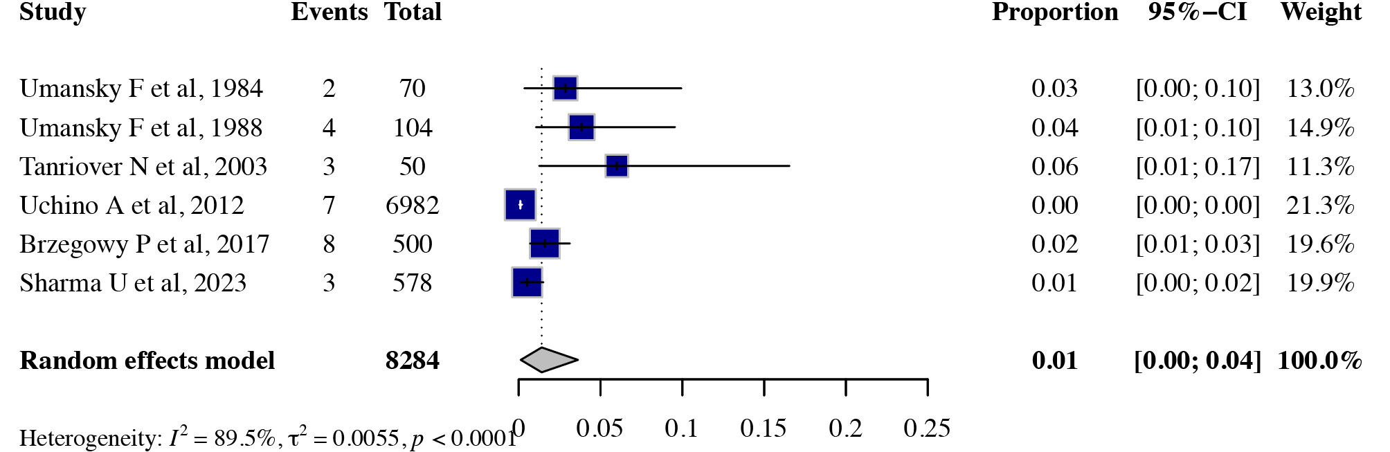
The observed presence of anomalies was quantified at 1.4% (heterogeneity [I²]= 89.5%; [τ²] = 0.0055; p < 0.0001), as shown in Figure 3. Heterogeneity was expected, particularly due to Uchino’s study [13] acting as an outlier. Application of the random effects model served to mitigate the influence of this disparity in the overall analysis.
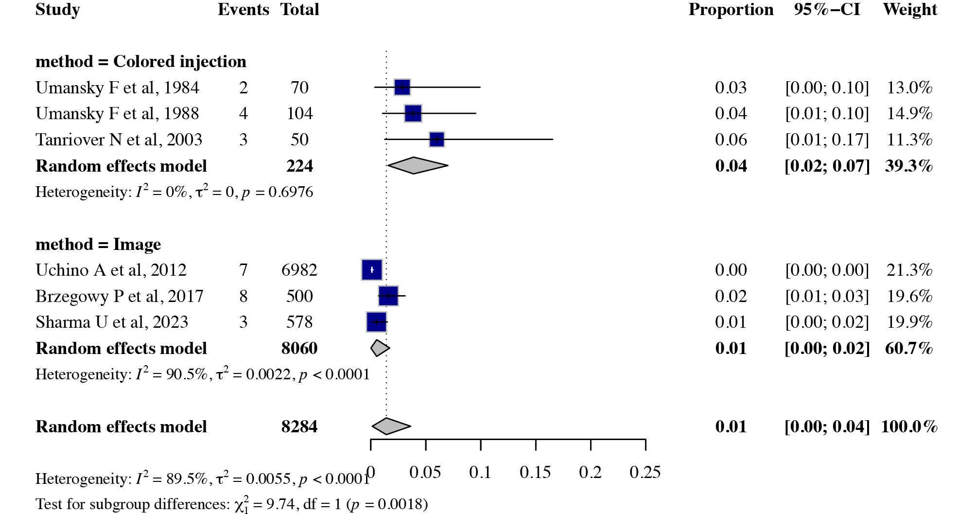
Subsequently, a subgroup meta-analysis was performed to compare two groups using the anomaly evaluation method (infusion of colored material versus imaging study). The findings indicated a statistically significant difference in prevalence between the two methods (χ²₁ = 9.74; df = 1; p = 0.0018).
Regarding the heterogeneity within each group, arteries assessed by the infusion of colored material exhibited an overall anomaly prevalence of 4.0% (I² = 0%; τ² = 0; p = 0.69), suggesting no heterogeneity among the studies. In contrast, arteries evaluated through imaging studies revealed a significantly lower prevalence of 1% (I² = 90.5%; τ² = 0.0055; p < 0.0001), indicating considerable heterogeneity and underscoring the discrepancies between the methodologies employed, as demonstrated in Figure 4.
7. Discussion
Detecting anatomical variations through the MCA is essential to ensure the safety of neurosurgical interventions, particularly in the context of aneurysm management. A thorough understanding of these variations contributes to more precise surgical planning and minimizes intraoperative risks.
This systematic review emphasizes the prevalence of various branching patterns, with bifurcation being the most common, occurring in approximately 84% of cases. Trifurcation represents the second most frequent variation, found in approximately 19% of cases. Additionally, less common variations such as monofurcation and tetrafurcation have been documented, although they appear in smaller proportions. Understanding whether different types of presentation of this artery are a risk factor in the development of some pathology remains necessary.
Anomalies, including duplications, accessory arteries, and fenestrations, can alter and potentially cause damage to blood flow. In cases with fenestrations, some evidence suggests that increased hemodynamic stress causes a increased prevalence of intracranial aneurysms in patients [11].
Among all selected studies, the estimated prevalence of these anomalies was 1.4%. However, a subgroup meta-analysis focusing exclusively on studies utilizing imaging methods indicated a prevalence of 0.5%. In contrast, studies that used colored injection techniques reported a higher prevalence of 3.9%.
This discrepancy (6.8 times higher in the colored injection technique) raises important questions regarding the capacity of imaging methods to capture more subtle anatomical details since this study indicates that traditional imaging methods may underestimate the prevalence of these variations. While imaging techniques such as magnetic resonance imaging (MRI) and computed tomography are commonly used to assess the MCA, they may not adequately detect the full range of anatomical variations . Although these imaging methods are convenient and broadly applicable, their limitations in identifying the complete spectrum of MCA anomalies need to be considered.
8. Acknowledgements
The author would like to thank all patients who somehow collaborated with scientific research and Editage for English language editing.
9. References
[1] Gibo H, Carver CC, Rhoton AL Jr, Lenkey C, Mitchell RJ. Microsurgical anatomy of the middle cerebral artery. J Neurosurg. 1981 Feb;54(2):151-69. https://doi.org/10.3171/jns.1981.54.2.0151. PMID: 7452329.
[2] Fauzi AA, Gunawan R, Suroto NS. Neuroangiography patterns and anomalies of middle cerebral artery: A systematic review. Surgical Neurology International, v. 12, p. 235, 25 maio 2021.
[3] Cilliers K, Page BJ. Anatomy of the middle cerebral artery: cortical branches, branching pattern and anomalies. Turkish Neurosurgery, v. 27, n. 5, p. 671-681, 2017. https://doi.org/10.5137/1019-5149.JTN.18127-16.1.
[4] Okahara M, Kiyosue H, Mori H, Tanoue S, Sainou M, Nagatomi H. Anatomic variations of the cerebral arteries and their embryology: a pictorial review. European Radiology, v. 12, n. 10, p. 2548–2561, 21 mar. 2002.
[5] Oo EM; Saw KEE, Oo HN, Than T, Thida K. Variable Anatomy of the Middle Cerebral Artery from Its Origin to the Edge of the Sylvian Fissure: A Direct Fresh Brain Study. The Scientific World Journal, v. 2021, p. 1–7, 9 mar. 2021. https://doi.org/10.1155/2021/6652676.
[6] Estevão IA, Camporeze B, Araujo Jr AS, Nery B, Antunes ÁCM, Smith TR, Aguiar PHP. Middle cerebral artery aneurysms: aneurysm angiographic morphology and its relation to pre-operative and intra-operative rupture. Arquivos de Neuro-Psiquiatria, v. 75, n. 8, p. 523–532, ago. 2017.
[7] Da Cunha CEG, Da Cunha Correia C. Middle cerebral artery extension and the risk for aneurysmal disease. Journal of the Neurological Sciences, v. 390, p. 219–221, 15 jul. 2018.
[8] Galvão J, Lima DD, Haas LJ. Prevalência de aneurismas cerebrais incidentais entre homens e mulheres. Saúde e Pesquisa, v. 13, n. 2, p. 309–316, 12 jun. 2020.
[9] Zhang W, Wang J, Li T, Mei M. Morphological parameters of middle cerebral arteries associated with aneurysm formation. Neuroradiology, v. 63, n. 2, p. 179–188, fev. 2021. https://doi.org/10.1007/s00234-020-02521-w.
[10] Ring BA. Middle cerebral artery: Anatomical and radiographic study. Acta Radiologica, v. 57, n. 4, p. 289–300, 1962. https://doi.org/10.3171/jns.1981.54.2.015110.3109/00016926209171758.
[11] Kashtiara A, Beldé S, Schollaert J, Menovsky T. Anatomical Variations and Anomalies of the Middle Cerebral Artery. World Neurosurg. 2024 Mar;183:e187-e200. https://doi.org/10.1016/j.wneu.2023.12.052. Epub 2023 Dec 14. PMID: 38101539.
[12] Teal JS, Rumbaugh CL, Bergeron RT, Segall HD. Anomalies of the middle cerebral artery: accessory artery, duplication, and early bifurcation. Am J Roentgenol Radium Ther Nucl Med. 1973 Jul;118(3):567-75. https://doi.org/10.2214/ajr.118.3.567. PMID: 4723180.
[13] Uchino A., Saito N, Okada Y, Nakajima R. Duplicate origin and fenestration of the middle cerebral artery on MR angiography. Surgical and Radiologic Anatomy, v. 34, p. 401–404, 2012.
[14] Page MJ, McKenzie JE, Bossuyt PM, Boutron I, Hoffmann TC, Mulrow CD, Shamseer L, Tetzlaff JM, Akl EA, Brennan SE, Chou R, Glanville J, Grimshaw JM, Hróbjartsson A, Lalu MM, Li T, Loder EW, Mayo-Wilson E, McDonald S, McGuinness LA, Stewart LA, Thomas J, Tricco AC, Welch VA, Whiting P, Moher D. The PRISMA 2020 statement: an updated guideline for reporting systematic reviews. BMJ. 2021 Mar 29;372:n71. https://doi.org/10.1136/bmj.n71. PMID: 33782057; PMCID: PMC8005924.
[15] Ouzzani M, Hammady H, Fedorowicz Z, Elmagarmid A. Rayyan—a web and mobile app for systematic reviews. Systematic Reviews, v. 5, n. 1, dez. 2016..
[16] Higgins JPT, Morgan RL, Rooney AA, Taylor KW, Thayer KA, Silva RA, Lemeris C, Akl EA, Bateson TF, Berkman ND, Glenn BS, Hróbjartsson A, LaKind JS, McAleenan A, Meerpohl JJ, Nachman RM, Obbagy JE, O’Connor A, Radke EG, Savović J, Schünemann HJ, Shea B, Tilling K, Verbeek J, Viswanathan M, Sterne JAC. A tool to assess risk of bias in non-randomized follow-up studies of exposure effects (ROBINS-E). Environ Int. 2024 Apr;186:108602. https://doi.org/10.1016/j.envint.2024.108602. Epub 2024 Mar 24. PMID: 38555664; PMCID: PMC11098530.
[17] Ministério da saúde. Diretrizes Metodológicas: Sistema GRADE – manual de graduação da qualidade da evidência e força de recomendação para tomada de decisão em saúde. Brasília: Ministério da Saúde, Secretaria de Ciência, Tecnologia e Insumos Estratégicos, Departamento de Ciência e Tecnologia, 2014.
[18] Ministério da saúde. Diretrizes metodológicas: elaboração de revisão sistemática e meta-análise de ensaios clínicos randomizados. Brasília: Ministério da Saúde, Secretaria de Ciência, Tecnologia, Inovação e Insumos Estratégicos em Saúde, Departamento de Gestão e Incorporação de Tecnologias em Saúde, 2021.
[19] Umansky F, Juarez SM, Dujovny M, Ausman JI, Diaz FG, Gomes F, Mirchandani HG, Ray WJ. Microsurgical anatomy of the proximal segments of the middle cerebral artery. J Neurosurg. 1984 Sep;61(3):458-67. https://doi.org/10.3171/jns.1984.61.3.0458. PMID: 6747682.
[20] Umansky F, Dujovny M, Ausman JI, Diaz FG, Mirchandani HG. Anomalies and variations of the middle cerebral artery: a microanatomical study. Neurosurgery. 1988 Jun;22(6 Pt 1):1023-7. https://doi.org/10.1227/00006123-198806010-00008. PMID: 3047592.
[21] Rogge A, Doepp F, Schreiber S, Valdueza JM. Transcranial color-coded duplex sonography of the middle cerebral artery: more than just the M1 segment. J Ultrasound Med. 2015 Feb;34(2):267-73. https://doi.org/10.7863/ultra.34.2.267. PMID: 25614400.
[22] Tanriover N, Kawashima M, Rhoton AL Jr, Ulm AJ, Mericle RA. Microsurgical anatomy of the early branches of the middle cerebral artery: morphometric analysis and classification with angiographic correlation. J Neurosurg. 2003 Jun;98(6):1277-90. https://doi.org/10.3171/jns.2003.98.6.1277. PMID: 12816276.
[23] Sharma U, Verma S, Aditham S. Variations in branching pattern of middle cerebral artery using CT angiography in South Indian population. European Journal of Anatomy, v. 27, n. 2, p. 155-164, 2023. https://doi.org/10.52083/EPCW5127.
[24] Brzegowy P, Polak J, Wnuk J, Łasocha B, Walocha J, Popiela TJ. Middle cerebral artery anatomical variations and aneurysms: a retrospective study based on computed tomography angiography findings. Folia Morphol (Warsz). 2018;77(3):434-440. https://doi.org/10.5603/FM.a2017.0112. Epub 2017 Dec 13. PMID: 29235088.
[25] Pai SB, Varma RG, Kulkarni RN. Microsurgical anatomy of the middle cerebral artery. Neurology India, v. 53, n. 2, p. 186-190, 2005. https://doi.org/10.4103/0028-3886.16406.