https://doi.org/10.36253/ijae-16011
Technical update of Delalande’s hemispherectomy
1 School of Medicine of Pontifical Catholic University of São Paulo (PUC-SP), Brazil
2 School of Medicine, Federal University of São Paulo (UNIFESP), Brazil
3.Faculty of Medical Sciences, ABC Medical School (FMABC), Brazil
4 Department of Neurosurgery, Hospital Santa Paula, São Paulo, Brazil
*Corresponding author. E-mail:danilotdequeiroz@gmail.com
Abstract. Introduction: Epilepsy is a common neurological disorder characterized by unprovoked seizures, affecting patients socially, psychologically, economically, and cognitively. When pharmacological treatment is insufficient, surgical intervention becomes necessary. Among hemispherectomy procedures, the most notable are anatomical (classic) and functional approaches, including Rasmussen, Delalande, Villemure, and Schramm. Other techniques described in the literature include cerebral hemicorticectomy, hemispherical deafferentation, transcortical subinsular hemispherectomy, and transopercular hemispherectomy. This study aims to analyze surgical methods and their effects while introducing a modified Delalande procedure and evaluating its efficacy through experiments on postmortem brains. Material and methods: The study used magnifying lenses (4.5x and 2.5x) and microsurgical instruments, funded by a PIBIC-CNPq scholarship. The proposed modification maintains the original disconnections but starts in the pericallosal area, offering a direct view of the corpus callosum. The procedure was tested on two formalin-fixed brains, divided into two groups of six coronal sections. Results were analyzed using the Student’s t-test. Results: The number of pericallosal artery lesions was statistically similar in both procedures. However, the modified technique resulted in a smaller neurosurgical incision. Conclusion: While the risk of pericallosal artery injury remains unchanged, a smaller incision may lead to shorter procedure time, better prognosis, and increased safety for both physician and patient. Further studies with larger samples and additional variables are necessary to fully assess its effectiveness.
Keywords: hemispherectomy, epilepsy, seizure, neurosurgery.
Contents
Functional Hemispherectomy (Rasmussen Variation)
Functional Transsylvian Hemispherectomy (Schramm Variation)
Lateral Perinsular Hemispherectomy (Villemure Variation)
Vertical Parasagittal Hemispherectomy (Delalande Variation)
3.2. Brains in coronal section
1. Introduction
Epilepsy is a disease defined by excessive or synchronous abnormal activity of neurons, even in the absence of external and reversible causative factors, such as toxins, metabolic substances, or fevers. It involves neuronal cells predisposed to generating recurrent seizures; with cognitive, neurobiological, psychological, and social consequences resulting from these episodes. Epilepsy itself cannot be understood as a single disease; it is, in fact, a multifaceted pathology with various etiological elements, including structural, genetic, infectious, metabolic, autoimmune, or even unknown factors.
Worldwide, it presents as one of the most common neurological diseases, alongside with chronic diseases such as migraine, stroke, and Alzheimer’s disease. According to the World Health Organization, an estimated 68 million people worldwide have epilepsy, with an active prevalence of 6.38 per 1,000 people and an incidence of almost 61.44 new cases per 100,000 population, a mortality rate 2 to 3 times higher than the general population (mostly when related to refractory crises and symptomatic etiology) and also high morbidity contributing to 0.7% of disability-adjusted life years, either through direct actions such as the risk of injuries and traumas or through indirect actions, such as the use of medications and negative impact on the quality and life experience of individuals with the condition.
Although anticonvulsant medications have the potential to suppress and even control seizures in up to two-thirds of all individuals with the disease, it does not alter its long-term prognosis. Therefore, epilepsy surgery is the most effective way to achieve long-term seizure freedom in selected individuals with drug-resistant focal epilepsy (also known as refractory epilepsy); however, it is currently underutilized. Thus, a better understanding of the gradual development of epilepsy, along with its epigenetic and pharmacogenomic determinants, gives hope for better pharmacological and non-pharmacological treatment strategies in the future, modifying the course of this disease or even, in the best cases, curing it.
Patients indicated for hemispherectomy are those who have:
- –Resistance to pharmacological treatment of seizure crises.
- –A remaining hemisphere suitable for good outcomes after seizures, noting that the spread of epileptiform discharges to the normal hemisphere, as seen on electroencephalogram, or even rare independent discharges on the normal side, do not indicate a poor response to surgery.
- –A contralateral hemisphere to the hemisphere with possible hemiplegia that can be identified with radiological and functional imaging as having diffuse abnormality.
Other indications for hemispherectomy are described in the literature, but with certain criteria sometimes controversial or relative, including patients with the following characteristics:
- –Contralateral hemiplegia, because if the procedure is done before total hemiplegia, the quality of distal lower limb movements (foot and digits) may be lost, even though the patient can walk and use the proximal muscles of the upper limb.
- –Delayed neurodevelopment is generally present due to the interference of seizures in the normal hemisphere’s development. Therefore, this would actually be a kind of relative prerequisite for hemispherectomy.
In pediatric patients, the use of hemyspherectomy presents itself as a favorable prognosis treatment, thanks to the high potential for neural plasticity present in this age range, offering many benefits for these young patients diagnosed with epilepsy:
- –Not having to deal with the harmful effects of frequent episodes of uncontrolled seizures, neither the use of high doses of antiepileptic medications during this period of neurological development.
- –Avoiding the debilitating social implications of the disease and the time lost/wasted in learning due to the disease and its symptoms must be considered.
- –Patients under 9 years of age, in a study, have shown better cognitive and motor postoperative outcomes, except for those who develop post-infarction sequelae.
On the other hand, it is also necessary to consider the morbidity of major surgeries in young patients and the possibility of increased neurological deficits in some cases, thus requiring careful and balanced consideration of hemispherectomy guidance against the substantial gains offered by the procedure for long-term seizure relief and functional outcomes; so that each family can weight out the potential benefits and risks in order to decide what is best for their child.
Nevertheless, there is also a discussion in the neurosurgery field about whether the presence of bilateral abnormalities in preoperative epileptogenic evaluation is actually associated with a worse postoperative outcome in hemispherectomy. It should also be noted that there are studies suggesting that hemispherectomy surgery can sometimes be used as a palliative exclusive focus procedure for severe cases with the onset of bilateral seizures predominating in one cerebral hemisphere. Additionally, surgery may be performed in cases of bilateral epilepsy with the attempt that, after its procedure, the use of antiepileptic medications may control seizures in the contralateral hemisphere.
Regarding the types of hemispherectomy, we will review:
Anatomical hemispherectomy
In this approach, the procedure begins by cautiously opening the Sylvian fissure, with great caution, as errors at this stage can cause severe damage to contralateral vessels. After ensuring access through the fissure, it is necessary to identify, dissect, clip, and divide the lateral branches of the lenticulostriate arteries of the ipsilateral basal ganglia. Similarly, it is also necessary to divide the proximal portion of the origin of the callosomarginal artery from the ipsilateral anterior cerebral artery.
In the second step, a cottonoid is placed in the foramen of Monro to protect the underlying choroid plexus and prevent blood and debris from entering the ventricular system through which the interhemispheric callostomy is performed.
Thus, for the implementation of callosotomy, microdissection, coagulation, and aspiration of the knee, the anterior portion, up to the splenium, which defines the posterior limit, can be used
Lastly, the frontobasal white matter is divided through the anterior part of the lateral ventricle. Then, the temporal stem is dissected, while the posterior communicating arteries are clipped and divided at segment P3 [34].
It is worth noting that the amygdala and hippocampus are removed by applying subpial dissection with special care to preserve the oculomotor nerve [34,35,36,38].
Regarding the choroid plexus, it is worth mentioning that it may be coagulated or left intact according to the surgeon’s preference, while the ipsilateral basal nuclei and the thalamus are left in situ for better motor outcomes [34,35,36,38].
Functional Hemispherectomy (Rasmussen Variation)
In this modification, the temporal lobe is removed with two cortical incisions, one in the superior temporal gyrus, running parallel to the Sylvian fissure, and the second located in the dorsal part of the temporal lobe, below the temporal lobe, perpendicular to the first and located 8cm from the pole of the temporal lobe [34]. The hippocampus, parahippocampal gyrus, medial part of the uncus, and lateral part of the amygdala are removed with an ultrasonic aspirator after opening the temporal pole, emphasizing that the ipsilateral third cranial nerve must be protected.
The next step involves providing access to the ipsilateral lateral ventricle through the resection of the suprasylvian cortex via two incisions parallel and perpendicular to the Sylvian fissure [34,35,39,40,41]. Finally, this stage ends with the transection of the corona radiata [34].
The following step is the completion/conclusion of the parasagittal transventricular callosotomy, followed by the removal of this cortical portion [34]. The pericallosal artery constitutes the medial border of the resection, given that it vascularized the knee of the corpus callosum. The fibers of the remaining posterior and anterior callosal tracts are disconnected by the ependymal surface towards the cingulate gyrus [34,35,39,41].
Lastly, the resection of the anterior and posterior connections of the frontal lobe with the parietal and occipital lobes is necessary [34,39]. Thus, the anterior cerebral artery, the superior circular sulcus, and the M1 segment of the medial cerebral artery are the borders of the transection of the corona radiata. The posterior disconnection occurs upon the full opening of the Sylvian fissure and the immediate elevation of the parietal operculum [35,41].
Highlighting the final part of this procedure, the disconnection line extends from the posterior part of the opening of the lateral ventricle to the trigone of the temporal pole cavity [34,35,41].
Functional Transsylvian Hemispherectomy (Schramm Variation)
Regarding the skin incision, it is characterized as curved from the anterior portion of the tragus to the incision in the upper frontal area. The temporal fascia is opened in the same manner [10,34,38].
A bone flap, measuring 4x5 cm, is used over the Sylvian fissure with the use of neuronavigation. The inferior and anterior edges are formed by the temporal operculum and the limen of the insula, respectively. The anterior edge is located 5cm anterior to the posterior edge, represented by the pulvinar projection [10,34,42].
After this step, the Sylvian fissure is extensively opened to expose the circular sulcus and the insula, as well as to identify and properly expose and skeletonize all branches of the middle cerebral artery. For the purpose of performing uncus-amygdalo-hippocampectomy, the temporal horn is opened through the inferior circular sulcus [10,42].
Thus, the next step involves the transection of the long fibers of the corona radiata, as a consequence of opening the ipsilateral lateral ventricle along its entire length. With this, the insular cortex becomes visible and can be safely resected [34].
Finally, the mesial disconnection is performed, which involves the frontobasal disconnection of the white matter fibers followed by the disconnection of the corpus callosum, along with the disconnection of the white matter fibers of the occipital and parietal lobes [10,34,42].
Lateral Perinsular Hemispherectomy (Villemure Variation)
The Villemure variation is a lateral disconnection procedure of the fronto-parieto-temporal opercular cortices. The skin incision is made centered topographically on the insula, with a bony window from the coronal suture, about 3-4 cm posterior to the external auditory canal. The lower part should be just above the middle fossa and ideally should extend upward to the mid convexity to provide access to the suprasylvian circular sulcus. Adequate exposure should allow access to the brain 2-2.5 cm below and above the Sylvian fissure. The dura mater is reflected either caudally or rostrally.
This technique is divided into three stages: the supra-insular, infra-insular, and insular phases. Subpial resection technique is employed during all phases of this procedure.
First, in the supra-insular phase, the resection of the frontal and parietal operculum is performed, leaving the adjacent insular cortex completely exposed.
The transection of the corona radiata is achieved by opening the lateral ventricle from the frontal horn to the trigone. All tissue entering the corpus callosum from the medial wall is sectioned, aiming to perform a transventricular parasagittal callosotomy. Orientation and location are confirmed by the cerebral falx, pericallosal vessels, and cingulate gyrus. At the level of the splenium, extending the anterior medial incision to reach the choroidal fissure will interrupt the fimbria-fornix and disconnect the hippocampus. The final step of this stage involves disconnecting the frontal lobe just anterior to the basal ganglia, from the rostrum toward the direction of the sphenoidal wing, while maintaining the frontal horn.
During the infra-insular phase, a temporal lobectomy is performed, including resection of the temporal operculum, transection of the temporal stalk, uncus, and removal of the amygdala and the anterior portion of the hippocampus. At this stage, with maximal resection, the optic tract becomes visible.
Finally, during the insular phase, the insula can be resected by subpial aspiration or undermined with an incision at the level of the claustrum/external capsule.
Vertical Parasagittal Hemispherectomy (Delalande Variation)
The first step of this approach is to make a linear transverse incision, which will allow for a small frontoparietal parasagittal craniotomy measuring 3x5 cm located 1-2 cm from the midline and 1/3 anterior and 2/3 posterior to the coronal suture [10,34,46].
After the skin incision, it is necessary to reach the ependyma of the lateral ventricle through cortical resection in the frontal cortex, measuring 3x2 cm [10,34,46]. After opening the lateral ventricle, the foramen of Monro and the posterior aspect of the thalamus must be identified, while the corpus callosum is found following the roof of the lateral ventricle medially [10,34,46].
With this, the body and the splenium are resected up to the roof of the third ventricle, and the arachnoid cisterns are exposed [10]. Subsequently, disconnection of the hippocampus is achieved by cutting the posterior column of the fornix at the level of the ventricular trigone [10,56]. A vertical incision is made laterally to the thalamus, guided by the choroid plexus of the temporal horn, and then follows the temporal horn of the trigone to the most anterior portion of the ventricle, keeping the incision in the white matter [10,34,46].
The callosotomy is then completed with the resection of the knee and rostrum of the corpus callosum up to the anterior commissure [34,46]. The next step is the resection of the posterior part of the straight gyrus, which allows visualization of the anterior cerebral artery and the optic nerve, as well as providing enough space for the final step of disconnection – a narrow anterolateral incision through the caudate nucleus of the straight gyrus to the anterior temporal horn [10,34].
The objectives of this work are to describe a new technique, within the variation of the functional hemispherectomy surgery described by Delalande, to open up more possibilities for performing the surgical intervention for epilepsy and seizure conditions. This variation aims to minimize the risk of involvement of the pericallosal artery, which can be damaged during Delalande’s hemispherectomy.
We also aim to review the main hemispherectomy techniques known and discussed in scientific articles, as well as their indications and risks, and to create a new possibility for surgical intervention for epilepsy and seizure disorders from an already used and documented technique, but with modifications to reduce the possibility of risk and harmful consequences to the patient and procedure.
Therefore, the objectives of this work are to approach the technical details, types of surgical methods and their effects (including benefits and harms to the patient), as well as point out, describe and test (in formalinized brains) a deepened technique variation of the Delalande procedure
2. Material and methods
The work was carried out under the auspices of the Institute of Medical Assistance of the Public Service of the State of São Paulo (IAMSPE-SP), with the co-participation of the Faculty of Medical and Health Sciences of the Pontifical Catholic University of São Paulo – Sorocaba Campus (FCMS-PUC-SP), where the practical work was performed. The project received a PIBIC-CNPq scholarship for the duration it was conducted, without specifications regarding the destination of the funds.
The materials used were: a 4.5x Loupe (Designs for Vision, Inc. – USA), 2.5x Loupe (Univet – Italy), and microsurgery materials (Aesculap). These materials were already available at the location where the practical part of the procedure was conducted (the autopsy lab of the Faculty of Medical and Health Sciences of the Pontifical Catholic University of São Paulo (PUC-SP) – Sorocaba Campus).
The modification proposed here to Delalande’s hemispherectomy variation is based on a different order and means of performing callosotomy, given that, due to the sequence of manipulations and physical processes of the procedure, rupture and/or injury to the pericallosal artery may occur.
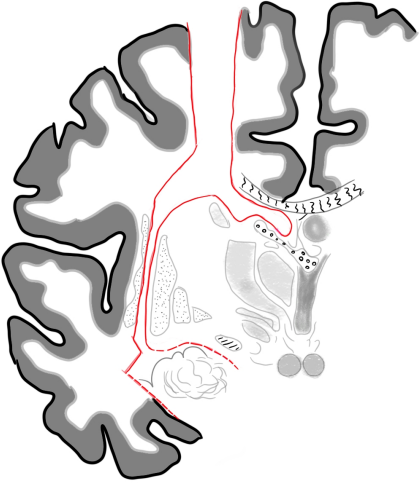
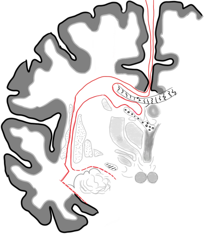
In the planned technique, the process of craniotomy will be the same, with modifications starting from the identification of the corpus callosum. Instead of the resection process proceeding laterally to the thalamus and then ascending for callosotomy, the process begins with an incision made directly on the corpus callosum, and only then are the structures of the basal ganglia disconnected. This choice is made because, through this sequence, the pericallosal artery is less at risk of being injured, as previously mentioned.
The procedure was performed on 14 formalin-fixed brains for illustration of the technique, and their results are present at the end of the article. The used brains were collected through the Death Verification Service (SVO), which performs the procedure in the autopsy room of the Faculty of Medical and Health Sciences of the Pontifical Catholic University of São Paulo – Sorocaba Campus, located on the ground floor of the building. All the brains undergo cranial structure removal in a procedure performed by the responsible service technicians. It is important to note that there is absolutely no transportation and/or removal of this material from the premises of the Faculty of Medical and Health Sciences of the Pontifical Catholic University of São Paulo – Sorocaba Campus.
It should also be noted that, as needed, the brains were collected after filling out a free and informed consent form, ensuring the awareness of close family members and/or responsible parties who claim full knowledge of the destination of the material for research; guaranteeing social, physical, and other integrity for the family and for the deceased. No brain was obtained from prisoners and/or prisoners of conscience. All personal information present will be duly protected and kept in total confidentiality in compliance with the General Data Protection Law (LGPD) – Brazilian Law No. 13.709, of August 14, 2018.
Of the 14 brains used in this project, one was subjected to the original procedure and another to the procedure suggested by this work without being subjected to a coronal sectioning beforehand. The other 12 were subjected to a coronal cut, to enable observation, measurement, and assessment of the differences obtained between the two surgical cuts, with this sample of 12 pieces being therefore those submitted to statistical analysis and discussed in the “Results” and “Discussion” sections.
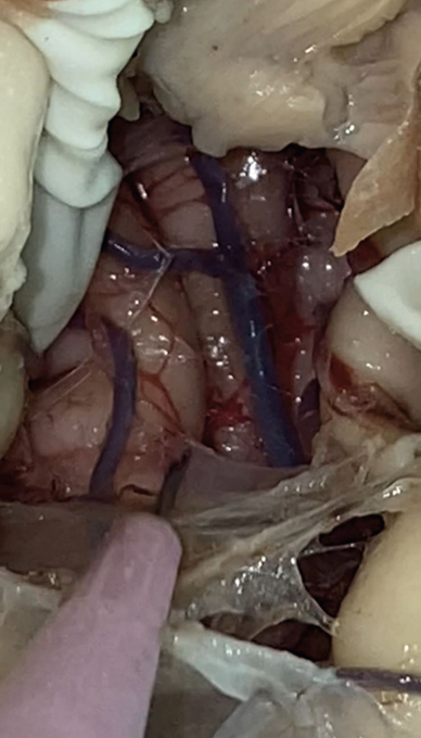
By performing the procedures on these brains that were not subjected to coronal cuts (whole brains), it was possible to obtain a clear visualization of the anatomical structures involved during the surgical procedure, as well as an important qualitative assessment of the operation itself. Through these experiments, it was really perceived how vulnerable the pericallosal artery is to being injured/perforated by the scalpel during the procedure (during the partial resection stage of the corpus callosum).
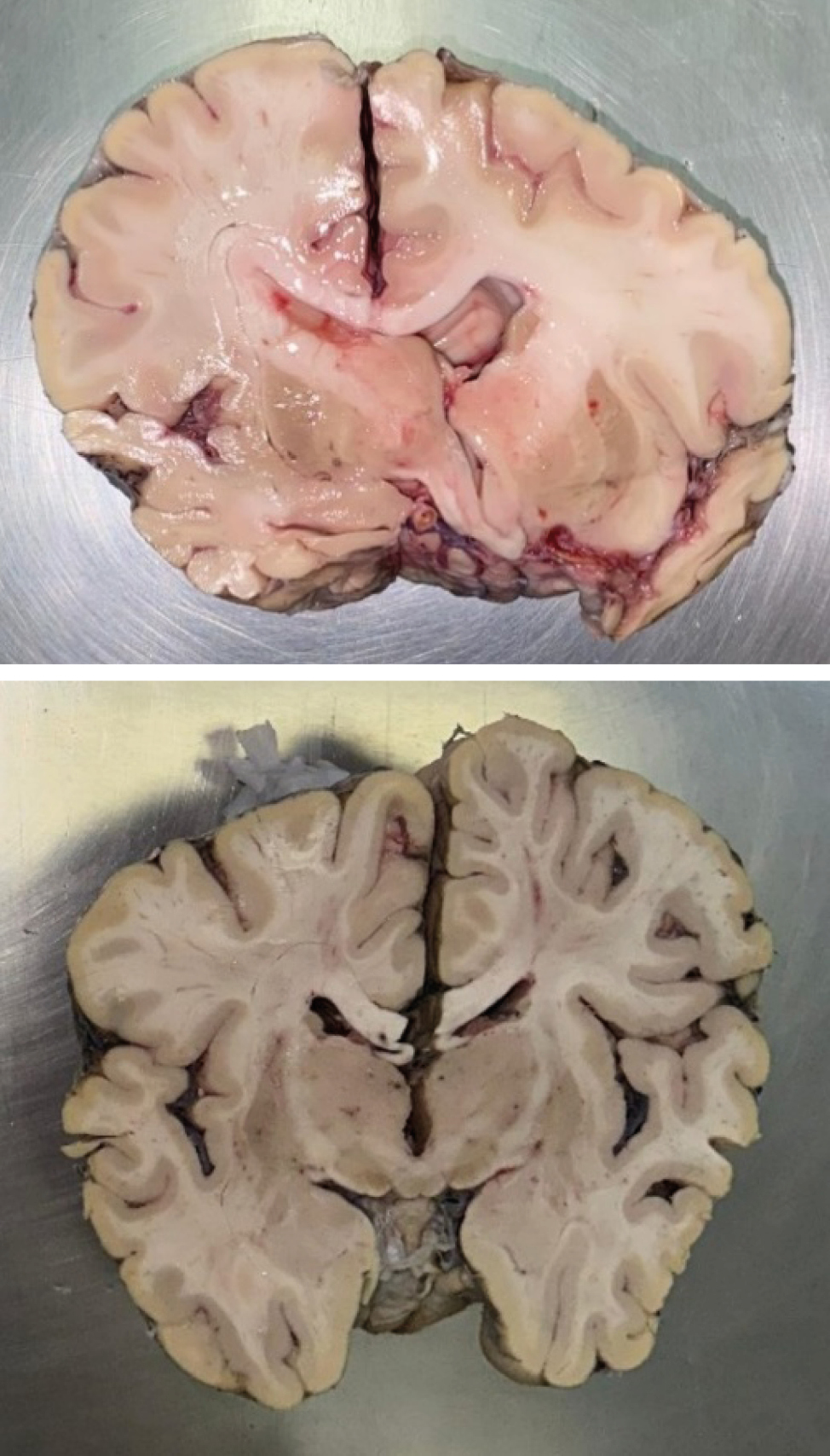
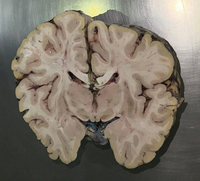
Even so, the majority of experiments were performed on brains subjected to a coronal cut due to the possibility to collect much more measurement data from the procedures, a greater and better anatomical assessment over the traditional technique and the proposal, as well as a better understanding, through the photos, of what this work aims to achieve.
For the evaluation of both methods, 5 items were proposed to be assessed after the completion of all the procedures on all the selected pieces (12):
- –Thickness of the corpus callosum
- –Distance from the initial incision to the pericallosal artery
- –In a straight line
- –Continuously
- –Distance from the initial incision to the insula
- –In a straight line
- –Continuously
- –Total distance traveled by the procedure
- –Presence or absence of lesion in the pericallosal artery
In the following figures, authored by us but based on the illustration by Peter A. Winkler [54], on the left (Fig. 1), it is possible to identify the locations covered by the hemispheric disconnection proposed by Delalande in its traditional form. Immediately to the right (Fig. 2) is the method proposed by our work to modify the technique.
2.1. Exclusion criteria
For the use of formalin-fixed brains, some exclusion criteria were defined and adopted, including brains with the following characteristics:
- –Hemorrhages
- –Cause of death from serious infectious diseases
- –Tumors
- –Malformations
- –Obtained from cadavers who died under the age of 18 or over the age of 60.
2.2. Statistical evaluation
Finally, for the purpose of better statistical understanding and the impact of the work, a comparative analysis between two groups was conducted. One group consisted of 6 formalin-fixed brains using the traditional technique and another group, also composed of 6 formalin-fixed brains, using the alteration proposed in this work, making a total of 12 brains for the analytical power of this study. The results will be presented and evaluated using the Student’s t-test method, which will allow for a clearer and more reliable assessment of the effect and consequence of the work on the addressed procedure.
Regarding the literature review, articles were selected from a bibliographical search of databases such as PUBMED, using terms such as “hemispherectomy,” “epilepsy,” and “convulsion” to construct this systematic review.
This work was approved by the research ethics committee of both IAMSPE-SP and FCMS-PUC-SP.
3. Results
In total, fourteen procedures were performed; two of them were conducted on whole brains, and twelve were performed on coronal sections of brains (which allowed clear visualization of the structures involved – also enabling more anatomically and surgically accurate procedures). Simultaneously, conducting procedures on whole brains allowed for significant learning regarding the procedures themselves (through both the traditional Delalande technique and the variation proposed in this work). In both scenarios – with whole brains and those with coronal sections – the procedures were conducted using both the Delalande method and the one proposed by this work.
3.1. Whole brains
Above, Fig. 3 shows the pericallosal artery via Delalande’s original procedure on a brain without a coronal cut, which was not used in the statistical analysis.
3.2. Brains in coronal section
Above it’s possible to see, in Figs 4 and 5, some brains used for data collection that we will present. Below, it is possible to analyze the table with the results of both methods comparing the 5 items addressed (Table 1), with measurements in millimeters.
| Procedure | Corpus Callosum thickness | Straight incision – pericallosal distance | Continuous incision-pericallosal distance | Straight incision-insula distance | Continuous incision-insula distance | Total distance | |||||||||||||
|---|---|---|---|---|---|---|---|---|---|---|---|---|---|---|---|---|---|---|---|
| N | Present work | 6 | 6 | 6 | 6 | 6 | 6 | ||||||||||||
| Delalande | 6 | 6 | 6 | 6 | 6 | 6 | |||||||||||||
| Mean | Present work | 5,83 | 7,67 | 7,67 | 29,8 | 41,2 | 59,0 | ||||||||||||
| Delalande | 5,83 | 46,2 | 66,8 | 43,3 | 51,3 | 74,3 | |||||||||||||
| Median | Present work | 5,50 | 8,00 | 8,00 | 31,5 | 42,00 | 63,0 | ||||||||||||
| Delalande | 5,50 | 45,00 | 66,00 | 41,5 | 53,00 | 68,5 | |||||||||||||
| Standard deviation | Present work | 1,47 | 1,51 | 1,51 | 3,82 | 3,54 | 9,23 | ||||||||||||
| Delalande | 1,17 | 6,05 | 9,60 | 7,28 | 7,55 | 14,5 | |||||||||||||
| Minimum | Present work | 4 | 5 | 5 | 25 | 36 | 45 | ||||||||||||
| Delalande | 5 | 39 | 54 | 34 | 40 | 62 | |||||||||||||
| Maximum | Present work | 8 | 9 | 9 | 33 | 45 | 68 | ||||||||||||
| Delalande | 8 | 53 | 80 | 53 | 59 | 98 | |||||||||||||
| Caption: Corpus callosum – thickness of the corpus callosum; Pericallosal artery (straight) – straight distance measured between the incision and the pericallosal artery; Pericallosal artery (continuous) – distance following the procedure’s path between the incision and the pericallosal artery; Insula (straight) – straight distance measured between the incision and the insula; Insula (continuous) – distance following the procedure’s path between the incision and the insula; Total distance – total length of the procedure’s path. All measurements are in millimeters (mm). | |||||||||||||||||||
As the number of lesions to the pericallosal artery was equal in both procedures (Table 2), it is impossible to associate lesser or greater safety with either procedure, which precludes evaluating this data to conclude our initial hypothesis. Therefore, we delved into the analysis of the distance traveled, in millimeters, in each procedure, using the statistical methodology of the Student’s t-test, promoting reliability and applicability to the collected results (Table 3).
| Procedure | No lesion | With lesion | Total |
|---|---|---|---|
| Present work | 5 | 1 | 6 |
| Delalande | 5 | 1 | 6 |
| Total | 10 | 2 | 12 |
| Group | N | Mean | Median | Standard deviation | Standard error |
|---|---|---|---|---|---|
| Distance in present work | 6 | 59,0 | 63,0 | 9,23 | 3,77 |
| Distance in Delalande | 6 | 74,3 | 68,5 | 14,5 | 5,91 |
Below is the table showing the application of the Student’s t-test, which reinforces and reveals the good association of the values with the conclusion, as the p-value is below 0.05 (Table 4).
| Estatística | gl | p | |||||||||||||||||
|---|---|---|---|---|---|---|---|---|---|---|---|---|---|---|---|---|---|---|---|
| Distance traveled on Student’s T test | -2,19 | 10,0 | 0,027 | ||||||||||||||||
| Score: Ha µ proposed < µ Delalande. | |||||||||||||||||||
Finally, we have the normality test revealing that the analysis meets the assumption of normality (since the p-value is above 0.05), a fact that, together with the data from the study, which are quantitative results, suggests a better indication for the use of the Student’s t-test (Table 5).
| W | p | ||||||||||||||||||
|---|---|---|---|---|---|---|---|---|---|---|---|---|---|---|---|---|---|---|---|
| Distance traveled | 0,931 | 0,387 | |||||||||||||||||
| Note: A small p-value suggests a violation of the normality assumption. | |||||||||||||||||||
4. Discussion
As observed, there was not a decreased likelihood of injury to the pericallosal artery when six procedures of each version (traditional/Delande and the one proposed by this work) were performed, as this study proposed in one of its initial objectives. However, the analysis revealed a shorter surgical cut, in terms of extension, than the original Delalande procedure, which may indicate a possible shorter surgical time and better clinical-surgical outcomes, given that a longer surgical procedure time is linked to higher risks to the patient and a worse prognosis, as previously seen in this work.
It is important to emphasize that there was no separation, choice, or determination regarding the sex, gender, and/or age of the individuals whose brains were used in the project. Only the application of the exclusion criteria was used, which makes it impossible to evaluate possible differences that may arise in the results and analyses due to these variations.
The fact that it was performed on post-mortem brains limits the associations and correlations that can be made about the results obtained and a real scenario of neurosurgery. It is not possible to predict whether there are better clinical repercussions in the procedure presented by this work, even though it is of notable probability, since there is less brain tissue affected by the proposed method. Another point to note is that as the work was done on brains subjected to coronal cuts, an element that will not be present in a real procedure, it is not possible to discuss the differences in the surgical characteristics of both procedures.
The results obtained and analyzed regarding the procedure proposed by this project demonstrate the same disconnections as the original Delalande procedure, indicating and ensuring similarity between the procedures, maintaining comparable characteristics between the two interventions.
5. Conclusion
In conclusion, the procedure proposed with this work can indicate a new approach over the hemispherectomy proposed by Delalande that, even though it does not show, in the number of procedures performed, a decreased likelihood of injury to the pericallosal artery (as initially theorized), it does in fact provide increased benefits to the patient as it has a smaller incision extent and, therefore, has a decreased procedure time and consequently a superior prognosis and safety, both to the physician and the patient.
On another note, further studies are needed with a larger number of brains, greater similarity to a real neurosurgical scenario, and a greater number of characteristics to be analyzed and compared, such as variations between age groups, ethnicities, and sex, and the possible repercussions these elements may have on the results. WIth that, it will be possible to fully test this new approach’s potential, and its efficacy on future surgeries to come, so that patients with refractory epilepsy can have an even safer surgical treatment option, with better recovery and prognosis.
Acknoledgments
Work funded by the PIBIC-CNPq scholarship, without specific areas of investment and have no declarations of interest.
References
[1] Fisher RS, Acevedo C, Arzimanoglou A, Bogacz A, Cross JH, Elger CE, et al. ILAE Official Report: A practical clinical definition of epilepsy. Epilepsia. 2014 Apr;55(4):475–82. https://doi.org/10.1111/epi.12550.
[2] Guerreiro CAM, Guerreiro MM, Cendes F, Lopes-Cendes I. Epilepsia. São Paulo: Lemos Editorial; 2002.
[3] Aguiar PH, Leal A, Ramina R. (EDS.). Tratado de neurologia clínica e cirúrgica. 1ª edição ed. [s.l.] Atena Editora, 2022.
[4] Fiest KM, Sauro KM, Wiebe S, Patten SB, Kwon CS, Dykeman J, et al. Prevalence and incidence of epilepsy. Neurology. 2017 Jan 17;88(3):296–303.
[5] Murray CJL, Vos T, Lozano R, Naghavi M, Flaxman AD, Michaud C, et al. Disability-adjusted life years (DALYs) for 291 diseases and injuries in 21 regions, 1990–2010: a systematic analysis for the Global Burden of Disease Study 2010. The Lancet [Internet]. 2012 Dec https://doi.org/10.1016/s0140-6736(12)61689-4
[6] Tedrus GM de AS, Fonseca LC, Carvalho RM. Epilepsy and quality of life: socio-demographic and clinical aspects, and psychiatric co-morbidity. Arquivos de Neuro-Psiquiatria. 2013 Jun;71(6):385–91. https://doi.org/10.1590/0004-282x20130044
[7] Bell GS. Predictive value of death certification in the case ascertainment of epilepsy. Journal of neurology, neurosurgery and psychiatry. 2004 Dec 1;75(12):1756–8. https://doi.org/10.1136/jnnp.2003.029918.
[8] Thijs RD, Surges R, O’Brien TJ, Sander JW. Epilepsy in adults. The Lancet [Internet]. 2019 Feb;393(10172):689–701. https://doi.org/10.1016/s0140-6736(18)32596-0
[9] Almeida AN de, Marino R, Marie SK, Aguiar PH, Teixeira MJ. Factors of morbidity in hemispherectomies: Surgical technique×pathology. Brain and Development. 2006 May;28(4):215–22. https://doi.org/10.1016/j.braindev.2005.08.005
[10] De Ribaupierre S, Delalande O. Hemispherotomy and other disconnective techniques. Neurosurgical Focus. 2008 Sep;25(3):E14. https://doi.org/10.3171/foc/2008/25/9/e14
[11] Costa JC da. Tratamento Cirúrgico das Epilepsias na Criança. Journal of Epilepsy and Clinical Neurophysiology. 2006 Mar;12(1 suppl 1):5–5. https://doi.org/10.1590/s1676-26492006000200001
[12] Daniel, R.T., Villemure J. Hemispherotomy Techniques. Journal of Neurosurgery, v. 98, n. 2, 1 fev. 2003.
[13] Fonseca LF, Melo RP de, Cukiert A, Burattini JA, Mariani PP, Brandão R, et al. Hemisferectomia funcional precoce na hemimegalencefalia associada à epilepsia refratária. Arquivos de Neuro-Psiquiatria. 2004 Dec;62(4):1063–7. https://doi.org/10.1590/s0004-282x2004000600024
[14] Lew SM, Koop JI, Mueller WM, Matthews AE, Mallonee JC. Fifty Consecutive Hemispherectomies. Neurosurgery. 2013 Oct 30;74(2):182–95. https://doi.org/10.1227/neu.0000000000000241
[15] Moosa ANV, Gupta A, Jehi L, Marashly A, Cosmo G, Lachhwani D, et al. Longitudinal seizure outcome and prognostic predictors after hemispherectomy in 170 children. Neurology. 2012 Dec 5;80(3):253–60. https://doi.org/10.1212/wnl.0b013e31827dead9
[16] Obeid M, Wyllie E, Rahi AC, Mikati MA. Approach to pediatric epilepsy surgery: State of the art, Part I: General principles and presurgical workup. European Journal of Paediatric Neurology. 2009 Mar 1;13(2):102–14. https://doi.org/10.1016/j.ejpn.2008.05.007
[17] Cats EA, Kuan Hua Kho, Onno Van Nieuwenhuizen, Veelen V, Gosselaar PH, Van PC. Seizure freedom after functional hemispherectomy and a possible role for the insular cortex: the Dutch experience. Journal of neurosurgery. 2007 Oct 1;107(4):275–80. https://doi.org/10.3171/ped-07/10/275
[18] Lew, S. M. Hemispherectomy in the treatment of seizures: a review. Translational pediatrics, v. 3, n. 3, p. 208–17, 2014.
[19] Lew, S. M. et al. Posthemispherectomy hydrocephalus: Results of a comprehensive, multiinstitutional review. Epilepsia. 2012 Oct 25; v. 54, n. 2, p. 383–389.
[20] Chandra, S. P. et al. Endoscopic-Assisted (Through a Mini Craniotomy) Corpus Callosotomy Combined With Anterior, Hippocampal, and Posterior Commissurotomy in Lennox-Gastaut Syndrome: A Pilot Study to Establish Its Safety and Efficacy. Neurosurgery. 2016 May 1; v. 78, n. 5, p. 743–751.
[21] Chandra S, Tripathi M. Endoscopic epilepsy surgery: Emergence of a new procedure. Neurology India. 2015;63(4):571. https://doi.org/10.4103/0028-3886.162056
[22] Chandra S, Tripathi M. Epilepsy surgery: Recommendations for India. Annals of Indian Academy of Neurology. 2010;13(2):87. https://doi.org/10.4103/0972-2327.64625
[23] Chandra PS, Kurwale N, Garg A, Dwivedi R, Malviya SV, Tripathi M. Endoscopy-Assisted Interhemispheric Transcallosal Hemispherotomy. Neurosurgery. 2015 Feb 12;76(4):485–95. https://doi.org/10.1227/neu.0000000000000675
[24] Tripathi M, Chandra P, Padma V, Shailesh G, Chandreshekar B, Sarkar C. Hemispherotomy for intractable epilepsy. Neurology India. 2008;56(2):127. https://doi.org/10.4103/0028-3886.41988
[25] Jonas R, Nguyen S, Hu B, Asarnow RF, Lopresti C, Curtiss S, et al. Cerebral hemispherectomy: Hospital course, seizure, developmental, language, and motor outcomes. Neurology. 2004 May 25;62(10):1712–21. https://doi.org/10.1212/01.wnl.0000127109.14569.c3
[26] Kossoff EH, Vining EPG, Pillas DJ, Pyzik PL, Avellino AM, Carson BS, et al. Hemispherectomy for intractable unihemispheric epilepsy Etiology vs outcome. Neurology. 2003 Oct 13;61(7):887–90. https://doi.org/10.1212/01.wnl.0000090107.04681.5b
[27] Limbrick DD, Narayan P, Powers AK, Ojemann JG, Tae Sung Park, Bertrand ME, et al. Hemispherotomy: efficacy and analysis of seizure recurrence. Journal of neurosurgery. 2009 Oct 1;4(4):323–32. https://doi.org/10.3171/2009.5.peds0942
[28] Michael Anthony Ciliberto, Limbrick D, Powers A, Titus JB, Munro R, Smyth MD. Palliative hemispherotomy in children with bilateral seizure onset. Journal of neurosurgery. 2012 Apr 1;9(4):381–8. https://doi.org/10.3171/2011.12.peds11334
[29] Greiner HM, Park YD, Holland K, Horn PS, Byars AW, Mangano FT, et al. Scalp EEG does not predict hemispherectomy outcome. Seizure. 2011 Dec;20(10):758–63. https://doi.org/10.1016/j.seizure.2011.07.006
[30] Salamon N, Andres M, Chute DJ, Nguyen ST, Chang JW, Huynh MN, et al. Contralateral hemimicrencephaly and clinical–pathological correlations in children with hemimegalencephaly. Brain. 2005 Nov 16;129(2):352–65. https://doi.org/10.1093/brain/awh681
[31] Vera Cristina Terra-Bustamante, Luciana Midori Inuzuka, Maria R, Escorsi-Rosset S, Lauro Wichert-Ana, Alexandre V, et al. Outcome of hemispheric surgeries for refractory epilepsy in pediatric patients. Childs Nervous System. 2006 Nov 7;23(3):321–6. https://doi.org/10.1007/s00381-006-0212-6
[32] Wyllie E, Deepak Lachhwani, Gupta A, Anil Chirla, Cosmo G, Worley S, et al. Successful surgery for epilepsy due to early brain lesions despite generalized EEG findings. 2007 Jul 24;69(4):389–97. https://doi.org/10.1212/01.wnl.0000266386.55715.3f
[33] Lupashko S, Malik S, Donahue D, Hernandez A, M. Scott Perry. Palliative functional hemispherectomy for treatment of refractory status epilepticus associated with Alpers’ disease. Childs Nervous System. 2011 Jun 1;27(8):1321–3. https://doi.org/10.1007/s00381-011-1495-9
[34] Brotis, A. G. Hemispherectomy: Indications, Surgical Techniques, Complications, and Outcome. Journal of Neurology & Neurophysiology. 2015; v. 06, n. 04.
[35] Fountas KN, Smith JR, Robinson JS, Tamburrini G, Pietrini D, Di Rocco C. Anatomical hemispherectomy. Child’s Nervous System. 2006 Jun 30;22(8):982–91. https://doi.org/10.1007/s00381-006-0135-2
[36] Krynauw RA. Infantile hemiplegia treated by removing one cerebral hemisphere. Journal of Neurology, Neurosurgery & Psychiatry. 1950 Nov 1;13(4):243–67. https://doi.org/10.1136/jnnp.13.4.243
[37] Dandy WE. Removal of right cerebral hemisphere for certain tumors with hemiplegia. Journal of the American Medical Association. 1928 Mar 17;90(11):823. https://doi.org/10.1001/jama.1928.02690380007003
[38] Nogueira A, Marino R, Aguiar PH, Teixeira MJ. Hemispherectomy: a schematic review of the current techniques. Neurosurgical Review. 2006 Feb 7;29(2):97–102. https://doi.org/10.1007/s10143-005-0011-7
[39] Villemure, JG. Anatomical to functional hemispherectomy from Krynauw to Rasmussen. Epilepsy research Supplement. 1992 Jan 1; v. 5, p. 209–215.
[40] Villemure J, Rasmussen Th. Functional Hemispherectomy in Children. Neuropediatrics. 1993 Feb;24(01):53–5. https://doi.org/10.1055/s-2008-1071514
[41] Rasmussen T. Hemispherectomy for Seizures Revisited. Canadian Journal of Neurological Sciences / Journal Canadien des Sciences Neurologiques. 1983 May;10(2):71–8. https://doi.org/10.1017/s0317167100044668
[42] Binder DK, Schramm J. Transsylvian functional hemispherectomy. Child’s Nervous System. 2006 Jun 9;22(8):960–6. https://doi.org/10.1007/s00381-006-0131-6
[43] Villemure JG, Daniel RT. Peri-insular hemispherotomy in paediatric epilepsy. Child’s Nervous System. 2006 Jun 29;22(8):967–81. https://doi.org/10.1007/s00381-006-0134-3
[44] Martínez, F. et al. Bases anatómicas de la hemisferotomía periinsular. Rev. méd. Urug. 2004; p. 208–214.
[45] Villemure JG, Mascott CR. Peri-insular Hemispherotomy. Neurosurgery. 1995 Nov 1;37(5):975–80. https://doi.org/10.1227/00006123-199511000-00018
[46] De Ribaupierre, S. et al. Contralateral frontal and cerebellar haemorrhages after peri-insular hemispherotomy. Acta Neurochirurgica. 2004 Jul 1; v. 146, n. 7, p. 743–744.