https://doi.org/10.36253/ijae-15973
Effects of streptozocin-induced diabetes on the histomorphometry of the small and large intestines of male Sprague Dawley rats
1 Department of Anatomy, School of Medicine, Sefako Makgatho Health Sciences University, Ga-Rankuwa, Pretoria, South Africa
2 Department of Physiology and Environmental Health, University of Limpopo, Sovenga, 0727, South Africa
*Corresponding author. Email: patiencenziyane@gmail.com
Abstract. Gastrointestinal tract (GIT) disorders affecting intestinal tissues are common among diabetes mellitus (DM) patients. GIT side effects of antidiabetic medication are a reason for an increase in use of antidiabetic herbal medicine, prompting research that uses 50mg/kg of streptozocin (STZ) to induce DM in animal experiments prior to testing safety and efficacy of the herbs over 21 days in most experiments. As DM is a progressive disease, it is not clear whether 21 days of DM is enough to induce intestinal tissue changes. We investigated effects of 50mg/kg STZ induced DM in intestines of male Sprague Dawley rats observed over 21 days. Intestinal tissues of 16 (control, n=8, STZ diabetic=8) rats stained with Masson’s Trichrome, hematoxylin and eosin were used. Villi height, width, number of goblet cells (GC), mucosa, submucosal collagen fibre (SCF) and muscularis externa (ME) were measured using ImageJ software in photomicrographs taken from slides with a microscope camera. Means were analysed using SPSS software. In DM animals, there was a significant increase in width of ileal villi (p < 0.001), reduction in number of GC in jejunum (p = 0.033) and ileum (p <0.001). SCF thickness increased in duodenum (p < 0.001), jejunum (p = 0.003), ileum (p = 0.005), and colon (p = 0.004). ME only increased in ileum (p = 0.004). 50mg/kg STZ DM induces significant changes across the intestines over 21 days, suggesting that this duration is effective for experimental modelling for future DM studies on the intestines.
Keywords: gastrointestinal tract, diabetes mellitus, Sprague Dawley rats, streptozocin (STZ), antidiabetic herbal medication.
Contents
Specimen processing and staining
Histomorphometric measurements
Villi height, width, and goblet cells
Thickness of the mucosa, muscularis externa and submucosal collagen fibre bundles
Induction of diabetes, weights and glucose level changes
Morphological changes in the villi of the duodenum
Morphological changes in the Jejunum
Morphological changes in the ileum
Morphological changes in the Colon
Introduction
Diabetes Mellitus (DM) is a group of metabolic disorders characterized by increased blood glucose levels commonly known as hyperglycemia (Cho et al., 2018; Lovic et al., 2020). It develops as a result of a deficiency in insulin secretion by pancreatic beta cells or insulin action (Cho et al., 2018). It is predicted that by 2040, 693 million people aged 18-99 years old will have diabetes, a number accounting for 9.9% of the world’s population (Lovic et al., 2020). The gastrointestinal tract (GIT) disorders are common among DM patients and have recently been identified as one of the most common DM complications (Zhao et al., 2017). Animal and human studies have shown that DM has negative effects on intestinal health, including impaired healing and cellular damage to the intestinal mucosa (Gotfried et al., 2017). The above could be partly due to the fact that hyperglycemia causes an overproduction and accumulation of advanced glycation end products (AGEs) in tissues which affect the structure and function of proteins and other intestinal wall components like collagen fibres (Zhao et al., 2017).
Metformin is a commonly used antidiabetic medication, but it has been shown to slow gastric emptying, reduce pyruvate dehydrogenase activity, and cause lactic acidosis in typer 2 diabetes mellitus patients (Borg et al., 2020). It can also cause abdominal pain, vomiting, diarrhoea, agitation, confusion, tachypnoea, and hypotension (Kalsi et al., 2017). Due to the above side effects of conventional medication, several diabetic patients are increasingly using antidiabetic herbal medication which is perceived to be safe (Atinga et al., 2018; Mekuria et al., 2018). However, the safety and efficacy of herbal medication lacks scientific validation in most cases (Mukherjee et al., 2022). Consequently, there is an increase in studies investigating the safety and efficacy of the antidiabetic herbal medication. These studies use streptozocin (STZ) to induce diabetes in experimental animals followed by treatment intervention (Eleazu et al., 2013; Goyal yet al., 2017). The dosage and duration of STZ used to induce diabetes is inconsistent across different experiments, with those that are interested in physiological changes employing shorter duration of between 7 to 21 days, while the dosage of STZ used to induce diabetes ranges from 20mg/kg to as high as 200mg/kg (Kooti et al., 2016). The short duration of diabetes may induce physiological changes but not necessarily tissue changes as diabetes is a progressive disease (Zhao et al., 2017). A dose of 50mg/kg of STZ with animals kept for 21 days either with or without treatment intervention is one of the most common experimental designs.
There is currently a lack of information on the pathologic profile of STZ-induced diabetes in the small intestines and the colon. While some studies have examined specific regions of the intestines after 21 days period of STZ induced diabetes, a more comprehensive investigation of the complete intestinal pathologic profile is lacking to fully comprehend the effects of STZ induced diabetes in the intestinal tissues. The current study has been designed against this backdrop to investigate the effects of 50mg/kg STZ induced diabetes on the histomorphometry of the small and large intestines of the male Sprague Dawley rats observed over 21 days after induction of diabetes. Such a study is important for accurate interpretation of the results in studies that use this experimental design in drug testing.
Materials and methods
Animals
Sixteen three months old male Sprague Dawley rats weighing 220–300 g that were purchased from Northwest University, Potchefstroom were used in the current study. Ethics approval (SMUAEC 03/2021) was obtained from the animal ethics committee at Sefako Makgatho Health Sciences University prior to conducting the study. The animals were kept in the Department of Physiology’s Laboratory Animal Center at Sefako Makgatho Health Sciences University. Each animal was kept in a separate cage in a temperature-controlled environment with a 12-hour light/12-hour dark cycle with free access to rat chow and water.
Diabetes induction
After a week of acclimation, animals were fasted for 18 hours before receiving a once-off intraperitoneal injection of 50mg/kg bodyweight of pancreatic-cell toxin STZ, 1ml/kg dissolved in 0.1M sodium citrate buffer (pH = 4.5) to induce type 1 DM. To avoid hypoglycemia, the STZ injected animals received a 5% glucose solution in their drinking water overnight. After 72 hours, blood glucose levels were measured in both normal control and STZ injected animals using blood samples obtained from the tail vein and analyzed with the accu-chek active glucometer (Roche Diabetes Care South Africa) to confirm diabetic status. For STZ injected animals, fasting blood glucose values exceeding 10.0 mM were considered indicative of hyperglycemia. All animals with the above blood glucose levels were included in the study and those animals that had glucose levels below 10mM were excluded from the study. The measurement of blood glucose levels in the normal control group was done for control purposes. After confirmation of diabetic status, both the normal control and STZ diabetic animals were put on unlimited food and water and observed for 21 days with glucose levels measured in days 14 and 21 of the experimental period and body weights measured on a daily basis.
Experimental design
Animals were randomly assigned to one of two experimental groups at random as follows: Group 1 (n=8) were normal rats that were not exposed to any treatment and used as a control, while Group 2 (n=8) were 50mg/kg STZ-induced diabetic rats. After induction of diabetes, the animals were kept for 21 days with no intervention. At the end of the 21 days (day 22) all animals were euthanised with an intraperitoneal injection of a mixture (1.4 ml/kg.bw) of Anaket V (Ketamine) 40-80mg/kg and Rompun 2% (Xylazine) 5-10mg/kg. The intestinal tissues were collected and preserved in 10% buffered formalin before processing and staining.
Specimen processing and staining
Four 1cm segments were harvested from the duodenum, jejunum, ileum and the colon. A segment of the duodenum was taken in its second part, 1cm from the pylorus. Jejunum was sampled 5cm below the ligament of Treitz, the ileum sampled 5cm proximally to the ileocecal valve and the colon sampled 5cm distal to the ileocecal valve as per the sampling method adopted by Zhao et al., (2017). The blocks of the intestinal tissues were processed, embedded, and serially sectioned at 5μm thickness with a rotary microtome, and one in two sections were mounted on glass slides for Masson’s Trichrome (MT) and Hematoxylin and Eosin (H&E) staining (Bancroft and Gamble, 2008). Hematoxylin and Eosin stains were used for tissue architecture to evaluate thickness of the layers and quantifying goblet cells, while the MT stain was used for connective tissue assessment of submucosal collagen fiber bundles. Photomicrographs of stained and mounted sections were taken with Leica software (Version 3.0), a Leica ICC50 HD camera, and a Leica microscope DM500 at 4X, 10X, and 40X.
Histomorphometric measurements
Villi height, width, and goblet cells
Twenty serial sections of the duodenum, jejunum and ileum were used in the current study to measure the height and width of the small intestines for each animal at 4X (duodenum) and 10X (jejunum and ileum) magnification. The length of the villi was measured from the tip of the villi to the base at the junction with the intestinal crypts, whereas the width was measured halfway between the tip and base of the villi. These measurements were randomly taken from the longitudinal sections of 5 well-oriented villi per tissue section in 20 sections adding up to 100 villi measured for each segment of the small intestine per animal (Lerkdumnernkit et al., 2022). Goblet cells characterised by visible wine glass shape cells with clear cytoplasm and round or oval shaped nucleus at the base were also counted in the above villi. The measurements and counts were done using the line and multi-point feature tools of the ImageJ software (Schneider et al., 2012) and the means were recorded for statistical analyses.
Thickness of the mucosa, muscularis externa and submucosal collagen fibre bundles
Twenty serial sections of the colon were used to measure the mean thickness of the mucosa, muscularis externa and in H&E-stained sections at 4X, and collagen fibres in MT-stained sections at 10X magnification using the line tool of the ImageJ software. Measurements of the mucosa were taken at two random points that were three mucosal glands apart in each of the twenty sections per animal from the tips of the mucosa to the muscularis mucosae, making it a total of forty points measured per animal in the colon. The muscularis externa of the colon was measured at two points in line with the mucosa measurement described above. For the small intestines, the muscularis externa was measured at two points that were two villi apart, while the submucosal collagen fibre thickness was measured at 40X (MT) magnification from the point where the muscularis mucosae ends to where the muscularis externa begins at two random points two intestinal crypts apart.
Data analysis
A student t-test was used to compare the means of villi parameters, thickness of intestinal layers and counts of goblet cells for normal control and STZ diabetic group in a Statistical Package for the Social Sciences (SPSS) software (Version 28). Data was normally distributed in Shapiro Wilk test. All variables were expressed as mean ± standard deviation and a p-value of ≤ 0.05 was considered significant.
Results
Induction of diabetes, weights and glucose level changes
Prior to the induction of diabetes with STZ injection, all animals had normal blood glucose levels ranging from 4.8 to 5.8 mmol/L. After 72 hours of diabetic induction, there STZ diabetic induced animals (DM) had a drastic increase of blood glucose levels to an average of 31.83 mmol/L to 33.20 mmol/L, which persisted throughout the 21 days of the experiment while the control group maintained normal blood glucose levels ranging from 4.8-5.5 mmol/L (Figure 1A).
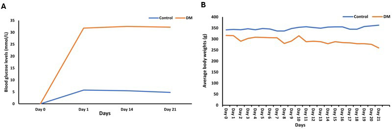
In this current study, animals with approximately the same body weights were used with an average weight of 310 g for the DM group, and an average weight of 338 g for control. The body weight of the control group significantly increased when comparing the initial and terminal body weights (28.00 ± 17.23; p = 0.003). The diabetic group had a significant weight loss by the end of 21 days when comparing the initial and terminal body weights (65.00 ± 38.60; p = 0.002; Figure 1B). The small and large intestine’s mean weight of the diabetic group (60.59 ± 9.25 g) was significantly higher than that of the control group (23.81 ± 2.51 g) (p <0.001; Table 1).
| Control | DM P-values | ||||||||||||||||||
|---|---|---|---|---|---|---|---|---|---|---|---|---|---|---|---|---|---|---|---|
| Initial BW (g) Terminal BW(g) Weight of small & large intestines (g) |
338.71 ± 7.57 364.00 ± 15.28 23.81 ± 2.51 |
310.71 ± 14.78 0.003 299.00 ± 36.82 0.002 60.59 ± 9.25 <0.001 |
|||||||||||||||||
| Glucose levels (mmol/L) | 5.26 ± 0.48 | 31.83 ± 0.59 <0.001 | |||||||||||||||||
| Data of all variables expressed as mean ± standard deviation. P <0.05. | |||||||||||||||||||
Histological findings
Morphological changes in the villi of the duodenum
The duodenum of the control group showed intact villi, and the Paneth cells (PC) had densely packed eosinophilic secretory granules (Figure 2A). In the DM group, the villi appeared to be short and mildly deformed (Figure 2B). The duodenal villi height of the DM group was slightly lower (1216.272 ± 274.59 µm) than that of the control group (1244.558 ± 04 µm; p = 0.425; Figure 2C) in the t-test. Similarly, the duodenal villi width of the DM group (182.155 ± 28.10 µm) was slightly lower than that of the control group (190.819 ± 26.11 µm; p = 0.311; Figure 2D). The number of goblet cells in the villi of the duodenum of the DM group was significantly lower (7.883 ± 2.16) than that of the control group (13.667 ± 1.85; p < 0.001; Figure 2E).
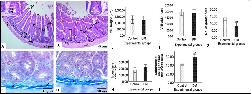
There were no statistically significant differences in the thickness of the muscularis externa (ME) of the duodenum of the DM group (214.809 ± 39.04 µm) compared to the control group (175.116 ± 36.60 µm, p = 0.090). However, the ME of the DM group was slightly thicker than that of the control group (Figure 2F).
In MT-stained sections, the control group had moderate amount of submucosal collagen fibre bundles (Figure 2G), and the DM group had a large amount of submucosal collagen fibre bundles (Figure 2H). The submucosal collagen fibre thickness of the duodenum of the DM group was significantly thicker (57.865 ± 6.69 µm) when compared to that of the control group (40.542 ± 2.06 µm; p < 0.001; Figure 2I).
Morphological changes in the Jejunum
Normal histological structures were observed in the jejunum of the control group such as finger-like villi and normal shaped intestinal crypts (Figure 3A). The jejunum of the DM group however showed distortion of the villi, cell infiltration in the lamina propria as well as disrupted crypts (Figure 3B). The jejunal villi height of the DM group (615.623 ± 184.644 µm) was slightly lower than that of the control group (655.603 ± 58.99 µm; p = 0.321; Figure 3C), while the jejunal villi width of the DM group (177.547 ± 26.14 µm) was slightly higher than that of the control group (147.594 ± 32.91 µm; p = 0.090; Figure 3D). The number of goblet cells in the villi of the jejunum of the DM group (8.033 ± 2.35 µm) was significantly lower compared to that of the control group (13.217 ± 4.03 µm; p = 0.033; Figure 3E).
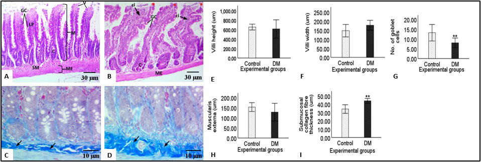
The t-test further showed that the thickness of the muscularis externa of the jejunum (ME) of the DM group (127.914 ± 41.63 µm) was not significantly different from that of the control group (152.991 ± 20.78 µm; p = 0.117; Figure 3F). However, the thickness of the ME of the DM group was slightly lower than that of the control group.
The MT-stained sections of the control group (Figure 3G) showed moderate amounts of collagen fibre bundles while the DM group had a large amount of submucosal collagen fibre bundles (Figure 3H). The submucosal collagen fibre thickness of the DM group was significantly higher (43.975 ± 2.69 µm) than that of the control group (34.101 ± 4.911 µm, p = 0.003; Figure 3I).
Morphological changes in the ileum
The ileum of the control group showed the normal histological structure with intact villi and epithelial lining (Figure 4A). In the DM group, the villi of the ileum appeared to be blunt and distorted (Figure 4B). The t-test showed that the ileal villi height of the DM group (6548.225 ± 38.44 µm) was not statistically significant when compared to that of the control group (536.481 ± 66.92 µm; p = 0.354). However, the villi height of the DM group was slightly higher than that of the control group (Figure 4C). Additionally, the ileal villi width of the DM group (183.283 ± 15.51 µm) was significantly higher compared to that of the control group (114.905 ± 8.03 µm; p < 0.001; Figure 4D). There were no significant differences in the number of goblet cells of the ileal villi of the DM (13.000 ± 1.25) and control groups (12.850 ± 1.34) in the t-test (p = 0.384). However, the number of goblet cells of the DM group was slightly higher than that of the control group (Figure 4E). The t-test also showed that the thickness of the muscularis externa of the DM group (178.227 ± 21.85 µm) was significantly higher when compared to that of the control group (142.501 ± 18.36 µm; p = 0.004; Figure 4F).
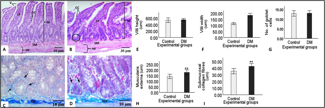
In MT-stained sections, the collagen fibre bundles showed a continuous layer around the epithelial cells in the control group, indicating an intact basement membrane (Figure 4G). In the DM group, collagen fibre bundles were spars around the epithelial cells, indicating the destruction of the basement membrane (Figure 4H). The submucosal collagen fibre thickness of the DM group (42.467 ± 4.06 µm) was significantly thicker when compared to that of the control group (35.331 ± 3.79 µm; p = 0.005; Figure 4I).
Morphological changes in the Colon
The colon of the group showed normal shaped mucosal crypts with intact epithelial lining on the luminal surface and narrow lumens (Figure 5A). In the DM group, some crypts had mildly distorted shape and the ME appeared thicker (Figure 5B). The colonic mucosal layer of the DM group (446.264 ± 81.97 µm) was significantly higher than that of the control group (318.528 ± 34.39 µm; p = 0.008; Figure 5C). The t-test also showed that the thickness of the colonic muscularis externa (ME) of the DM group (172.546 ± 79.22 µm) was not significantly different from that of the control group (152.434 ± 15.70 µm; p = 0.291; Figure 5D). However, the thickness of the ME of the DM group was higher than that of the control group (Figure 5D).
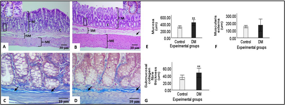
The MT-stained tissue sections of the control group showed moderate amount of submucosal collagen fibre bundles (Figure 5E) while the DM group (Figure 5F) had abundant submucosal collagen fibre bundles when compared to the control group. The colonic submucosal collagen fibre thickness of the DM group (47.87 ± 12.27 µm) was significantly higher than that of the control group (35.348 ± 6.00 µm; p = 0.004; Figure 5G).
Discussion
The diabetic group exhibited mildly distorted and blunt villi in the small intestines compared to the normal control group. This observation may be attributed to an increased enterocyte proliferation rate reported in the diabetic animals (Boudry et al.,2007). The migration of immature cells to the villus surface is a possible explanation for the observed blunting and distortion of the villi (Boudry et al., 2007).
The present study revealed a decrease in villi height in the duodenum and jejunum of the diabetic group, consistent with the findings of previous authors (Mesgari-Abbasi et al., 2019). This reduction is likely attributed to chronic inflammation associated with diabetes, as previously suggested (Garci et al., 2010). In the ileum, an increase in villi height was observed in the diabetic group compared to the normal group, in line with the previous studies (Chen et al., 2012; Hvid et al., 2016). Diabetes has been shown to significantly affect the terminal intestinal segment of the small intestine, potentially increasing villi height (Chen et al., 2012).
It must be noted that conflicting findings have been reported regarding the effect of diabetes on specific intestinal segments. One study found an increase in the villi height of the duodenum (Lerkdumnernkit et al., 2022), while another study reported an increase in the villi height of the jejunum (Hvid et al., 2016), both of which contradicts the findings of the current study. In the studies mentioned above, animals were kept for 28 days with dosages of 60mg/kg and 54mg/kg respectively, contrary to the 21 days and 50mg/kg of STZ of the current study. It is known that longer durations of diabetes are associated with more severe effects on the tissues (American Diabetes Association, 2014). Therefore, differences in the duration of the experiments can be a reason for conflicting findings.
In the present study, a decrease in villi width was observed in the duodenum of the DM group compared to the control group as previously reported (Mesgari-Abbasi et al., 2019), while in the jejunum and ileum, the villi width increased in DM group. The reduction in villi width can be attributed to alterations in the production of growth factors, particularly insulin-like growth factor-1 and 2 (IGF-1 and IGF-2), which play a crucial role in the growth and maintenance of the villi (Dube et al., 2006). The observed increase in villi width in the jejunum and ileum of diabetic rats can be attributed to villous hypertrophy (Zhao et al., 2003; Zhao et al., 2017).
The number of goblet cells significantly decreased in the duodenum and jejunum of the diabetic group compared to the control group, in agreement with previous reports (Mesgari-Abbasi et al., 2019; Lerkdumnernkit et al., 2022). The reduction in the number of goblet cells is due to increase in tumor necrosis factor alpha (TNF-α) secretion by intestinal macrophages in the diabetic intestines, which ultimately cause production of active caspase-8 which activates caspase-3, resulting in apoptosis of goblet cells (Lau et al., 2012).
In the ileum of diabetic rats, the number of goblet cells increased consistent with the view that the terminal portion of the intestinal tract is less affected by diabetes (Chen et al., 2012). Contrary to these findings, Akinola et al., (2009) reported a decrease in the number of goblet cells in the ileum of diabetic rats, probably due to longer period of 50 days and the STZ dosage of 70mg/kg in their experiment.
In the current study, an increase in the thickness of the muscularis externa was recorded in the duodenum and ileum of the diabetic group compared to the control group in line with previous reports (Chen et al., 2012; Zhao et al., 2017). The increase may be due to hyperplasia, a condition in which the number of cells in the muscularis externa increases, contributing to a rise in overall thickness (Zhao et al., 2003; Zhao et al., 2017). The current study also reported a reduction in the ME of the jejunum in the diabetic group compared to the control group. The decrease of ME noted in the jejunum could be due to the sensitivity of smooth muscle to oxidative stress which increase cell death (Kashyap and Farrugia, 2011). A significant increase in the thickness of the submucosal collagen fibre bundles of the diabetic group was recorded in the duodenum, jejunum and ileum compared to the control group in agreement with previous reports (Zhao et al., 2017; Lerkdumnernkit et al., 2022). The above could be indicative of fibrosis caused by inflammation and tissue damage in diabetic animals (Lenti and Di Sabatino, 2019).
The current study recorded a significant increase in the thickness of the mucosa, muscularis externa and submucosal collagen fibre bundles compared to the control group as previously reported (Zhao et al., 2017; D’arpino et al., 2018). The increase in thickness of the layers of the colon is due to inflammation (Kashyap and Farrugia, len2011). Chronic inflammation can lead to fibrosis, muscular hypertrophy and smooth muscle hyperplasia leading to increased muscle thickness and reduced flexibility (Gromova et al., 2021). Table 2 summarises the comparisons of the findings of the effects of STZ diabetes by different authors. It is apparent from the table that the current study is the first to report on the effects of STZ diabetes on the entire intestinal tissue and that the longer duration the diabetic animals are kept in experiments is a contributing factor in the contradiction in the results by different authors
| Author(s) | Dosage | Duration | Duodenum | Jejunum | Ileum | Colon | ||||||||||||||||||
|---|---|---|---|---|---|---|---|---|---|---|---|---|---|---|---|---|---|---|---|---|---|---|---|---|
| VH | VW | GC | ME | SCFT | VH | VW | GC | ME | SCFT | VH | VW | GC | ME | SCFT | M | ME | SCFT | |||||||
| Lerkdumnernkit et al., (2022) | 60mg/kg | 28 days | ↑ | ↓ | ↑ | |||||||||||||||||||
| Hvid et al., (2016) | 54mg/kg | 28 days | ↑ | ↑ | ↑ | |||||||||||||||||||
| Zhao et al., (2017) | - | 56 days | ↑ | ↑ | ↑ | ↑ | ↑ | ↑ | ↑ | ↑ | ↑ | |||||||||||||
| Mesgari-Abbasi et al., (2019) | 55mg/kg | 21 days | ↓ | ↑ | ↓ | |||||||||||||||||||
| Akinola et al., (2009) | 70mg/kg | 50 days | ↓ | |||||||||||||||||||||
| Chen et al., (2012) | - | - | ↑ | |||||||||||||||||||||
| Current study | 50mg/kg | 21 days | ↓ | ↓ | ↓ | ↑ | ↑** | ↓ | ↑ | ↓** | ↓ | ↑** | ↑ | ↑** | ↑ | ↑** | ↑** | ↑** | ↑ | ↑** | ||||
| Villi height (VH), villi width (VW), goblet cells (GC), muscularis externa (ME), submucosal collagen fibre thickness (SCFT), mucosa (M). ↑: increase, ↓: decrease/reduction. ** represents significant changes. | ||||||||||||||||||||||||
Conclusion
In this study, we conclude that a dosage of 50mg/kg of streptozocin (STZ) administered over a period of 21 days is sufficient to induce diabetes and produce observable tissue changes in various parts of the intestines. This timeframe is deemed appropriate for future research focused on analysing the effects of diabetes mellitus on intestinal health. The findings also provide a solid foundation for subsequent studies exploring the therapeutic effects of antidiabetic treatments on intestinal tissue.
Author contributions
Conceptualization: ML Mpholwane, PN Nziyane and NK Xhakaza. Manuscript drafting: PN Nziyane. Critical review of the manuscript: ML Mpholwane and NK Xhakaza. Final manuscript approval: Both authors.
Acknowledgements
The authors of current study declare no conflict of interests. Data used in the current study may be shared by the corresponding author upon reasonable request. The authors would like to thank Dr Du Plessis for valuable advises. Authors would also like to acknowledge Ms Thuso Valencia Mudau for her contribution in handling of the live animals during the experiment and Ms Phindi Matsebane for her technical assistance.
Funding
This work was supported by the National Research Foundation (NRF) grant number 14159.
References
AKINOLA, O.B., ZATTA, L., DOSUMU, O.O., AKINOLA, O.S., ADELAJA, A.A., DINI, L., CAXTON-MARTINS, E.A., 2009. Intestinal lesions of streptozotocin-induced diabetes and the effects of Azadirachta indica treatment. Pharmacology, 3:872-881.
AMERICAN DIABETES ASSOCIATION, 2014. Diagnosis and classification of diabetes mellitus. Diabetes care, 37(Suppl. 1): S81-S90, https://doi.org/10.2337/dc14-S081.
ATINGA, R.A., YARNEY, L., GAVU, N.M., 2018. Factors influencing long-term medication non-adherence among diabetes and hypertensive patients in Ghana: a qualitative investigation. PloS one,13(3): e0193995, https://doi.org/10.1371/journal.pone.0193995.
BANCROFT, J.D., GAMBLE, M., 2008. Theory and practice of histological techniques. Elsevier health sciences.
BORG, M.J., RAYNER, C.K., JONES, K.L., HOROWITZ, M., XIE, C., WU, T., 2020. Gastrointestinal mechanisms underlying the cardiovascular effect of metformin. Pharmaceuticals, 13(11):410, https://doi.org/10.3390/ph13110410.
BOUDRY, G., JURY, J., YANG, P.C., PERDUE, M.H., 2007. Chronic psychological stress alters epithelial cell turn-over in rat ileum. American Journal of Physiology-Gastrointestinal and Liver Physiology, 292(5): 1228-1232, https://doi.org/10.1152/ajpgi.00358.2006.
CHEN, P., ZHAO, J., GREGERSEN, H., 2012. Up-regulated expression of advanced glycation end-products and their receptor in the small intestine and colon of diabetic rats. Digestive diseases and sciences, 57:48-57, https://doi.org/10.1007/s10620-011-1951-0.
CHO, N.H., SHAW, J.E., KARURANGA, S., HUANG, Y., DA ROCHA FERNANDES, J.D., OHLROGGE, A.W., MALANDA, B.I., 2018. IDF Diabetes Atlas: Global estimates of diabetes prevalence for 2017 and projections for 2045. Diabetes research and clinical practice, 138:271-281, https://doi.org/10.1016/j.diabres.2018.02.023.
D’ARPINO, M.C., FUCHS, A.G., SÁNCHEZ, S.S., HONORÉ, S.M., 2018. Extracellular matrix remodelling and TGF‐β1/Smad signalling in diabetic colon mucosa. Cell biology international, 42(4):443-456, https://doi.org/10.1002/cbin.10916.
DUBÉ, P.E., FORSE, C.L., BAHRAMI, J., BRUBAKER, P.L., 2006. The essential role of insulin-like growth factor-1 in the intestinal tropic effects of glucagon-like peptide-2 in mice. Gastroenterology, 131(2):589-605, https://doi.org/10.1053/j.gastro.2006.05.055.
ELEAZU, C.O., ELEAZU, K.C., CHUKWUMA, S., ESSIEN, U.N., 2013. Review of the mechanism of cell death resulting from streptozotocin challenge in experimental animals, its practical use and potential risk to humans. Journal of diabetes & metabolic disorders, 12(60):1-7, https://doi.org/10.1186/2251-6581-12-60.
GARCIA, C., FEVE, B., FERRE, P., HALIMI, S., BAIZRI, H., BORDIER, L., GUIU, G., DUPUY, O., BAUDUCEAU, B., MAYAUDON, H., 2010. Diabetes and inflammation: fundamental aspects and clinical implications. Diabetes & metabolism, 36(5):327-338, https://doi.org/10.1016/j.diabet.2010.07.001.
GOTFRIED, J., PRIEST, S., SCHEY, R., 2017. Diabetes and the small intestine. Current treatment options in gastroenterology, 15:490-507, https://doi.org/10.1007/s11938-017-0155-x.
GOYAL, S.N., REDDY, N.M., PATIL, K.R., NAKHATE, K.T., OJHA, S., PATIL, C.R., AGRAWAL, Y.O., 2016. Challenges and issues with streptozotocin-induced diabetes–a clinically relevant animal model to understand the diabetes pathogenesis and evaluate therapeutics. Chemico-biological interactions, 244:49-63, https://doi.org/10.1016/j.cbi.2015.11.032.
GROMOVA, L.V., FETISSOV, S.O., GRUZDKOV, A.A., 2021.Mechanisms of glucose absorption in the small intestine in health and metabolic diseases and their role in appetite regulation. Nutrients,13(7):2474, https://doi.org/10.3390/nu13072474.
HVID, H., JENSEN, S.R, WITGEN, B.M., FLEDELIUS, C., DAMGAARD, J., Pyke, C., RASMUSSEN, T.B., 2016. Diabetic phenotype in the small intestine of Zucker diabetic fatty rats. Digestion, 94(4):199-214, https://doi.org/10.1159/000453107.
KALSI, A., SINGH, S., TANEJA, N., KUKAL, S., MANI, S., 2017. Current treatments for Type 2 diabetes, their side effects and possible complementary treatments. International Journal, 10(3):13-18.
KASHYAP, P., FARRUGIA, G., 2011. Oxidative stress: key player in gastrointestinal complications of diabetes. Neuro-gastroenterology & Motility, 23(2):111-114, https://doi.org/10.1111/j.1365-2982.2010.01659.x.
KOOTI, W., FAROKHIPOUR, M., ASADZADEH, Z., ASHTARY-LARKY, D., ASADI-SAMANI, M., 2016. The role of medicinal plants in the treatment of diabetes: a systematic review. Electronic physician, 8(1):1832-1842, https://doi.org/10.19082/1832.
LAU, K.S, CORTEZ-RETAMOZO, V., PHILIPS, S.R, PITTET, M.J., LAUFFENBURGER, D.A., HAIGIS, K.M., 2012. Multi-scale in vivo systems analysis reveals the influence of immune cells on TNF-α-induced apoptosis in the intestinal epithelium. PLOS Biology, 10(9): e1001393, https://doi.org/10.1371/journal.pbio.1001393.
LENTI, M.V, DI SABATINO, A., 2019. Intestinal fibrosis. Molecular aspects of medicine, 65:100-109, https://doi.org/10.1016/j.mam.2018.10.003.
LERKDUMNERNKIT, N., SRICHAROENVEJ, S., LANLUA, P., NIYOMCHAN, A., BAIMAI, S., CHOOKLIANG, A., PLAENGRIT, K., PIANRUMLUK, S., MANOONPOL, C., 2022. The Effects of Early Diabetes on Duodenal Alterations in the Rats. International Journal of Morphology, 40(2):389-395, doi:10.4067/S0717-95022022000200389.
LOVIC, D., PIPERIDOU, A., ZOGRAFOU, I., GRASSOS, H., PITTARAS, A., MANOLIS, A., 2020. The growing epidemic of diabetes mellitus. Current vascular Pharmacology, 18(2):104-109, https://doi.org/10.2174/1570161117666190405165911.
MEKURIA, AB., BELACHEW, S.A., TEGEGN, HG., ALI, DS., NETERE, AK., LEMLEMU, E., ERKU, D.A., 2018. Prevalence and correlates of herbal medicine use among type 2 diabetic patients in Teaching Hospital in Ethiopia: a cross-sectional study. BMC complementary and alternative medicine,18(article 85):1-8, https://doi.org/10.1186/s12906-018-2147-3.
MESGARI-ABBASI, M., GHADERI, S., KHORDADMEHR, M., NOFOUZI, K., TAYEFI-NASRABADI, H., MCINTYRE, G., 2019. Enteroprotective effect of Tsukamurella inchonensis on streptozotocin induced type 1 diabetic rats. Turkish Journal of Biochemistry, 44(5):683-91, https://doi.org/10.1515/tjb-2018-0309.
MUKHERJEE, P.K., BANERJEE, S., GUPTA, B.D., KAR, A., 2022. Evidence-based validation of herbal medicine: Translational approach. In Evidence-Based Validation of Herbal Medicine, (pp. 1-41). Elsevier, doi:10.1016/B978-0-323-85542-6.00025-1.
SCHNEIDER, C.A., W.S. RASBAND, & ELICIERI K.W., 2012. NIH Image to ImageJ: 25 years of image analysis. Nature Methods, 9(7): 671.
ZHAO, J., YANG, J., GREGERSEN, H., 2003. Biomechanical and morphometric intestinal remodelling during experimental diabetes in rats. Diabetologia, 46:1688-1697, https://doi.org/10.1007/s00125-003-1233-2.
ZHAO, M., LIAO, D., ZHAO, J., 2017. Diabetes-induced mechano-physiological. changes in the small intestine and colon. World journal of diabetes, 8(6):249-269, https://doi.org/10.4239/wjd.v8.i6.249.