https://doi.org/10.36253/ijae-15942
Accessory diaphragm: Uncommon structure in partial anomalous pulmonary venous return (PAPVR) donor
1 Department of Biomedical Sciences, Cooper Medical School of Rowan University, Camden, New Jersey, 08103, USA
2 Medical Education Research Collaborator, Winston-Salem, North Carolina 27104, USA
3 Department of Biomedical Sciences, Cooper Medical School of Rowan University, Camden, New Jersey 08103, USA
*Corresponding author. E-Mail: Melovitz-Vasan@rowan.edu
Abstract. This study builds upon an earlier case report, where the authors briefly mentioned the accessory diaphragm. This report provides a more detailed analysis of the embryological origins of the accessory diaphragm malformation, its clinical features, and, importantly, its impact on lung development. Duplication of the diaphragm, a rare congenital anomaly typically found on the right side and often associated with lobar agenesis-aplasia complex, is commonly referred to as an “accessory diaphragm”. The exact cause of diaphragm duplication is not fully understood. However, it is believed that this condition may result from a lack of coordination between the downward migration of the septum transversum and the development of the bronchial system. The accessory diaphragm is characterized as a delicate fibromuscular membrane lined with serosa that attaches to the anterior part of the diaphragm. This membrane extends in a posterosuperior direction to connect with the posterior chest wall, effectively dividing the right hemithorax into two distinct regions. Early formation of the bronchial system may lead to the developing septum transversum being cleaved as it encounters the developing lung. Literature on accessory diaphragms indicates that the associated pulmonary vascular malformations occur during this embryonic period. In all reported cases, aplasia or varying degrees of pulmonary hypoplasia occurred on the affected side. Consequently, the accessory diaphragm divides the right pleural cavity into two sections, trapping part or all of the right middle or lower lobes beneath it.
Keywords: accessory diaphragm, pseudo horseshoe lung, congenital cardiovascular and pulmonary anomaly, partial anomalous pulmonary venous return (PAPVR), embryology of diaphragm development.
Contents
Respiratory system: right lung
Introduction
A previous case report, briefly stated the presence of an ‘accessory diaphragm’ in a donor body [12]. This follow-up report includes a more detailed analysis of the embryological reasons behind this malformation, its clinical features, and, its effect on the lung’s development.
Duplication of the diaphragm, also known as ‘accessory diaphragm,’ is a rare congenital anomaly that is almost always located on the right side and frequently associated with lobar agenesis-aplasia complex [9]. In the normal development of the diaphragm, the ‘central hiatus’ allows the passage of blood vessels and bronchial structures [15]. When the central hiatus is considerably narrowed and the trapped lung is not aerated, it may appear as a solid mass. In contrast, when the trapped lung is aerated, the accessory diaphragm can be seen as a fissure-like structure in the right base, extending from the anterior aspect of the hemidiaphragm toward the posterior chest wall [15].
The exact cause of diaphragm duplication is not fully understood. However, it has been postulated that this anomaly may arise from a lack of synchronization between the caudal migration of the septum transversum and the development of the bronchial system [1,5]. In gross pathology, the accessory diaphragm is described as a delicate fibromuscular membrane lined with serosa that is connected to the anterior part of the diaphragm. It extends in a posterosuperior direction to connect with the posterior chest wall, dividing the right hemithorax into two distinct regions. It often features a central hiatus medially, allowing the passage of vessels and bronchial structures [17]. While some patients may remain asymptomatic, most present with varying degrees of respiratory difficulties. Radiographic image features include a small lung with a shift of the mediastinum and a hazy border to the mediastinum.
Very early formation of the bronchial system may result in the developing septum transversum being cleaved as it contacts the developing lung. The associated pulmonary vascular malformations reported in cases of accessory diaphragm in the literature suggest that the pathogenic event occurs during the same embryonic period [2,6]. An accessory diaphragm is a malformation more common in males, and most often right sided [1]. It can occur in isolation or in association with lung hypoplasia or other vascular malformations. Hashida and Sherman reported that the accessory diaphragm is not always lethal in the neonatal period and may be associated with chronic pulmonary infection in later life [11]. Priyadarshi et al. reported an accessory diaphragm associated with non-immune hydrops fetalis [14].
In all the reported cases, aplasia or some degree of pulmonary hypoplasia was evident on the affected side, as seen in our earlier report [12]. The accessory diaphragm thus separates the right pleural cavity into two parts, trapping part or all of the right middle or lower lobes beneath it [4], as seen in this case. If the lung trapped beneath the accessory diaphragm is aerated, as the previous study revealed [12], it will move with respiration [7]. Furthermore, a branch of the pulmonary artery supplied the ‘sequestered’ segments. Most interestingly, an accessory diaphragm separating the fused anterior and lateral basal segments could be aerated using an air pump, thus providing evidence that it was functional [12].
Materials and methods
The cadaveric specimen reported on previously and here was obtained from the willed body program intended for dissection by medical students. This case report is based on the cadaveric dissection of a 71-year-old Caucasian male donor who died of chronic obstructive pulmonary disease and hypertension [12]. Observation revealed what appeared to be surgical interventions of the heart, lung, and vasculatures, possibly due to malformations and anomalous venous drainage. Since students performed most of the dissection, some vascular and other structures were not optimally preserved. Additionally, the surgical corrective procedures disturbed the natural architecture of the gross morphology.
Observations and results
On the right side of the thoracic wall, we observed an incision extending from the parasternal region at the fourth intercostal level to the midaxillary line, apparently to address anomalies surgically. Considering the shape of the incision, the surgery was probably performed during the donor’s childhood to correct problems in the thoracic viscera [12].
Respiratory system: right lung
The right lung seemed functional, but its physical morphology was abnormal. In the hypoplastic right lung, the oblique fissure separated the upper lobe from the lower lobe, and a horizontal fissure defining the small middle lobe was also observed. The inferior lobe was vastly disorganized and severely hypoplastic and showed uncharacteristic morphology. The superior bronchopulmonary segment was markedly hypoplastic; the posterior basilar and medial basal segments (P/M basal) were not only hypoplastic and fused but also formed a slender “tail” that extended to the left pulmonary cavity behind the heart/pericardium and in front of the esophagus and aorta. However, the fused, tail-like right segments did not fuse with the similar segments of the left lung, as seen in the “scimitar” lung [8]. Closer examination of all the thoracic viscera, vasculatures, and surgical interventions revealed a hypogenetic right lung with partial anomalous pulmonary venous return (PAPVR) and a defined “accessory diaphragm.” (Fig. 1).
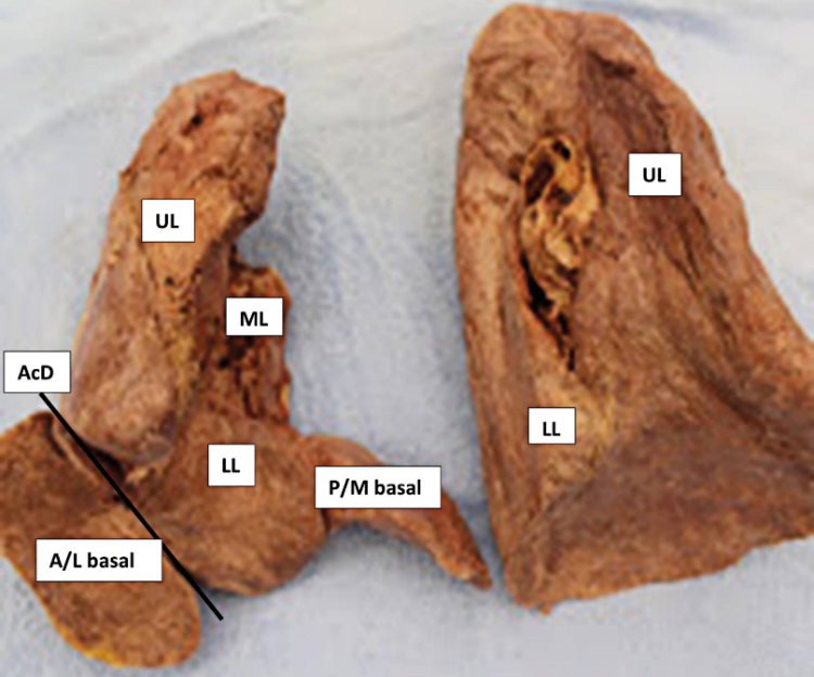
The accessory diaphragm separated the fused anterior and lateral basal segments (Fig. 1). Additionally, the fused anterior and lateral basal segments, which were situated between the actual diaphragm and the accessory diaphragm, were inflatable. Although hypoplastic, the bronchi remained patent; we could inflate the superior and middle lung segments using an air pump. Notably, we could also inflate all the bronchopulmonary segments of the lower lobe and successfully pass a soft probe (such as a pipe cleaner) into the pulmonary arterial branches of these lower lobe segments.
The accessory diaphragm described and reported here is a rare congenital anomaly associated with pediatric patients having an atrial septal defect [13,16]. It occurs mainly on the right side [3] and comprises fibromuscular tissue with a serosal lining [5]. In all documented cases, aplasia or some degree of pulmonary hypoplasia was evident on the affected side [3].
Heart and Vasculatures
The heart’s morphology appeared normal; however, the heart and mediastinum were displaced more to the right side, as observed in previous studies [10,12]. Evidence pointed to surgical procedures to correct the atrial septal defect and right superior pulmonary vein that reached into the superior vena cava.
Discussion and analysis
A person like the one in this report who has scimitar syndrome or partial anomalous pulmonary venous return (PAPVR), is often asymptomatic and may be unaware of the condition. However, the person (donor) in this report had surgical interventions at a young age to correct anomalous venous return and cardiac anomalies such as ASD.
Accessory diaphragm
An accessory diaphragm is a thin fibromuscular membrane that appears to be related to the incomplete descent of the septum transversum [5]. This anomaly mainly occurs on the right side. Its cause remains unclear, but the timing difference in the growth of the two lung buds, as previously suggested, may provide an explanation [5]. In all the reported cases, aplasia or varying degrees of pulmonary hypoplasia were evident on the affected side, as also seen in the case presented here (Fig. 1). The accessory diaphragm separates the right pleural cavity into two parts, which can trap part or all of the right middle or lower lobes beneath it [4], as observed in the case presented here. If the lung located beneath the accessory diaphragm is aerated, as demonstrated in this report, it will move with respiration [7]. Interestingly, an accessory diaphragm separated the fused anterior and lateral basal segments; these segments could be aerated using a handheld cycle pump, thus providing evidence that it was functional. Additionally, the ‘sequestered’ segments received blood supply from a branch of the pulmonary artery.
Conclusion
In conclusion, in the follow-up of the case of the hypogenetic right lung, another unusual feature, the accessory diaphragm, is reported here. Very early formation of the bronchial system may result in the developing septum transversum being cleaved as it makes contact with the developing lung, which likely results in an accessory diaphragm. The associated pulmonary vascular malformations reported in cases of accessory diaphragm in the literature suggest that the pathogenic event occurs during this embryonic period.
Acknowledgements
The authors sincerely thank the donor and his family for their generosity, which made this study possible and facilitated scientific and medical innovations in patient care.
Funding
This study was supported by the Cooper Medical School of Rowan University research and innovation grant.
Disclosure
Drs. Melovitz-Vasan and Vasan participated in the dissections. All authors participated in the article preparation. Dr. Melovitz-Vasan envisioned the project and edited the final manuscript along with Ms. Huff. All the authors approved the final form of the paper.
References
[1] Becmeur F, Horta P, Donato L, Christmann D, Sauvage P. (1995) Accessory diaphragm – review of 31 cases in the literature. Eur J Pediatr Surg. 5: 43–7, indexed in Pubmed: 7756236 https://doi.org/10.1055/s-2008-1066162.
[2] Bellini C, Hennekam RCM. (2012) Non-immune hydrops fetalis: a short review of etiology and pathophysiology. Am J Med Genet A. 158A: 597–605, indexed in Pubmed: 22302731, https://doi.org/10.1002/ajmg.a.34438.
[3] Cooper G. (1836) Case of malformation of the thoracic viscera consisting of imperfect development of the right lung and transposition of the heart. London Medical Gazette. 18: 600–602.
[4] Currarino G, Williams B. (1985) Causes of congenital unilateral pulmonary hypoplasia: a study of 33 cases. Pediatr Radiol. 15(1): 15–24, indexed in Pubmed: 3969292, https://doi.org/10.1007/BF02387847,.
[5] Davis WS, Allen RP. (1968) Accessory diaphragm. Duplication of the diaphragm. Radiol Clin North Am. 6(2): 253–263, indexed in Pubmed: 5667501.
[6] Drake EH, Lynch JP. (1950) Bronchiectasis associated with anomaly of the right pulmonary vein and right diaphragm. J Thorac Surg. 19:433–37, indexed in Pubmed: 15410515.
[7] Felson B. (1973) Chest roentgenology. Philadelphia, Saunders. 81–92.
[8] Frank JL, Poole CA, Rosas G. (1986) Horseshoe lung: clinical, pathologic, and radiologic features and a new plain film finding. AJR Am J Roentgenol. 146(2): 217–226, indexed in Pubmed: 3484566, https://doi.org/10.2214/ajr.146.2.217.
[9] Hart JC, Cohen IT, Ballantine TV, Varrano LF. (1981) Accessory diaphragm in an infant. J Pediatr Surg. 16; 947-949, indexed in Pubmed: 7338778, https://doi.org/10.1016/s0022-3468(81)80851-2.
[10] Hartman T. Case 9 – Horseshoe lung. (2011) In: Hartman T (ed.). Pearls and Pitfalls in Thoracic Imaging: Variants and Other Difficult Diagnoses. Cambridge University Press, Cambridge. 22–23.
[11] Hashida Y, Sherman FE. (1961) Accessory diaphragm associated with neonatal respiratory distress. J Pediatr. 59; 529-532, indexed in Pubmed: 13905178, https://doi.org/10.1016/s0022-3476(61)80236-9.
[12] Melovitz-Vasan C, White A, Huff S, Vasan N. (2023) Hypogenetic right lung with partial anomalous pulmonary venous return and accessory diaphragm: a case of “scimitar lung.” Folia Morphol (Warsz). 82(4): 980-987. https://doi.org/10.5603/FM.a2022.0098. Epub 2022 Dec 6. PMID: 36472398.
[13] Olson MA, Becker GJ. (1986) The Scimitar syndrome: CT findings in partial anomalous pulmonary venous return. Radiology. 159(1): 25–26, https://doi.org/10.1148/radiology. 159.1.3952313, indexed in Pubmed: 3952313.
[14] Priyadarshi A, Sugo E, Challis D, Bolisetty S. (2014) Accessory diaphragm associated with non-immune hydrops fetalis. BMJ Case Rep. Jul 4;2014:bcr2014204090. https://doi.org/10.1136/bcr-2014-204090. PMID: 25100808; PMCID: PMC4091454.
[15] Restrepo R, Lee EY. (2008) The diaphragm. In: Slovis TL (ed.). Caffey’s Pediatric Diagnostic Imaging (11th Edition). Mosby Elsevier, Philadelphia. 587-592.
[16] van den Hoven AT, Chelu RG, Duijnhouwer AL, Demulier L, Devos D, Nieman K, Witsenburg M, van den Bosch AE, Loeys BL, van Hagen IM, Roos-Hesselink JW. (2017) Partial anomalous pulmonary venous return in Turner syndrome. Eur J Radiol. 95: 141–146, https://doi.org/10.1016/j. ejrad.2017.07.024, indexed in Pubmed: 28987660.
[17] Wille L, Holthusen W, Willich E. (1975) Accessory diaphragm. Report of 6 cases and a review of the literature. Pediatr Radiol. 4; 14-20, indexed in Pubmed: 1233449, https://doi.org/10.1007/BF00978814.