https://doi.org/10.36253/ijae-15941
Corpus callosum motor fibers dissection using Klingler’s method and methylene blue
1 Federal University of São Paulo, (UNIFESP) São Paulo, Brasil
2 Anatomy Department of ABC Medical School, Santo André, Brazil
3 Neurosurgery resident at Santa Paula Hospital, São Paulo, Brazil
4 Medical Student at Pontifical Catholic University of São Paulo (PUC/SP), São Paulo, Brazil
5 Neurosurgery resident at ABC medical school, Santo André, Brazil
6 Neurosurgery resident at Heliopólis Hospital, São Paulo, Brazil
7 Chief of Epilepsy Department at Federal University of São Paulo (UNIFESP), São Paulo, Brasil
*Corresponding author. E-mail: carolina.smartini@gmail.com
Abstract. Background: The corpus callosum (CC) is the largest white matter bundle in the brain, connecting the left and right cerebral hemispheres. It plays a crucial role in integrating information and facilitating somatosensory, motor, and cognitive function. This study examined the anatomy of the CC using white matter dissection and explored a posterior callosotomy approach for treating drop attacks in Lennox-Gastaut syndrome (LGS). LGS is a rare form of childhood-onset epilepsy, characterized by multiple seizure types, abnormal electroencephalograms, and progressive mental retardation. The authors proposed that fibers from the medial and posterior CC regions converge on the precentral gyrus, and dissection was performed to investigate this theory. Methods: This study employed the Klingler method to examine white matter fibers in four adult brains. Dissection involved removing the cortex from the superior frontal, precentral, and postcentral gyri, followed by the cingulate gyrus. Results: The CC is divided into five parts: the rostrum, genu, body, isthmus, and splenium. Each part has distinct anatomical relationships with the lateral ventricles and connects to different cortical regions Conclusions: Our findings are consistent with the evidence that the motor fibers of the brain traverse the posterior region of the corpus callosum, suggesting that selective posterior callosotomy may be a more effective treatment for refractory drop attacks in Lennox-Gastaut Syndrome.
Keywords: Lennox Gastaut Syndrome, corpus callosum, white matter.
Contents
Introduction
The corpus callosum (CC), the largest commissural white matter bundle, is a crucial structure of the brain. It connects the left and right cerebral hemispheres and plays a vital role in integrating the information between the [1,2]. The corpus callosum comprises myelinated nerve fibers that extend across the midline of the developing brain and connect the two hemispheres. This structure facilitates the transmission of sensory, motor, and cognitive functions between the homologous regions of the brain [3,4].
It is divided into regions, from anterior to posterior, the rostrum, genu, body isthmus, splenium, and tapetum, each with fibers in different cerebral regions [2]. The CC is intimately related to the fornix and lateral ventricles and represents the anterior, superior, and posterior border of the septum pellucidum, which together with the fornix separates the lateral ventricles from each other [1].
Lennox-Gastaut syndrome (LGS) is a severe form of epilepsy that affects children. It was first identified by Henri Gastaut in 1966. In 1950, Dr. William G. Lennox characterized the electroencephalogram features of this condition. LGS represents approximately 2-5% of all childhood epilepsies [5]. It is one of the most intractable epilepsies and is characterized by multiple types of seizures, electroencephalographic (EEG) characteristics such as generalized slow sharp and wave discharges (GSW), generalized paroxysmal fast activities, and progressive mental retardation. GSW with bilateral synchronization in secondary generalized epileptic encephalopathy, such as LGS, can originate from the primary epileptogenic zone through the transcallosal pathway [6]. Due to the difficulty of identifying and localizing single epileptogenic lesions and the presence of multifocal characteristics, most patients with LGS are ineligible for resective surgery [5]. Palliative surgical procedures, including corpus callosotomy, for refractory epilepsy were first introduced in the 1940s by Van Wagenen and Herren, and have since become an established treatment option for patients with intractable epilepsy who are not suitable candidates for resective surgery [5,7]. The most common indication for corpus callosotomy is a drop attack. Patients who undergo total corpus callosotomy show a higher reduction in seizures than patients who undergo partial corpus callosotomy [7]. Epileptic drop attacks are usually refractory to medication, and their association with the sudden bilateral synchronization of ictal discharges provides a rationale for callosotomy. The procedure is indicated when focal resection is not feasible and specifically targets sudden falls irrespective of the underlying etiology. Thus, the clinical heterogeneity of the candidates is the norm [8]. Although a 1-stage total corpus callosotomy can be performed in patients younger than 16 years, a 2-stage corpus callosotomy is preferred for patients older than 16 years, given its lower risk for disconnection syndrome [9].
This article delves into the physiopathology of LGS-related drop attacks and posterior callosotomy employed as a treatment, with a foundation in the anatomy of the corpus callosum and its constituent fibers. The authors proposed that fibers from the medial and posterior regions of the corpus callosum converge on the precentral gyrus. To investigate this theory, dissection of the corpus callosum and a study of its fibers were conducted.
Materials and methods
This study employed the Klingler method to examine white matter fibers. It comprises of four (4) phases: fixation, freezing, thawing, and dissection.
Four normal adult brains (eight hemispheres) were fixed in a 10% formalin solution for at least three months. The superficial veins and arachnoid membranes were carefully removed before the brains were frozen at 0–5°C for three weeks. This step allowed formalin to crystallize between the fibers, causing them to swell and separate for further preservation and observation. Samples were thawed and stored in diluted formalin. A ZEISS EyeMag with 2-2,5x amplification, a medical loupe, was used to better visualize the fibers during dissection, and a wooden spatula was used to remove the cortex and other regions during the procedure.
Following dissection of the zone of interest (forceps major and minor), the brains were stained with methylene blue 5% aqueous solution using a painting brush. After one hour, the hemispheres were rinsed, and the fibers emerging from the forceps major were traced to determine their endings. In addition, fibers from the medial and posterior regions of the CC that were not stained were studied.
Results
The dissection involved the removal of the cortex from the superior frontal gyrus, precentral gyrus, and postcentral gyrus. Subsequently, the white fibers were extracted to access the cingulate gyrus, which was also removed. The anterior fibers of the corpus callosum were traced into the forceps minor, situated under the frontal lobe, whereas the posterior fibers were located to lead to the forceps major, situated under the occipital lobe (Figure 1 and 2).
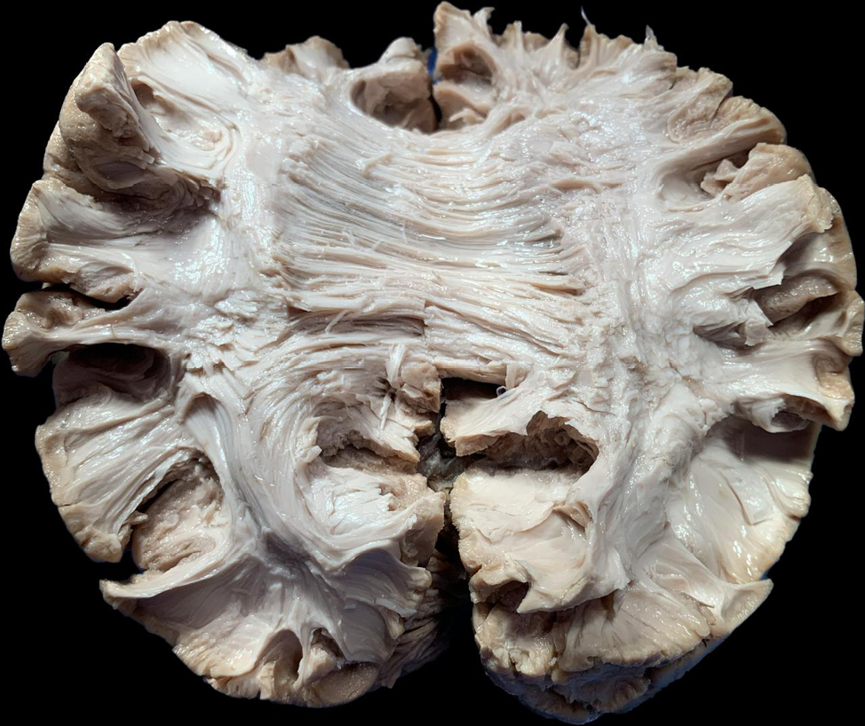
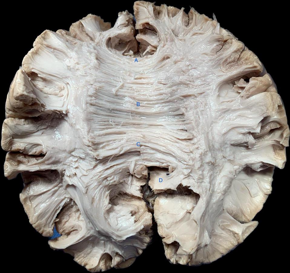
Subsequently, the major and minor forceps were exposed and stained with methylene blue to make their paths more visible (Figure 3).
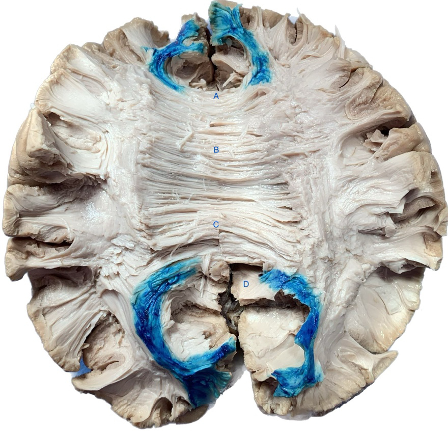
The use of methylene blue 5% to staining the major and minor forceps (as shown in Figure 3) enabled researchers to more clearly observe the path of the fibers emanating from these areas.
Regarding the medial portion of the CC, in all eight hemispheres examined, the fibers were traced from this region to the precentral gyrus as well as connecting the two occipital hemispheres. In the posterior region of the CC, the fibers were traced to the precentral and postcentral gyri in all eight hemispheres (Table 1).
| Structure | Number of hemispheres | White Fibers ending point | |||||||||||||||||
|---|---|---|---|---|---|---|---|---|---|---|---|---|---|---|---|---|---|---|---|
| Medial region | 8 (100%) | Precentral gyrus | |||||||||||||||||
| Posterior region | 8 (100%) | Precentral and postcentral gyrus | |||||||||||||||||
| The percentage in parentheses indicates the proportion of findings found compared to the total number of cases (n=8). | |||||||||||||||||||
Discussion
Anatomy and physiology
The corpus callosum is the largest bundle of white fibers in the human brain, with over 200 million axons communicating with the two cerebral hemispheres. It participates in connections in motor, sensory pathways, and cognitive functions [10,11]. The corpus callosum appears to be a “C” shape structure, viewed from above the interhemispheric fissure [12], and lies contouring almost all walls of the lateral ventricles [13,14].
The corpus callosum has two different embryogenic origins, dividing it into an anterior portion, which comprises the genu, rostrum, and body, and a posterior portion, represented by the splenium. The anterior portion, originating from the glial sling, merges with fibers from the hippocampal commissure and parieto-temporo-occipital cortex [15].
Histologically, the corpus callosum has a higher density of larger-diameter fibers in the isthmus and posterior splenium, while finer fibers are more abundant in the genu and anterior splenium. The splenium is the thickest part of the corpus callosum [15,16].
It is divided in five parts: rostrum, genu, body, isthmus, splenium – anterior to posterior extent, respectively [14]. The rostrum forms the floor of the frontal horn the genu forms its anteromedial portion, the body forms the roof of the body of the lateral ventricle, the isthmus forms the medial wall of the atrium of the lateral ventricle, and the splenium forms the medial wall of the atrial and occipital horn of the lateral ventricle [14,17].
The rostrum begins at the anterior commissure and extends to the genu, which is the anterior limb of the septum pellucidum, and has fibers that connect the prefrontal cortex and anterior cingulate area, forming the forceps minor. The callosal body begins in the genu and extends to the fornix. The isthmus ends at the point where the fornix meets the inferior callosal surface, the splenium forms the forceps major, and the callosal body, together with the splenium, forms the tapetum, arising in the posterior part of those structures [14,18].
Above the corpus callosum, acting as a thick layer, the indusium griseum covers its surface, and on each side, there is a fine line of myelinated fibers and the medial and lateral longitudinal striae of Lancisi [18].
The internal carotid artery system and pericallosal artery provide arterial supply to the cingulate cortex (CC), with the exception of the splenium, which is vascularized by the terminal and choroidal branches of the posterior cerebral artery. Additionally, the anterior communicating artery, a part of the carotid system, contributes to the arterial supply of the CC by removing its median artery. The callosal and callosal cingular veins are responsible for facilitating venous drainage of the CC towards the internal cerebral veins [1].
Superior/dorsal callosal radiation arises from the corpus callosum and runs medially into brain hemispheres. It courses below the cingulate gyrus and medial superior longitudinal fasciculus (SLF), and laterally to the SLF. It ascends and forms the corona radiata, which connects the motor and supplementary motor areas of both hemispheres [17]. Dorsal callosal radiation is divided into anterior and posterior components, which originate from the genu and anterior body of the CC and form the roof of the frontal horn of the lateral ventricle; the posterior component, also known as the tapetum, originates from the posterior region of the CC and forms the roof and lateral wall of the atrium as well as the lateral wall of the temporal horn of the lateral ventricle [17]. The anterior callosal radiations originate from the genu and rostrum, with the majority of fibers bending anteriorly to reach the medial frontal-orbital region, also known as the forceps minor (Figure 2), forming the lateral ventricle frontal horn. Additionally, other fibers emerge from the rostrum and curve laterally on the basal surface of the frontal lobe, running along the medial surface of the uncinate fasciculus, ultimately reaching the temporal stem in close association with the external capsule, and connecting the insular regions of both hemispheres [17]. Posterior callosal radiations originating from the CC splenium extend posteriorly to the medial occipital region. This structure is known as forceps major, as it connects the occipital lobes of both hemispheres. It creates a protuberance in the medial wall of the atrium of the lateral ventricle known as the atrium bulb [17].
With human cadaveric dissection and a study of primate brains, Witelson in 1989 proposed that the human corpus callosum should be divided into seven segments, according to geometric landmarks, associated with a cortical region: rostrum, genu, rostral body, anterior midbody, posterior midbody, isthmus and splenium [19]. Each of these subregions sends and receives fibers from the cortical regions, basal ganglia, thalamus, and the brainstem [20].
The genu and anterior third of the body of the corpus callosum are associated with the prefrontal cortex; the central part is linked to the premotor and supplementary motor cortex; the posterior third is associated with the primary motor area (precentral gyrus); the posterior third and splenium are associated with the primary sensory cortex (postcentral gyrus); and the splenium is associated with the parieto-temporo-occipital cortex [2,11,21]. Moreover, the posterior part of the corpus callosum (posterior midbody, isthmus, and splenium) contains fibers projecting from the posterior parietal cortex to the hippocampus through the para-hippocampal and entorhinal areas [22].
With the development of tractography and functional magnetic resonance imaging studies, it is known that the organization of the white matter of the corpus callosum is more complex, showing a spatial distribution pattern of its fibers. There is also an overlap between the communicated regions, and the fibers are not only related to cortical regions, but can also connect with other white fiber bundles (Wang et al., 2021).
An investigation conducted by Küçükyürük et al. in 2020 revealed that motor fibers originating from the motor cortex traverse the posterior section of the corpus callosum, primarily in the posterior body and isthmus, a conclusion that aligns with the research conducted by Shah et al. in 2021. This finding was also documented in a study led by Hofer and Frahm in 2006, who utilized DTI to highlight the corpus callosum white matter tracts [17,23,24]. The results of our dissection are consistent with literature findings, which indicate that motor fibers from the brain connect to the posterior area of the corpus callosum.
Due to the complex connections of the corpus callosum with various areas of the brain, it is the main interhemispheric pathway for the spread of epileptic activity, and therefore, is of interest in Epilepsy Surgery [20].
Callosotomy x Epilepsy
Lennox-Gastaut Syndrome (LGS) is one of the most difficult epileptic syndromes to control and accounting for 1-10% of childhood epilepsies. It consists of a triad of different types of seizures, a characteristic interictal electroencephalogram with a slow spike-wave pattern or generalized paroxysms of fast activity, and cognitive deficits. It can be caused by various factors, such as traumatic or hypoxic brain injuries, tuberous sclerosis complex, congenital infections, brain malformations, and hereditary metabolic diseases. There is no common epileptogenic zone (EZ) for all patients, and in most cases, the EZ is multiple or diffuse. Hence, in addition to being drug-resistant, there is a wide range of responsiveness to different surgical modalities. Furthermore, the maintenance of seizures is related to a worse cognitive prognosis, in addition to the risk of direct injury during seizures like generalized tonic-clonic and drop attacks, so delaying surgical treatment in an attempt to control the disease with medication is not advisable [5,6,21,25].
EZ resection surgery with curative potential is the treatment of choice when the syndrome is caused by a focal and well-defined epileptogenic zone. However, not all patients achieve seizure freedom, as LGS is considered a disease of intercortical connections and white fiber pathways [6,20]. Studies have shown that animal models with epileptogenic lesions in the neonatal period exhibit a decrease in the corpus callosum surface area, indicating alterations in its microstructure, and hence, its connections [22]. Furthermore, Andrade et al. demonstrated diffusion alterations in the corpus callosum of patients with cortical developmental anomalies, which is probably a combined entity with defects in white matter development [21].
Therefore, when the EZ is diffuse, multiple or poorly defined palliative procedures, such as callosotomy, are employed. Callosotomy was introduced in 1940 and aims to interrupt the propagation of the epileptic pathway. It mainly affects tonic seizures, atonic seizures, such as drop attacks, and tonic-clonic seizures. There are different modalities of callosotomy: resection of the anterior third, anterior half, anterior two-thirds, preservation only of the splenium, total callosotomy, and selective posterior callosotomy [6,8,25].
Total callosotomy and anterior callosotomy were similar in technique. Disconnection in anterior callosotomy starts at the genu/rostrum of the corpus callosum and ends at the transition between the body and the isthmus. In total callosotomy, disconnection ends at the splenium of the corpus callosum. In posterior callosotomy, disconnection starts at the posterior end of the splenium and ends at the isthmus.
Disconnection syndrome, severity of cognitive deficit, and developmental delay should be considered when deciding the volume of resection of the corpus callosum. Total callosotomy tends to be more effective in seizure control. However, they have a higher risk of developing disconnection syndrome, especially in elderly patients. In cases where anterior callosotomy did not effectively reduce the number of seizures, complementary posterior callosotomy might be considered [26].
The most common complication of callosotomy is the disconnection syndrome. They can be classified as either acute or chronic. Acute syndrome comprises severe decreases in spontaneous speech, paresis of the non-dominant leg, and incontinence. Several patients may require tube feeding during the postoperative period. These symptoms tended to disappear after a few months. On the other hand, the chronic syndrome is characterized by alien hand syndrome, dichotic listening suppression, tactile dysnomia, hemispatial neglect, nondominant hand agraphia, and tachistoscopic visual suppression [26]. Other possible complications include temporary postoperative disturbance of consciousness and chemical meningitis [27].
Selective posterior callosotomy (SPC) is mainly performed for cases of drop attacks, such as fibers from the premotor cortex, primary motor cortex, and sensorimotor area responsible for axial and lower limb control crossing from the midbody to the posterior half of the corpus callosum. Therefore, SPC interrupts the pathway that leads to atonic seizures without disconnecting the frontal pathways responsible for aspects of cognition, volition, and sphincter control. In a study by Paglioli et al., 83% of patients experienced complete reduction or >90% reduction in drop attacks after SPC, along with improved functionality scores, and none experienced mutism, ataxia, or sphincter incontinence. SPC has a higher incidence of disconnection syndromes than selective anterior callosotomies, but in patients with LGS, who already have significant cognitive impairment, these changes do not result in noticeable deficits [8,9].
Our work demonstrates, using the white matter dissection technique combined with methylene blue staining, the path of motor and sensitive fibers via the posterior portions of the corpus callosum. This corroborates with the statement made by Pagliogli et al. that selective posterior callosotomy is sufficient to treat refractory epilepsy cases that lead to falls, especially drop attacks; therefore, we can provide the patient with a better treatment, with less risk of disconnection syndrome and greater control of the disease. To the best of our knowledge, this is the first anatomical study to show these connections enhanced by methylene blue staining.
Conclusions
Our research aimed to provide further evidence that the motor fibers of the brain traverse the posterior region of the corpus callosum, suggesting that selective posterior callosotomy may be a more effective treatment for refractory drop attacks in Lennox-Gastaut Syndrome. Our findings are consistent with those of the latest studies. The novel aspect of our work was the utilization of methylene blue to examine CC fibers.
References
[1] Fitsiori A, Nguyen D, Karentzos A, Delavelle J, Vargas MI. The corpus callosum: white matter or terra incognita. Br J Radiol. 2011 Jan;84(997):5–18.
[2] Radwan AM, Sunaert S, Schilling K, Descoteaux M, Landman BA, Vandenbulcke M, et al. An atlas of white matter anatomy, its variability, and reproducibility based on constrained spherical deconvolution of diffusion MRI. NeuroImage. 2022 Jul 1;254:119029.
[3] Pearce JMS. Corpus Callosum. Eur Neurol. 2007;57(4):249–50.
[4] Fabri M, Polonara G, Mascioli G, Salvolini U, Manzoni T. Topographical organization of human corpus callosum: An fMRI mapping study. Brain Res. 2011 Jan;1370:99–111.
[5] Alanazi GM, ALOsaimi TF, Alwadei AH, Al-Otaibi AD, Jad LA, Al-Attas AA. Efficacy and safety of corpus callosotomy versus vagus nerve stimulation as long-term adjunctive therapies in children with Lennox–Gastaut syndrome: Experience of a tertiary care center. Neurosciences. 2022 Apr;27(2):59–64.
[6] Hur YJ, Kim HD. The causal epileptic network identifies the primary epileptogenic zone in Lennox–Gastaut syndrome. Seizure – Eur J Epilepsy. 2015 Dec 1;33:1–7.
[7] Ueda R, Matsuda H, Sato N, Iwasaki M, Sone D, Takeshita E, et al. Alteration of the anatomical covariance network after corpus callosotomy in pediatric intractable epilepsy. Biagini G, editor. PLOS ONE. 2019 Dec 5;14(12):e0222876.
[8] Paglioli E, Martins WA, Azambuja N, Portuguez M, Frigeri TM, Pinos L, et al. Selective posterior callosotomy for drop attacks: A new approach sparing prefrontal connectivity. Neurology. 2016 Nov 8;87(19):1968–74.
[9] Ito H, Morino M, Niimura M, Takamizawa S, Shimizu Y. Posterior callosotomy using a parietooccipital interhemispheric approach in the semi-prone park-bench position. Abstract.
[10] Ruddy KL, Leemans A, Carson RG. Transcallosal connectivity of the human cortical motor network. Brain Struct Funct. 2017 Apr;222(3):1243–52.
[11] Wang P, Wang J, Tang Q, Alvarez TL, Wang Z, Kung YC, et al. Structural and functional connectivity mapping of the human corpus callosum organization with white-matter functional networks. NeuroImage. 2021 Feb 15;227:117642.
[12] Shah A, Goel A, Jhawar SS, Patil A, Rangnekar R, Goel A. Neural Circuitry: Architecture and Function—A Fiber Dissection Study. World Neurosurg. 2019 May;125:e620–38.
[13] Cumming WJK. An Anatomical Review of the Corpus Callosum. Cortex. 1970 Mar;6(1):1–18.
[14] Rhoton AL. THE CEREBRUM. Neurosurgery [Internet]. 2007 Jul;61(1). Available from: https://journals.lww.com/00006123-200707001-00004
[15] Fabri M, Pierpaoli C, Barbaresi P, Polonara G. Functional topography of the corpus callosum investigated by DTI and fMRI. World J Radiol. 2014;6(12):895.
[16] Ribas EC, Yağmurlu K, De Oliveira E, Ribas GC, Rhoton A. Microsurgical anatomy of the central core of the brain. J Neurosurg. 2018 Sep;129(3):752–69.
[17] Shah A, Jhawar S, Goel A, Goel A. Corpus Callosum and Its Connections: A Fiber Dissection Study. World Neurosurg. 2021 Jul;151:e1024–35.
[18] Raybaud C. The corpus callosum, the other great forebrain commissures, and the septum pellucidum: anatomy, development, and malformation. Neuroradiology. 2010 Jun;52(6):447–77.
[19] Witelson SF. Hand and sex differences in the isthmus and genu of the human corpus callosum. Brain. 1989;112(3):799–835.
[20] Asadi-Pooya AA. Lennox-Gastaut syndrome: a comprehensive review. Neurol Sci. 2018 Mar;39(3):403–14.
[21] Andrade CS, Leite CC, Otaduy MCG, Lyra KP, Valente KDR, Yasuda CL, et al. Diffusion abnormalities of the corpus callosum in patients with malformations of cortical development and epilepsy. Epilepsy Res. 2014 Nov;108(9):1533–42.
[22] Meng Y, Hu X, Zhang X, Bachevalier J. Diffusion tensor imaging reveals microstructural alterations in corpus callosum and associated transcallosal fiber tracts in adult macaques with neonatal hippocampal lesions. Hippocampus. 2018 Nov;28(11):838–45.
[23] Hofer S, Frahm J. Topography of the human corpus callosum revisited—Comprehensive fiber tractography using diffusion tensor magnetic resonance imaging. NeuroImage. 2006 Sep;32(3):989–94.
[24] Küçükyürük B, Uzan M, Avyasov R, Tahmazoğlu B, İşler C, Sanus GZ, et al. Evaluation of Ideal Extent of Corpus Callosotomy Based on the Location of Intracallosal Motor Fibers. World Neurosurg. 2020 Dec;144:e568–75.
[25] Thirunavu V, Du R, Wu JY, Berg AT, Lam SK. The role of surgery in the management of Lennox–Gastaut syndrome: A systematic review and meta‐analysis of the clinical evidence. Epilepsia. 2021 Apr;62(4):888–907.
[26] Uda T, Kunihiro N, Umaba R, Koh S, Kawashima T, Ikeda S, et al. Surgical Aspects of Corpus Callosotomy. Brain Sci. 2021 Dec 5;11(12):1608.
[27] Fujimoto A, Okanishi T. Corpus Callosotomy: Editorial. Brain Sci. 2022 Jul 29;12(8):1006.