https://doi.org/10.36253/ijae-15808
A case report of the right vertebral artery’s origin as a unique ‘trifurcation’ involving the brachiocephalic trunk, right common carotid artery, and the right vertebral artery
1 Department of Biomedical Sciences, Cooper Medical School of Rowan University, Camden, New Jersey, 08103 USA
2 Medical Education Research Collaborator, Winston-Salem, NC, USA
3 Department of Biomedical Sciences, Cooper Medical School of Rowan University, 401 South Broadway, Camden, New Jersey 08103, USA
*Corresponding author. E-mail: melovitz-vasan@rowan.edu
Abstract. While the anomalous origin of the left vertebral artery is more often reported, the atypical right vertebral artery arising from the aortic arch is sparsely recognized. During the dissection of a donor’s body, we recognized that the right vertebral artery arose as a ‘trifurcation’ from the aortic arch distal to the right subclavian artery. Intrigued, we explored the arterial branching further to realize a unique pattern not seen in earlier reports. The trifurcation involved the brachiocephalic trunk, right common carotid, and right vertebral artery. It is essential to recognize the possibility of people living normally without symptoms due to some anomalous arterial pattern.
Keywords: anomalous origin of right vertebral artery, right vertebral artery origin as a ‘trifurcation’, dilatation at the proximal and distal end of the right vertebral artery.
Contents
Introduction
Any observations related to anatomical structures identified during the dissection of a donor body or in patients following various radiographic modalities are to further our knowledge and or provide better treatment. However, minor vascular anomalies may or may not often cause clinical symptoms, and people could live normal asymptomatic lives. Prior articles about the vertebral arteries have been published since the beginning of the last century (Poynter, 1916; Congdon, 1922; Iyer, 1927; Adachi, 1928; Windle et al., 1928). These include how the vertebral arteries originate and from which part of the aortic origin, the number of vertebral arteries (up to six), and other details. Most importantly, knowledge of vascular anomalies is invaluable when surgical modalities are contemplated. In the case of the vertebral artery, prior knowledge of the region is vital because of its location, which is close to several other critical neurovasculatures. As presented in this report, the unique trifurcation of the right vertebral artery involving the brachiocephalic trunk, right common carotid artery, and right vertebral artery could significantly impact surgical planning and patient outcomes. To our knowledge, there are no reported cases in the literature where the right vertebral artery arises as a trifurcation from the brachiocephalic trunk, right common carotid artery, and right vertebral artery.
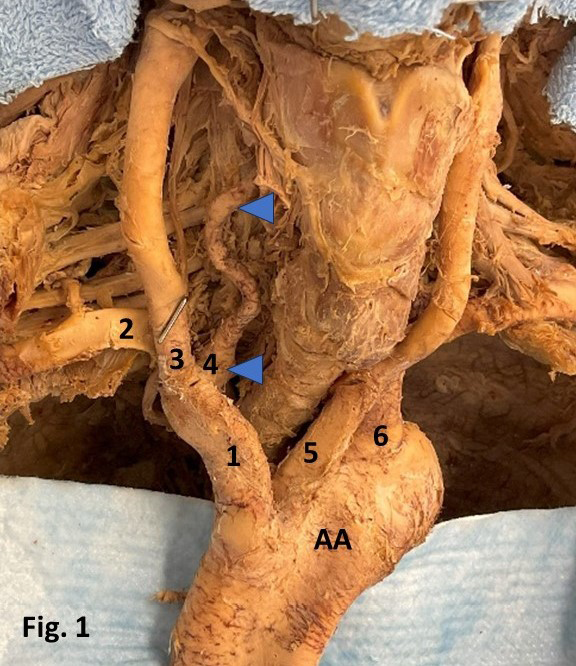
In 1999, Lemke et al. reviewed the literature on the anomalous origin of the right vertebral artery and sketched images of different variants of anomalous right vertebral artery origins, rate of occurrence, and their potential embryological development. This report describes an anomalous right vertebral artery origin as a ‘trifurcation’ with the brachiocephalic trunk, right common carotid artery, and right vertebral artery (Fig. 1).
Materials and methods
The cadaveric specimen in this study was obtained from the willed body program for medical student dissection. The donor was a 94-year-old Caucasian female who died of right femur fracture sequelae. During routine cadaveric dissection of the thorax, we observed that the right vertebral artery originated as a ‘trifurcation’ with the brachiocephalic trunk, right common carotid artery, and right vertebral artery (Fig. 1). Proximally and distally, before entering the foramen transversarium, the right vertebral artery also exhibited recognizable distension (Fig. 1). A plausible explanation for this feature will be further explained and discussed in the later section.
Results and observation
During the dissection of the donor thoracic structures, we observed that the right vertebral artery originated as a ‘trifurcation’ with the brachiocephalic trunk, right common carotid artery, and right vertebral artery (Fig. 1). Proximally at its origin and distally before entering the foramen transversarium, the right vertebral artery also exhibited recognizable distension (Fig. 1). A plausible explanation for this physical feature will be further explained and discussed in the subsequent section. The right vertebral artery displayed some natural tortuosity during its course through the cervical region before entering the foramen transversarium (Fig. 1).
Discussion
Since Poynter’s 1916 report that there were 4-6 vertebral artery branches from the aortic arch, there have been other similar studies (Matula et al., 1997; Lemke et al., 1999). To our knowledge, no case has been reported in the literature in which the right vertebral artery originates as a ‘trifurcation’ between the brachiocephalic trunk, right common carotid artery, and right vertebral artery (Fig. 1). Is there a clinical significance to our findings? Yes, in surgical reconstructions of the aortic arch and its branches, one must be aware of this ‘trifurcation’ anomaly.
It has been noted that the vertebral artery’s preforaminal (V1 segment) tends to have a tortuous path in about 39% of cases Matula et al. (1997) studied, which is consistent with our observation in this study (Fig. 1) and an earlier report (Freilich et al., 1986). Additionally, there is a difference in the twistiness of the right and left vertebral arteries, with 32% of the right vertebral artery being twisted compared to 68% of the left vertebral arteries. The V1 segment of both the right and left preforaminal vertebral arteries showed some natural tortuosity, with the left artery more tortuous than the right. The prevalence of transverse tortuosity was the highest compared to coronal and sagittal tortuosity (Matula et al., 1997; Russo et al., 2011; Uchino et al., 2013). While the twists in the proximal or preforaminal segment of the vertebral artery do not have any hemodynamic consequences (Wuttke et al., 1990), the loops of the proximal segments have been known to cause nerve root compressions, leading to radicular symptoms. Some experts believe that abnormalities in the origins and distribution of the large aortic arch vessels can cause changes in cerebral hemodynamics, which can result in cerebral abnormalities (Wuttke et al., 1990). According to Wuttke et al. (1990), the anomalous origins and distribution of the large vessels of the aortic arch can alter cerebral hemodynamics, which might result in cerebral abnormalities and pathology (Lemke et al., 1999). The diagnostic advantage prior to surgery of supraaortic arteries is the actual value of detecting anomalous origins (Flynn, 1968; Tardieu, 2017).
Furthermore, the right vertebral artery showed noticeable distension both proximally at its origin and distally before entering the foramen transversarium (see Fig. 1). A potential reason for the observed bulge in the vertebral artery is that, at its beginning, the flow of blood from the aorta (a larger vessel with high pressure) into the narrower vertebral artery most likely could have caused the distension observed. Similarly, the vertebral artery entering the narrower foramen transversarium might cause a distention because of the resistance encountered. Thus, the vascular pressure gradient is a natural physiological phenomenon likely to cause a bulge or distension.
Besides embryological, physical, and physiological studies, vertebral artery anomalies have clinical significance in birth defects such as Down syndrome. Research has shown a 40% occurrence rate of vertebral artery anomalies and a 36% occurrence rate of aberrant Right Subclavian Carotid Artery (RSCA) in individuals with Down syndrome (Roofthooft et al., 2008; Chen et al., 2023). Other case studies (Rathore et al., 1989; Mishra et al., 2012) also demonstrated both an aberrant RSCA and an anomalous origin of the right vertebral artery from the right common carotid artery in a patient with Down syndrome. The deletion of Chromosome 22q11, also known as CATCH 22, is commonly associated with DiGeorge syndrome, conotruncal anomaly face syndrome, and velocardiofacial syndrome. Patients with this deletion are more likely to have anomalies of the aortic arch, aortic branches, ductus arteriosus, and pulmonary arteries than those without the deletion (Momma et al., 1999).
Analysis
The following analysis, related to the case presented here, is based on our observations, findings, and published research studies on vertebral artery origin, malformations, and their effect on anatomy and physiology. In the analysis, we have considered the donor’s age and gender and the cause of death. Any conclusion drawn in the discussion is the authors’ assumption or based on available published materials. During routine cadaveric dissection of the thorax, we observed that the right vertebral artery originated as a ‘trifurcation’ with the brachiocephalic trunk, right common carotid artery, and right vertebral artery (Fig. 1).
In general, vertebral artery anomalies are due to how they begin during embryonic development, how they course through the cervical region of the neck, and the morphology of each of the four segments. The vertebral artery starts to form during weeks four to eight of embryonic development. At this stage, the horizontal part of the 1-6 intersegmental arteries (ISA) begins to recede. By developing longitudinal anastomoses that link the cervical ISA, the seventh ISA becomes the proximal subclavian artery. This artery is the starting point of the adult vertebral artery.
The anomalous origins of the right vertebral artery were divided into three categories: those originating directly from the aorta, carotid arteries, or the brachiocephalic artery. The variant of the right vertebral artery originating from the brachiocephalic trunk, right subclavian artery, and the right vertebral artery itself as a ‘trifurcation’ is unique and hitherto unreported.
In an earlier article, Lemke et al. (1999) used 11 drawings to schematically represent the anomalous origin of the right vertebral artery based on publications dating from 1927 to 1992. In the case currently presented, the origin of the right vertebral artery as a ‘trifurcation’ appears unique. As a result, this warrants that clinicians in cardiothoracic surgery are aware of this and other rare and atypical variations.
Conclusion
In the above paragraphs, this study provides an embryological explanation for how and why the right vertebral artery originates as a ‘trifurcation’ involving the brachiocephalic trunk, the right subclavian artery, and the right vertebral artery. In the analysis, we have provided some historical relevance and schematic diagrams showing earlier views of the origin of the right vertebral arteries. The study also captures how this anomalous origin of vertebral arteries is clinically relevant in surgeries that involve the cervical region and supraaortic arch. The study also explores how the tortuosity of the proximal vertebral artery, the aortic origin of the left vertebral artery, and their impact on the hemodynamics of cerebral circulation are related.
Our research goes beyond simply reporting the ‘trifurcation’ as a unique vertebral artery anomaly and delves into their clinical significance in relation to other birth defects, such as Down syndrome.
Acknowledgements
The authors sincerely thank the donor and her family for their generosity, which made this study possible and facilitated scientific and medical innovations in patient care. We are indebted to the families of our donors.
For research using human subjects, the American Association for Anatomy endorses the protections embodied in the Basic Principles of the Declaration of Helsinki and their expansion in the regulations governing research supported by the U.S. Government (45 CFR Part 46; 56 FR 28003). The authors state that every effort was made to follow all local and international ethical guidelines and laws regarding the use of human donors in anatomical research.
Declaration
The authors state that every effort was made to comply with all the local and international ethical guidelines and laws that pertain to the use of human cadaveric donors in anatomical research.
Funding
This study was supported by the Faculty Development for Research and Innovation grant administered by the Cooper Medical School of Rowan University.
Author contributions
All the authors were involved in the research, preparation, and final approval of the manuscript. Dr. Melovitz-Vasan was responsible for conceiving the project idea, creating the initial draft with Dr. Vasan, and revising the manuscript until the final version. Dr. Melovitz-Vasan and Dr. Vasan were responsible for dissection and collecting images. Ms. Huff and Dr. Vasan conducted the literature search, which was used to draft the manuscript and prepare the final format.
Abbreviations used
AA: Aortic Arch; RVA: Right vertebral artery; BrT: brachiocephalic trunk; RCC: Right common carotid artery.
References
Adachi, B. (1928) Das Arteriensystem der Japaner. Verlag der Kaiserl. Jap. Univ., Kyoto, Vol. 1.
Chen J., Liu L, Kou X., Wang C. (2023) Case report: Right vertebral and carotid artery anomalies with an aberrant right subclavian artery in two patients. Front Neurol. 14. https://doi.org/10.3389/fneur.2023.1282127.
Congdon B.D. (1922). Transformation of the aortic-arch system during the development of the human embryo. Cont Embryol. 14: 47–110.
Flynn R.E. (1968) External carotid origin of the dominant vertebral artery. Case report. J Neurosurg. 29: 300–301. PMID: 5684411 https://doi.org/10.3171/jns.1968.29.3.0300
Freilich M., Virapongse C., Kier E.L., Sarwar M., Bhimani S. (1986) Foramen transversarium enlargement due to tortuosity of the vertebral artery. Computed tomographic appearance. Spine. 11: 95–98. https://doi.org/10.1097/00007632-198601000-00030.
Iyer A.A. (1927) Some anomalies of origin of the vertebral artery. J Anat. 62 (Pt 1): 121–122.
Lemke A.J., Benndorf G., Liebig T., Felix R. (1999). Anomalous origin of the right vertebral artery: review of the literature and case report of right vertebral artery origin distal to the left subclavian artery. Am J Neuroradiol. 20: 1318–1321. PMID: 10472992; PMCID: PMC7055987.
Matula C., Trattni S., Tschabitscher M., Day J.D., Koos W.T. (1997) The course of the prevertebral segment of the vertebral artery: anatomy and clinical significance. Surg Neurol. 48: 125–131. PMID: 9242236 https://doi.org/10.1016/s0090-3019(97)90105-1
Mishra A., Pendharkar H., Jayadaevan E.R., Bodhey N. (2012) Anomalous origins of bilateral vertebral arteries in a child with Down syndrome and Moyamoya disease: A case report. Interv Neuroradiol. 18: 259-263. https://doi.org/10.1177/159101991201800303
Momma K., Matsuoka R., Takao A. (1999) Aortic arch anomalies associated with Chromosome 22q11 Deletion (CATCH 22). Pediatr Cardiol. 20: 97–102. PMID:9986884: https://doi.org/10.1007/s002469900414
Poynter, C.W.M. (1916) Arterial anomalies pertaining to the aortic arches and the branches arising from them. Nebr. Univ. Stud. 16: 229-345.
Rathore M.H., Sreenivasan V.V. (1989) Vertebral and right subclavian artery abnormalities in the Down syndrome. Am J Cardiol. 63: 1528– 1529. PMID: 252496 https://doi.org/10.1016/0002-9149(89)90023-4
Roofthooft M.T., van Meer H., Rietman W.G., Ebels T., Berger R.M. (2008) Down syndrome and aberrant right subclavian artery. Eur J Pediatr. 167(9): 1033-6. https://doi.org/10.1007/s00431-007-0637-2. Epub 2008 Jan 3. PMID: 18172685; PMCID: PMC2491432.
Russo V.M., Graziano F., Peris-Celda M., Russo A., Ulm A.J. (2011) The V(2) segment of the vertebral artery: Anatomical considerations and surgical implications. J. Neurosurg Spine. 15: 610–619. https://doi.org/10.3171/2011.7.spine1132
Tardieu G.G., Edwards B., Alonso F., Watanabe K., Saga T., Nakamura M., Motomura M., Sampath R., Iwanaga J., Goren O., Monteith S., Oskouian R.J., Loukas M., Tubbs R.S. (2017) Aortic arch origin of the left vertebral artery: An anatomical and radiological study with significance for avoiding complications with anterior approaches to the cervical spine. Clin Anat. 30: 811–816. https://doi.org/10.1002/ca.22923. Epub 2017. PMID: 28547783
Uchino A., Saito N., Takahashi M., Okada Y., Kozawa E., Nishi N., Mizukoshi W., Nakajima R., Watanabe Y. (2013) Variations in the origin of the vertebral artery and its level of entry into the transverse foramen diagnosed by CT angiography. Neuroradiology. 55: 585–594. PMID: 23344682. https://doi.org/10.1007/s00234-013-1142-0
Windle W.F., Zeiss F.R., Adamski M.S. (1928) Note on a case of anomalous right vertebral and subclavian arteries. J Anat. 62(Pt 4): 512-514. PMID: 17104207; PMCID: PMC1249992.
Wuttke V., Schmitt R., Pogan J., Clar H.E. (1990) Zervikales Wurzelkompressionssyndrom durch die Arteria vertebralis [Cervical nerve root compression syndrome caused by the vertebral artery]. Rofo. 152: 473-4. German. https://doi.org/10.1055/s-2008-1046908. PMID: 2160113.