https://doi.org/10.36253/ijae-15656
Radiographic detection of anatomical variations in the mental foramen position in a sample of Salahuddin province population
1 Department of Dental Anatomy, College of Dentistry, Tikrit University, Salahuddin Province, Iraq
2 Pharmacology Department, Faculty of Medicine, Universiti Sultan Zainal Abidin, 20400Kuala Terengganu, Malaysia
*Corresponding author. E-mail: mazzubaidi@unisza.edu.my
Abstract. The mental foramen (MF) is an important anatomical structure represented as a bilateral opening located on the buccal surface of the mandible, most often situated between the lower first and second premolars. This study was conducted to evaluate the size& position of mental foramen in the Iraqi population of Salahuddin province. A total of 146 OPG radiographs were included according to criteria of: Position 1: Situated anterior (medial) to the first premolar; Position 2: In line with the first premolar; Position 3: Between the first and second premolars; Position 4: In line with the second premolar; Position 5: Posterior (lateral) to the second premolar. The results revealed that the mental foramen was found most frequently at Position 4, in line with the second premolar (43.2%). Position 3, located between the first and second premolars, accounted for 31.5% of the cases. Positions 1 (anterior to the first premolar), 2 (in line with the first premolar), and 5 (posterior to the second premolar) were less common at 8.2%, 6.2%, and 11.0%, respectively. The average size of the mental foramen in the study population was 3.52 ± 0.56 mm. There was no statistically significant difference between males and females in the mental foramen size. Both groups demonstrated a similar mean size of 3.5 mm. This variation in the position of mental foramen may be due to several causes: developmental disturbances of the mandible during the fetal period, or position can be change as a consequence of dental loss and aging, as age advances and teeth are lost, the resorption of the alveolar ridges result in an apparent change in position of the mental foramen when measured from the superior border.
Keywords: mental foramen, Salahuddin, anatomical variation, radiography.
Contents
Anatomy of mental nerve and foramen
The significance of mental foramen
Mental Foramen Size according to Side and patient’s sex
Mental Foramen Size according to Position
Mental Foramen Size according to Position, sex, and side
Introduction
The mental foramen (MF) is an important anatomical structure represented as a bilateral opening located on the buccal surface of the mandible, most often situated between the lower first and second premolars [1].
The mental nerve – a branch of the inferior alveolar nerve from the mandibular division of the trigeminal nerve- together with corresponding arteries and veins, exit through the mental foramen (MF) [2].
The mental nerve supplies sensation to the lower lip, the labial mucosa, lower canines, and premolars, whereas blood vessels supply soft tissues of the lower jaw [3]. To anesthetize the anterior teeth, including the premolars and canines, it is possible to avoid using an inferior alveolar nerve block by injecting an anesthetic solution adjacent to the mental foramen [4].
Anatomy of mental nerve and foramen
The mandibular canal runs anteriorly along the internal surface of the mandible; it splits into the mental and incisive canals; the incisive canal continues anteriorly to the incisors & lower canine teeth, and the mental canal runs superior-laterally to the mental foramen [5].
The mandibular nerve (V3) originates from the trigeminal nerve (CN V). It enters the mandibular foramen at the medial aspect of the ramus and horizontally forward in the body as an inferior alveolar nerve (IAN) along with the inferior alveolar artery and vein [5].
The mental foramen is situated on the buccal cortex of the mandibular bone, just below the corner of the lip on either side and in close relation to the root of the 2nd mandibular premolar tooth. It moves in a posterior direction during the development of the mandible [6,7].
A very small percentage of the population (1%) has bifurcated mandibular canals. Therefore, the bifurcated mandibular canal will exit in two separate mental foramina. In this case, clinicians must be cautious since a panoramic or periapical film may not show it. However, there are some rare reports of anatomical variations, including accessory mental foramina [8,9].
The significance of mental foramen
A mental nerve block, performed in the mental foramen region, is a very useful method of achieving local anesthesia for carrying out painful procedures in its field of supply. Mental nerve blocks are frequently carried out by dentists, oral maxillofacial surgeons, emergency physicians, and plastic and reconstructive surgeons [10]. It is an important landmark to consider during surgical endodontic procedures [11]
Dentists carry out mental nerve blocks to facilitate the management of periodontal pathologies such as tooth extractions, root canal treatments, scaling, polishing, and the treatment of gingival disease. Oral maxillofacial surgeons need to accurately locate the mental foramen and nerve during complex intra-oral procedures such as implant surgery, periapical surgery, orthognathic procedures, and trauma to avoid injury to the nerve [12].
Plastic and reconstructive surgeons perform mental nerve blocks for the repair of lower lip and chin lacerations and reconstructive procedures involving the area of supply of the mental nerve [13].
Complications related to procedures involving the mental nerve include local anesthetic toxicity due to inadvertent intravascular administration or from using large toxic doses of local anesthetic agent, inadequate anesthesia, hematoma formation, and nerve injury [14].
The mental nerve may be injured by direct trauma from the injection needle, nerve compression as a result of inadvertent infiltration into the mental foramen, nerve compression secondary to hematoma formation, and neurotoxicity from the local anesthetic itself [15].
Mental foramen in radiograph
A precise size and location of mental foramen (MF) is important for different clinical dental procedures [16]. Lack of knowledge about the correct position of the mental foramen leads to repeated failure during injections and operative procedures [17]. Before dental surgery, high safety measures must be taken to avoid harming these vital structures using appropriate imaging techniques [18,19].
Injuries of the mental nerve via dental procedures such as curettage, root canal treatment, periapical surgery, orthognathic surgery, or local anesthetics injection in implant will lead to defects in the lower lip sensation, in addition, the surrounding skin and soft tissues [20,21].
The exact identification of the mental foramen area is still, even now, the most common challenge for many dentists arranging to operate on or close the mental foramen.
Panoramic radiograph allows us to get a clear image of layers where the maxilla and mandible are in, called focal trough, while other parts are blurred. In panoramic radiographs, the entire body of the mandible can be viewed, which allows a more accurate location of the mental foramen in buccolingual measurement and both the horizontal and vertical dimensions. Hence, there are no absolute anatomical landmarks for reference, and the foramen cannot be clinically visualized or palpated [15].
This study aims to evaluate the radiographical size and location of the mental foramen in the Iraqi population of Salah-Uddin province.
Materials and methods
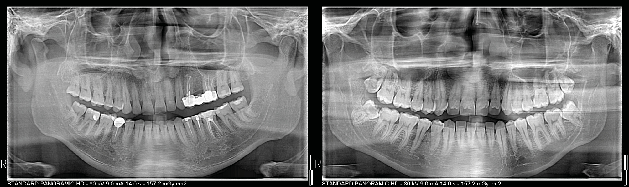
Radiographic determination of the position and size of mental foramen was done. Of 255 recruited OPG radiographs, 146 (96 males and 50 females) were deemed suitable and examined. The remaining 104 patients did not tally with the inclusion and exclusion criteria. OPG radiographs were initially decided for patients in Salahuddin province, Iraq, for various diagnostic reasons such as treatment planning before implant placement, assessment of relationships of teeth location with clinically important anatomical structures, dental surgery, and diagnosis of radiolucent lesions, as revealed in Fig. 1.
The OPG Radiographs were selected according to the following criteria:
| Inclusion criteria | Exclusion criteria |
|---|---|
| High quality with respect to angulations and contrast | Inadequate Radiograph quality. |
| Presence of mandibular teeth from the right first molar to the left first molar | Previous orthodontic treatment. |
| Visibility of mental foramen | There is no visualization of the mental foramen unilaterally and bilaterally on OPG. |
| No lesion in the apical area of premolars and MF, | Presence of pathological lesions in the mandible |
| No bone resorption. | Bone resorption or fractures in regions of examination. |
| Extraction of lower 1st molars, 1st or 2nd premolars unilaterally and bilaterally |
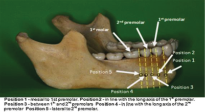
The position (P) of the MF was recorded according to the following:
P 1: MF situated anterior (medial) to the first premolar; P 2: MF is in line with the first premolar; P 3: MF is between the first and second premolars; P 4: MF is in line with the second premolar; P 5: Posterior (lateral) to the second premolar, as shown in Fig. 2 [22].
Results and discussion
Position of the Mental Foramen (see Table 1).
| Index | Frequency (n=146) | Percentage |
|---|---|---|
| Sex | ||
| Female | 50 | 34.2 % |
| Male | 96 | 65.8 % |
| Position of mental foramen | ||
| P1 | 12 | 8.2 % |
| P2 | 9 | 6.2 % |
| P3 | 46 | 31.5 % |
| P4 | 63 | 43.2 % |
| P5 | 16 | 11.0 % |
| Mental Foramen Size (Mean ± SD) (mm) | 3.52±0.56 | |
Mental Foramen Size according to Side and patient’s sex
A significant difference in the mental foramen size between the left and right sides was observed, with the mean foramen size being larger on the left (3.73 ± 0.4 mm) than the right (3.3 ± 0.6 mm), as per the Independent Samples Test (p < 0.01) (Table 2). There was no statistically significant difference between males and females in the mental foramen size. Both groups demonstrated a similar mean size of 3.5 mm. The Independent Samples Test yielded a p-value of 0.51, indicating that the difference was not statistically significant (Table 2).
| Mean ± SD (mm) | P value | ||||||||||||||||||
|---|---|---|---|---|---|---|---|---|---|---|---|---|---|---|---|---|---|---|---|
| Side | |||||||||||||||||||
| Left | 3.73±0.4 | <0.01** | |||||||||||||||||
| Right | 3.3±0.6 | ||||||||||||||||||
| Sex | |||||||||||||||||||
| Female | 3.5±0.7 | 0.51 NS | |||||||||||||||||
| Male | 3.5±0.4 | ||||||||||||||||||
| P value calculated by Independent Samples T Test* | |||||||||||||||||||
Mental Foramen Size according to Position
Notably, the size of the mental foramen varied significantly based on its position, as demonstrated by the One-way ANOVA Test (p < 0.01). The largest average size was recorded for P 2 (4 ± 0.63 mm), followed by P 5 (3.9 ± 0.55 mm), P 4 (3.6 ± 0.25 mm), P 1 (3.4 ± 0.43 mm), and P 3 (3.1 ± 0.64 mm) (Table 3).
| Position of mental foramen | Mean ± SD (mm) | ||||||||||||||||||
|---|---|---|---|---|---|---|---|---|---|---|---|---|---|---|---|---|---|---|---|
| P1 | 3.4±0.43 | ||||||||||||||||||
| P2 | 4±0.63 | ||||||||||||||||||
| P3 | 3.1±0.64 | ||||||||||||||||||
| P4 | 3.6±0.25 | ||||||||||||||||||
| P5 | 3.9±0.55 | ||||||||||||||||||
| P value | <0.01** | ||||||||||||||||||
| P value calculated by One-way ANOVA Test* | |||||||||||||||||||
Mental Foramen Size according to Position, sex, and side
In summary, the results indicate a broad variation in the size and position of the mental foramen in the examined population. Additionally, a significant difference in the foramen size was found between the left and right sides of the mandible. However, no significant differences were observed between males and females. These findings underscore the importance of carefully evaluating the mental foramen during dental surgical procedures and implant planning (Table 4).
| Position | Gender | Side | Number | Percentage (by Group) | Size (mm) |
|---|---|---|---|---|---|
| P1 | Male (n=96) | Right | 3 | 6% | 2.77 |
| Left | 3 | 6% | 3.23 | ||
| Female (n=50) | Right | 3 | 12% | 3.87 | |
| Left | 3 | 12% | 3.6 | ||
| P2 | Male | Right | 3 | 6% | 4 |
| Left | 2 | 4% | 3.4 | ||
| Female | Right | 1 | 4% | 3 | |
| Left | 3 | 12% | 4.7 | ||
| P3 | Male | Right | 16 | 33% | 2.68 |
| Left | 17 | 35% | 3.61 | ||
| Female | Right | 7 | 28% | 2.23 | |
| Left | 6 | 24% | 4.12 | ||
| P4 | Male | Right | 21 | 44% | 3.74 |
| Left | 21 | 44% | 3.89 | ||
| Female | Right | 9 | 36% | 3.47 | |
| Left | 12 | 48% | 3.2 | ||
| P5 | Male | Right | 5 | 11% | 3.88 |
| Left | 5 | 11% | 3.85 | ||
| Female | Right | 5 | 20% | 3.68 | |
| Left | 1 | 4% | 6 |
There is considerable debate regarding the normal position of mental foramen in different populations [23]. According to the available research findings, the mental foramen is usually located between the lower premolars [24,25]. However, some studies reported that the mental foramen most commonly lies near the apex of the second premolar [26].
The current study results for mental foramen position demonstrate that the commonest position for the right side in both male and female patients was P4 (n=30, 41%), followed by P3 (n=23, 31.5%), then P5 (n=10, 13.6%), then P1 (n=7, 8.3%), and then P2 (n=4, 5.6%).
For the commonest position of the mental foramen on the left side in male and female patients, the results were P4 (n=33, 45.2 %) followed by P3 (n=23, 31.5%), then P1 (n=6, 8.2%) and P5 and (n=6, 8.2%) respectively, then P2 (n=5, 6.9%).
These findings showed that the most common position for the mental foramen was in line with the second premolar in the given population, which comes in agreement with findings reported by Sankar et al. [21], Ukoha et al. [22], Gangotri et al. [27]. Contrastingly, a study on a Chinese population sample showed that the MF was most commonly located between the first and second premolars and its position was related to the height of the mandibular ramus. [28] In another study on a sample of the Korean population, the MF was below the second premolar. [29]
Regarding mental foramen size according to side, the current study findings revealed a significant difference between the left and right sides, with the left side having a larger mean size (3.73±0.4 mm) than the right side (3.3±0.6 mm). This finding is contrary to a study by Bello et al. [30], which found no significant difference in the size of mental foramen with respect to side; this is an interesting finding that could warrant further investigation.
In terms of mental foramen size according to position, the present study found that there was a significant difference between positions, with position 2 (in line with the first premolar) having the largest mean size (4±0.63 mm) and position 3 (between the first and second premolars) having the smallest mean size (3.1±0.64 mm). This is also an interesting finding that could be compared to other studies; for instance, a study by Shalash et al. on an Egyptian population found that the most common location of the mental foramen was below the apex of the second premolar, with a mean size of 3.32 mm in females and 3.60 mm in males [31].
In terms of differences in mental foramen size with regards to sex, the current study found no statistically significant difference between the males and females, in contrast to a study by Pelé et al. [32], which demonstrated that the mean diameter of the mental foramen was found to be larger in males than in females, with a difference that could reach up to 0.62 mm.
Another survey by Ngeow et al. [33] on the Malay population reported that the mental foramen was located under the second premolar in most cases and between the two premolars in 19.6%.
Although the outcomes of this study did not coincide with research on the Nigerian population by Olasoji et al. reported that the mental foramen was located between the first and second premolars in 34% of the cases and below the apex of the second molar in 24.5% of the cases [23].
This variation in the position of mental foramen may be due to several causes: developmental disturbances of the mandible during the fetal period, or position can be change as a consequence of dental loss and aging, as age advances and teeth are lost, the resorption of the alveolar ridges result in an apparent change in position of the mental foramen when measured from the superior border. The distance of the foramen from the alveolar margin is significantly reduced when edentulous [34].
Gender also influences the position of the mental foramen. Moreover, genetics, ethnics or race affect the position of the mental foramen, where it was in line & below the apex of lower second premolars but with a higher rate of 58.9% in Chinese population [25], of 52.9% in British mandibles [26], of 45.3% in Saudi population [35], and of 47.2% in Iranian population [18].
In addition, the food & feeding behaviors are important factors in the morphologic characteristics of dental structures; variability in mental foramen position may be related to different feeding habits, subsequently affecting mandibular development [34].
Conclusions
- –The mental foramen is an important anatomical landmark in the orofacial region, especially when administering dental anesthetics.
- –The position of the mental foramen varies among ethnic groups.
- –The present study found that the most common mental foramen position in a selected sample from the population of Salahuddin province in Iraq was below the second premolar, followed by between the two premolars.
Acknowledgments
The authors would like to thank Prof. Dr. Syed Hatim for contributing to the statistical evaluation of the results.
References
[1] S. Boopathi, S Chakravarthy Marx, S Dhalapathy, and S Anupa, “Anthropometric analysis of the infraorbital foramen in a south Indian population,” Singapore Medical Journal 2010; 51(9), 730–735.
[2] M. Concepcion and H. J. Rankow, “Accessory branch of the mental nerve,” Journal of Endodontics, 26(10), 619-620, 2000.
[3] J. Iwanaga, K. Watanabe, T. Saga et al., “Accessory mental foramina and nerves: Application to periodontal, periapical, and implant surgery,” Clinical Anatomy 2016, 29(4), 493– 501.
[4] M. Lipski, I. M. Tomaszewska, W. Lipska, G. J. Lis, and K. A. Tomaszewski, “The mandible and its foramen: Anatomy, anthropology, embryology, and resulting clinical implications,” Folia Morphologica (Poland), 2013; vol. 72, no. 4, pp. 285–292.
[5] I. Roa Henr´ı?quez and O. Arriagada, “Anatomical variations of Mandibular canal with clinical significance. Case Report,” International Journal of Morphology, vol. 33, no. 3, pp. 971–974, 2015.
[6] Chu RA, Nahas FX, Di Martino M, Soares FA, Novo NF, Smith RL, Ferreira LM (2014) The enigma of the mental foramen as it relates to plastic surgery. J Craniofac Surg 25:238–242. https://doi.org/10.1097/SCS.0000000000000445
[7] Wadu SG, Penhall B, Townsend GC (1997) Morphological variability of the human inferior alveolar nerve. Clin Anat 10: 82-87.
[8] Neves FS, Torres MGG, Oliveira C, Campos PSF, Crusoe´-RebelloI (2010) Lingual accessory mental foramen: a report of an extremely rare anatomical variation. J Oral Sci 52:501–503
[9] Al-Khateeb T, Al-Hadi Hamasha A, Ababneh KT (2007) Position of the mental foramen in a northern regional Jordanian population. Surg Radiol Anat 29:231–237. doi:10.1007/s00276-007-0199-z
[10] F. M. Fabian, “Position, shape and direction of opening of the mental foramen in dry mandibles of Tanzanian adult black males,” Italian Journal of Anatomy and Embryology, vol. 112, no. 3, pp. 169–177, 2007.
[11] Moiseiwitsch JR (1998) Position of the mental foramen in a North American, white population. Oral Surg Oral Med Oral Pathol Oral Radiol Endod 85: 457- 460.
[12] Aminoshariae, A., Su, A., Kulild, J.C., 2014. Determination of the location of the mental foramen: a critical review. J. Endodontics 40 (4), 471–475.
[13] Loudon J (2011) Beware the mental foramen. Br Dent J 210:293. doi:10.1038/sj.bdj.2011.249
[14] Smith MH, Lung KE (2006) Nerve injuries after dental injection: a review of the literature. J Can Dent Assoc 72, 559–564.
[15] Meechan JG (2011) The use of the mandibular infiltration anesthetic technique in adults. J Am Dent Assoc 142(Suppl):19S–24S
[16] Bou Serhal C, Jacobs R, Flygare L, Quirynen M, van Steenberghe D. Perioperative validation of localisation of the mental foramen. Dentomaxillofac Radiol 2002;31(1): 39–43.
[17] Haghanifar S, Rokouei M. Radiographic evaluation of the mental foramen in a selected Iranian population. Indian J Dent Res 2009; 20:1502.
[18] Seema S, Bhavana D, Kamlesh T. Morphometric analysis of mental foramen in human mandibles of Gujarat region. Int J Sci Res 2014; 1:367.
[19] Kquiku L, Weiglein A, Kamberi B, Hoxha V, Meqa K, Stadtler P. Position of the mental foramen in Kosovarian population. Coll Antropol 2013; 37:545-549.
[20] Sankar DK, Bhanu SP, Susan PJ. Morphometric and morphological study of mental foramen in dry dentulous mandibles of South Andhra population of India. Indian J Dent Res 2011; 22, 5426.
[21] Ukoha UU, Umeasalugo KE, Ofoego UC, Ejimofor OC, Nzeako HC, Edokwe CG. Position, shape and direction of the mental foramen in mandibles in South Eastern Nigeria. Int J Biomed Res 2013; 4: 499503.
[22] Abdullah Ebrahim Laher, Mike Wells, Feroza Motara, Efraim Kramer, Muhammed Moolla, Zeyn Mahomed. Finding the mental foramen. Surg Radiol Anat 2016; 38:469–476.
[23] Olasoji HO, Tahir A, Ekanem AU, Ab-ubakar AA. Radiographic and anatomic locations of mental foramen in northern Nigerian adults. Niger Postgrad Med J. 2004 Sep;11(3):230-3
[24] Akhilandeswari. B Priya Ranganath. A morphologic and morphometric study of incidence & position of mental foramen in south Indian dry mandibles. International Journal of Anatomy and Research, Int J Anat Res 2017, Vol 5(1):3470-73.
[25] Wang TM, Shih C, Liu JC, Kuo KJ. A clinical and anatomical study of the location of the mental foramen in adult Chinese mandible. Acta Anat 1986; 126:29-33.
[26] Santini A, Land M. A comparison of the position of the mental foramen in Chinese and British mandibles. Acta Anat 1990; 137:208-212.
[27] Gangotri S, Patni VM, Sathwane RS. Radiographic determination of position and symmetry of mental foramen in Central Indian population. J Indian Acad Oral Med Radiol 2011;23:1013.
[28] Guo JL, Su L, Zhao JL, Yang L, Lv DL, Li YQ, Cheng FB (2009) Location of mental foramen based on soft- and hard-tissue landmarks in a Chinese population. J Craniofac Surg 20:2235–2237. https://doi.org/10.1097/SCS.0b013e3181bf85f4
[29] Kim IS, Kim SG, Kim YK, Kim JD. Position of the mental foramen in a Korean population: a clinical and radiographic study. Implant Dent. 2006 Dec;15(4):404-11.
[30] Bello SA, Adeoye JA, Ighile N, Ikimi NU. Mental Foramen Size, Position and Symmetry in a Multi-Ethnic, Urban Black Population: Radiographic Evidence. Journal of Oral and Maxillofacial Research 2018;9.
[31] Shalash M, Khallaf ME, Ali AR. Position and dimensions of the mental foramen and presence of the anterior loop in the Egyptian population: a retrospective CBCT study. Bulletin of the National Research Centre 2020; 44.
[32] Pelé A, Berry P-A, Evanno C, Jordana F. Evaluation of Mental Foramen with Cone Beam Computed Tomography: A Systematic Review of Literature. Radiology Research and Practice 2021;2021:1–10.
[33] Ngeow WC, Yuzawati Y. The location of the mental foramen in a selected Malay population. J Oral Sci 2003; 45:171-5.
[34] Greenstein, G., Tarnow, D., 2006. The mental foramen and nerve: clinical and anatomical factors related to dental implant placement: a literature review. J. Periodontol. 77 (12), 1933–1943.
[35] Al Jasser NM, Nwoku AL. Radiographic study of the mental foramen in a selected Saudi population. Dento-maxillofacial Radiol 1998; 27, 341-3