https://doi.org/10.36253/ijae-15275
Use of figurative artwork in clinical anatomy as a valid teaching tool to train visual skills and critical thinking: a pictorial guide
Section of Human Anatomy, Department of Anatomy, Histological, Forensic, and Orthopedic Sciences (SAIMLAL), Sapienza University of Rome
E-mail: rose.heyn@uniroma1.it
Abstract. Observation has a key role in Anatomy; Art, in turn, is a powerful tool for training the “clinical eye” in medical undergraduate students. In the last years, different projects have been integrated into our three-semester course of human and clinical anatomy: webinars, conferences, videos, social media activities, and figurative artwork shown as clinical triggers. A practical guide to link artworks with body systems is herein proposed. Attention is given to choosing narrative paintings or sculptures suggesting anatomic details, malformations, and/or pathologies related to the several chapters treated along the anatomy course program. Good-resolution images can be uploaded from public-domain media file repositories. The teacher guides the students in trying to “read” and “decode” each artwork, not just looking at the main plane but also focusing on the background, color shades, textures, and body language. Free discussion, as well as critical and lateral thinking, should be encouraged. Students actively and enthusiastically participate, focusing on distinctive features of the artwork, thus rendering learning enjoyable and meaningful. The use of art positively impacts students’ analytical and diagnostic skills, empathy, humanness, and mindfulness. Moreover, it promotes problem-solving and lateral thinking, all of which are essential in forming the person and a better future clinical performance. The benefits of using art in the anatomy course are clear, enhancing students’ learning experience and preparing them for their future clinical practice.
Keywords: undergraduate medical education, medical humanities, artwork, lateral thinking, medical curriculum, visual skills.
Contents
Use of art in medical education
The concept of beauty, aging, and memento mori
Introduction
“Art teaches nothing except the significance of life” – Henry Miller.
Observation has a key role in Anatomy and is a key step preceding medical diagnosis, prognostication, and treatment (Bramstedt, 2016). Art, in turn, is a powerful tool for training the “clinical eye” in medical undergraduate students (Jasani and Saks, 2013; Srivastava et al., 2022). Clinical diagnosis includes observation, description, and interpretation of data coming from medical imaging (radiographic, magnetic resonance imaging, computerized tomographic imaging, angiographies, and three-dimensional volume renderings). Careful patient observation is a skill that may be learned, but that is rarely explicitly taught (Bramstedt, 2016). Skills can be stimulated and trained through the medical humanities, in this case, through the analysis of figurative artworks, i.e., paintings or sculptures (Heyn, 2018). Moreover, Art as a visual narrative can show scenes of social reality and relationships and can catch the essence of life, old age, childhood, or illness. Art can show the human body in all its forms and actions (Powley and Higson, 2013).
Use of art in medical education
Anatomy teaching is undergoing significant changes. Social media are becoming increasingly prevalent as student learning aids in anatomy education. Therefore, a change in anatomy teachers’ perceptions regarding the use of social media may be needed, given that most students consult online resources such as YouTube or Facebook rather than ask the educator to answer a question (Barry et al., 2016).
Many universities in the world offer -mostly elective- courses that focus on visual skills and the art of seeing (Naghshineh et al., 2008; Friedlaender and Friedlaender, 2013; Miller et al., 2013): Karolinska Institutet (“Visual Art Program”, for Nursing); Yale University (“Enhancing Observational Skills”); Weill Cornell Medical College (“The Art of Observation”); McMaster University (“The Art of Seeing”); Harvard University (“Training the Eye: Improving the Art of Physical Diagnosis”). However, arts/medical humanities in general have not traditionally been part of European medical education.
During the last ten years, Sapienza University of Rome has been developing a pilot project to introduce artworks within the medical curriculum. Art has been integrated into the three-semester course of Human and Clinical Anatomy of the Faculty of Medicine and Psychology (one out of four medical faculties of Sapienza University of Rome). Students organized webinars, either online during the pandemic (“Training the Eye During the Pandemics with an Art Webinar”) or hybrid after the pandemic (“Artists’ Diseases in Art and Anatomy”). Students themselves chose the teachers to participate in these activities within a project called “Art and Disease. The gift of Art is the first step to caring for someone.” Up until now, it has arrived in the second edition. In addition, asynchronous, pre-registered video tutorials covering clinical anatomy-related topics and subtopics have been uploaded to social media. These videos include short introductory remarks and legends. With this same aim, both a public, personally managed page of Facebook called L’Anatomia nell’Arte: l’Arte di Osservare; “Anatomy in Art: The Art of Observing” (https://www.facebook.com/arteanatomia/) as well as an academic channel of YouTube (https://www.youtube.com/channel/UCU1c-4GTyIXC8YH-1ZcicsA) are being used. Besides, the full video of a hybrid webinar entitled “Art and Genetics”, which was organized by the medical students in 2023, was uploaded on a personal channel on YouTube (https://www.youtube.com/c/RosemarieHeyn).
Finally, figurative artworks are shown as clinical triggers or enhancers during traditional in-presence lessons. Artworks are chosen throughout a long time span, including Prehistory, the Classical period, the Late Middle Ages, the Renaissance (in particular, Flemish and Italian periods), Romanticism, Impressionism, and Modern times (Heyn, 2018). Attention is given to choosing narrative paintings, installations (i.e., Salvador Dalí’s Mae West Room), or sculptures, suggesting anatomic details, malformations, and/or pathologies related to the main chapters treated along the course program.
Images of good resolution (possibly uploaded as a large format), mostly classical and representative paintings, sculptures, or photographs (especially if showing the artists), are chosen either directly from online world museums’ collections, Google Arts & Culture (an online platform through which images may be obtained from partner cultural organizations in the world), or from Wikimedia Commons (an online collection of freely usable media files). In the case of most modern artists, like Frida Kahlo and Pablo Picasso, whose artworks are under the laws of copyright and not in the public domain, low-resolution images may be used, being certain that the images can be projected in the classroom without pixelation or blurring. Even a natural monument may well fit in a lesson on the eye, for example, the rock called “The Eye of the Sun” in the Monument Valley (Utah, USA). Alternatively, scientific journals may include artworks. Such is the case of the nice cover shown by Thorax (referring to the article of Ambrogi, 2014), illustrating a bronchial branching hidden in the Baby Jesus coral pendant (a detail of Piero della Francesca’s artworks). In addition, the Journal of Endocrinological Investigation periodically publishes articles on Art, Anatomy, and health disorders.
Our course of Anatomy is divided into three semesters during the first two years as follows: musculoskeletal and cardiovascular systems (1st semester of the 1st year; Anatomy 1); splanchnology, including lymphatic; respiratory; digestive; urinary, and genital systems (2nd semester of the 1st year; Anatomy 2); lastly, nervous system (central and peripheral systems as well as sense organs); integumentary (skin and its adnexa), and endocrine systems (1st semester of the 2nd year; Anatomy 3). After the first two semesters, the students get partial marks for Anatomy 1 and 2; the final mark is obtained with Anatomy 3.
Students are asked about the image they are seeing; the teacher guides the students during the lesson in trying to “read” and “decode” each artwork, not just looking to the main plane but also focusing on the background: color shades (i.e., pale; congestive, cyanotic, etc.); textures (i.e., smooth; irregular; swollen; wrinkled, etc.); body shape (i.e., dwarf; short; tall; giant; thin; overweight; obese), and body language (figure position and/or expression; emotions). A few minutes are allowed to observe and analyze the artwork properly before commenting. Even if having more than two hundred students in class may be challenging, one asks them what they see in the picture, what the picture represents to them, and what signs they see according to what they have just learned. Free discussion, as well as critical and lateral thinking, are encouraged, emphasizing that there are no “silly” questions but curiosity to find a collective -no single- truth with reasoning and imagination. An arrow or a circle may be drawn afterward to mark the detail in the picture, to look at it, and to read it again. Students replied with clearer and more focused answers; some of them gave written feedback by mail.
Thanks to the rich information regarding anatomy, health, and disease, given by the paintings and related discussions, there is a skill enhancement, training the clinical eye. The same artwork may be used more times to discuss various aspects of anatomy. For example, “The Three Graces” by Peter Paul Rubens, may be especially useful for discussing scoliosis; hyperlordosis; overweight; lateral deviation of the nipple; hyperextension of the metacarpal joints; flat feet; hallux valgus; signs of rheumatoid arthritis; cellulite or panniculosis deformans, and even locally advanced breast cancer with lymphadenopathy.
Suggested artwork
General introductory topics
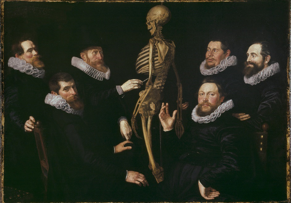
General topics, such as classical anatomy lessons (Figure 1), the figure of the physician in Art, classical surgery lessons, usually taking place in medical amphitheaters, and old-style dissections, may be used within any introductory lesson. Table 1a shows a list of artworks associated with general introductory topics.
| Topic | Period | Artist | Artwork |
|---|---|---|---|
| Beauty | |||
| Paleolithic (c. 25,000 -30,000 BCE) | Austria | Venus of Willendorf | |
| Mid 1480s | Sandro Botticelli | The Birth of Venus | |
| Aging | |||
| c. 1512 | Leonardo da Vinci (attributed) | Portrait of a Man in Red Chalk (self-portrait at about 60 years old) | |
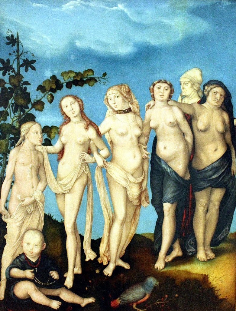
| 1544 | Hans Baldung (Grien) | Die Sieben Lebensalter des Weibes (“The Seven Ages of Woman”) | |
| 1626-1669 | Rembrandt | Self-portraits | |
| 1835 | Caspar David Friedrich | The Stages of Life | |
| 1905 | Gustav Klimt | The Three Ages of Woman | |
| Memento mori | |||
| c. 1485-1490 | Hieronymus Bosch | Death and the Miser | |
| c. 1509-1510 | Hans Baldung (Grien) | Three Ages of the Woman and the Death (The Three Phases of Life and Death) | |
| 1626-1628 | Frans Hals | Young Man with a Skull | |
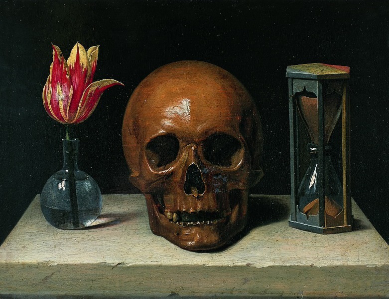
| c. 1671 | Philippe de Champaigne | Still-Life with a Skull | |
| 1885-1886 | Vincent van Gogh | Head of a Skeleton with a Burning Cigarette |
| Topic | Period | Artist | Artwork |
|---|---|---|---|
| Anatomy lessons | |||
| 1601-1603 | Aert Pietersz | Anatomy Lesson of Dr. Sebastiaen Egbertsz | |
| 1619 | Nicolaes Eliaszoon Pickenoy | The Osteology Lesson of Dr. Sebastiaen Egbertsz | |
| 1887 | Pierre Aristide André Brouillet | A Clinical Lesson at the Salpêtrière | |
| Physician | 1926 | Otto Dix | Portrait of Dr. Wilhelm Mayer-Hermann (laryngologist) |
| Surgery | |||
| c. 1494 | Hieronymus Bosch | The Extraction of the Stone of Madness | |
| 1628 | Rembrandt | The Foot Operation | |
| 1875 | Thomas Eakins | Portrait of Dr. Samuel D. Gross (The Gross Clinic; thigh surgery) | |
| 1889 | Thomas Eakins | The Agnew Clinic (partial mastectomy in a medical amphitheater) | |
| Dissection | |||
| 1493 | Johannes de’ Ketham | The Dissection (woodcut) | |
| 1632 | Rembrandt | The Anatomy Lesson of Dr. Nicolaes Tulp | |
| 1656 | Rembrandt | The Anatomy Lesson of Dr. Deijman (brain dissection) | |
| 1670 | Adriaen Backer | Anatomy Lesson by Dr. F. Ruysch | |
| 1683 | Jan van Neck | The Anatomical Lesson of Frederik Ruysch |
The concept of beauty, aging, and memento mori
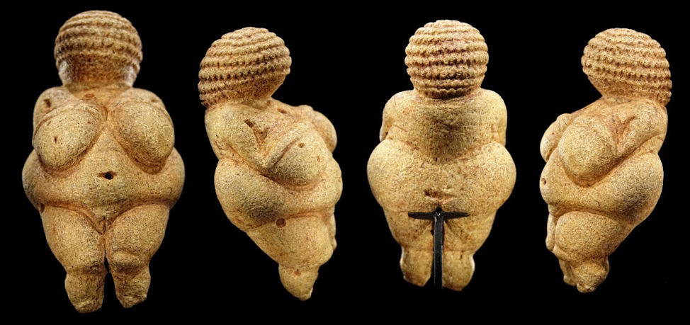
The concepts of beauty and aging may be part of any anatomical chapter. The former is an abstract concept, elaborated by artists of all time, but not univocal in its form of representation. The idea of beauty changes according to the historical, cultural, economic, and social context in which artists live. Nevertheless, universal beauty may converge over the centuries through the representation of Venus, a symbol of fertility and femaleness. Examples are the prehistoric figurines Venus of Hohle Fels (Germany, Upper Paleolithic period) and Venus of Willendorf (Austria, Paleolithic period; Figure 14). More recent examples are the Venus de Milo (Aphrodite of Melos, Hellenistic period, c. 160-110 BCE) and the one painted by Sandro Botticelli in the “Birth of Venus” (mid 1480s).
On the other hand, brief remarks on aging and how ephemeral life is may be useful at any time of the course. Picturing the different ages of men and women was a favorite subject of the Renaissance artists, as the symbol of evanescence (Figure 2). Moreover, memento mori and vanitas were extremely popular in the 17th century, i.e., artworks designed to remind the viewer of their mortality (Figure 3), of the shortness and fragility of human life, earthly achievements, and pleasures. These paintings usually consist of a portrait with a skull and/or other symbols of mortality (hour glasses, clocks, guttering candles). Interestingly, Rembrandt Harmenszoon van Rijn (referred to as simply Rembrandt) left a legacy of about 80 self-portraits, allowing us to notice how his face changed with aging, from 1628 – in his early 20s – up to months before his death, in 1669, at 63 years old. Viewers have a unique insight into the life, character, and psychological development of the man and the artist, a perspective of which the artist was profoundly aware and that he intentionally gave the viewer, as though a more thoughtful and studied precursor to the modern selfie (Marder, 2020). Table 1b shows a list of artworks associated with beauty, aging, and memento mori.
The musculoskeletal system
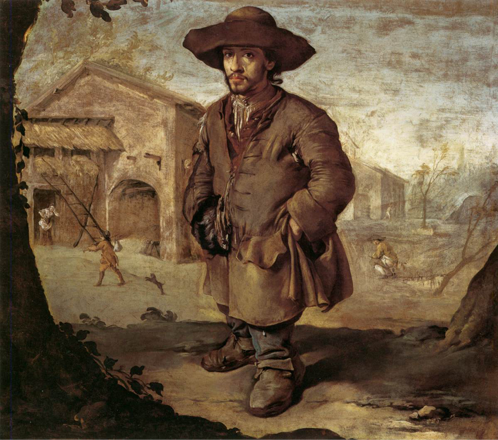
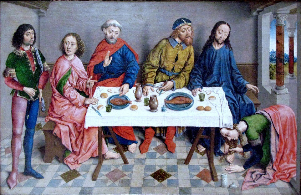
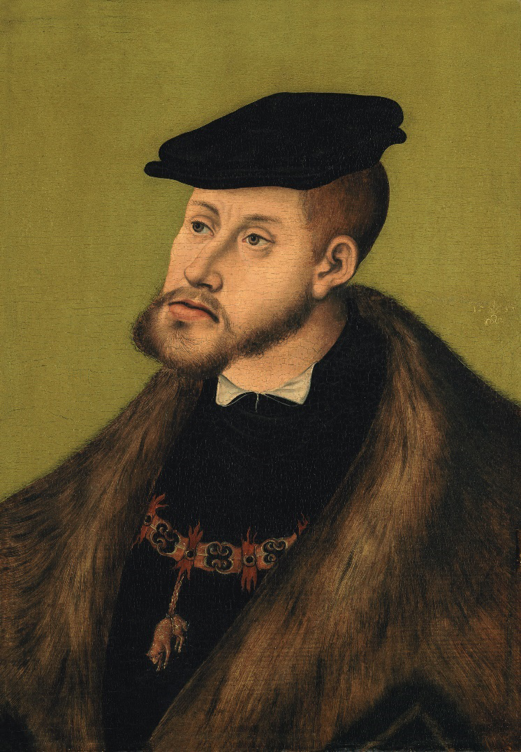
Examples of the skeletal anatomy as well as musculoskeletal disorders are easy to be found throughout Art history: achondroplasia in court dwarves (Figure 4); arachnodactyly (exaggerated length of ribs, extremities, and digits); polydactyly (supernumerary fingers); hallux valgus, i.e., lateral deviation of the hallux (Figure 5); prognathism due to consanguinity (Figure 6); connective tissue disorders, i.e., Marfan syndrome (Pyeritz, 2021); hand osteoarthritis and rheumatoid arthritis. A separate chapter is represented by genetic syndromes altering the musculoskeletal system. The famous large-format painting “The Family of King Philip IV” (better known as “Las Meninas”) by Diego de Velázquez is an excellent example of characters suffering different genetic syndromes with skeletal abnormalities. Table 2a shows a list of artworks associated with the musculoskeletal system.
| Topic | Period | Artist | Artwork |
|---|---|---|---|
| Proportions of the human body | c. 1492 | Leonardo da Vinci | The Proportions of the Human Figure (after Vitruvius) |
| Skull | |||
| Skull sectioned | c. 1489 | Leonardo da Vinci | The skull sectioned and the seat of the “senso comune” (drawing) |
| Skull carving | Contemporary | Gregory Raymond Halili | Mother-of-pearl shell hand-carving |
| Musculoskeletal disorders | |||
| Dwarfism | 1465-1474 | Andrea Mantegna | The Camera Picta |
| 1635-1645 | Diego de Velázquez | Francisco Lezcano (El Niño de Vallecas) | |
| c. 1720 | Giacomo Ceruti (“Pitocchetto”) | The Dwarf | |
| Gigantism | 1605 | Guido Reni | David with the Head of Goliath |
| 2001-2002 | Ron Mueck | The Mask II (self-portrait) | |
| Talipes equinovarus (clubfoot) | 1642 | José de Ribera (“Spagnoletto”) | El Patizambo (The Clubfoot) |
| Hallux valgus | c. 1490 | Aelbrecht Bouts | Christ at the Feast of Simon the Pharisee (the foot on the right) |
| Scoliosis | 1625-1630 | Pietro Paolini | Bacchic Concert |
| Scoliosis, hyperlordosis, and flat feet | 1636 | Peter Paul Rubens | The Three Graces |
| Mandibular prognathism | 1533 | Lucas Cranach the Elder | Portrait of the Emperor Charles V (consanguinity) |
| Connective tissue disorders | 1534-1540 | Parmigianino | Madonna with the Long Neck |
| Pectus excavatum, kyphosis, connective tissue disorders | 1929 | Christian Schad | Agosta, the Pigeon-Chested Man, and Rasha, the Black Dove |
| Paget’s disease of bone | c. 1513 | Quentin Matsys | The Ugly Duchess (aka “A Grotesque Old Woman”) |
| Genetic syndromes | 1656 | Diego de Velázquez | The Family of King Philip IV (aka “Las Meninas”) |
| Polydactyly | 1912-1913 | Marc Chagall | Self-portrait with Seven Digits |
| Hand osteoarthritis | c. 1540 | Jacopino Del Conte | Michelangelo’s portrait |
| Rheumatoid arthritis | 1483 | Sandro Botticelli | Portrait of a Young Man (Portrait of a Youth) |
| Topic | Period | Artist | Artwork |
|---|---|---|---|
| Studies of the major organs and vessels | c. 1480-1482 | Leonardo da Vinci | Drawing |
| Studies of the stomach and heart | c. 1508-1509 | Leonardo da Vinci | Drawings |
| Anatomy of a woman, showing the cardiovascular system and principal organs | c.1509-1510 | Leonardo da Vinci | Drawing |
| Studies of the chambers of the heart | c. 1513 | Leonardo da Vinci | Drawings |
| Heart | 1982 | Keith Haring | Untitled (painting of a radiating heart) |
| Bleeding heart | 1937 | Frida Kahlo | Memory, the Heart |
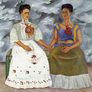
| 1939 | Frida Kahlo | The Two Fridas | |
| Heart, death, autopsy | 1890 | Enrique Simonet y Lombardo | Anatomy of the Heart (aka And She Had a Heart! or The Autopsy) |
| Dextrocardia and clubbing (“drumstick”) fingers | 1932 | Dick Ket | Self-portrait |
The cardiovascular system
The cardiovascular system may be triggered by the interesting self-portrait of Dick Ket, a Dutch painter who represents the New Realism in Art history. He suffered of cardiovascular problems and dextrocardia. In fact, a heart shadow is observed on the right part of his white t-shirt together with clubbing or “drumstick” fingers in his self-portrait of 1932). Besides, Frida Kahlo’s self-portraits “Memory, the Heart” and “The Two Fridas” (Figure 7) show bleeding hearts, a symbol of her life struggles (she was divorcing from his husband, the famous Mexican painter Diego Rivera). On the other hand, Leonardo da Vinci performed many anatomical studies during his life, either as sketches or as drawings. Most of these drawings reside in the Royal Library at Windsor Castle (UK). His drawings and writings on the heart’s anatomy and function are remarkably accurate, even if dated to the beginning of the 16th century. He described the heart’s central function, noting that the “blood from the heart receives freshness and air in the lungs” and described the muscle meshwork architecture of the endocardium (Sterpetti, 2019). Table 2b shows a list of artworks associated with the cardiovascular system.
Splanchnology
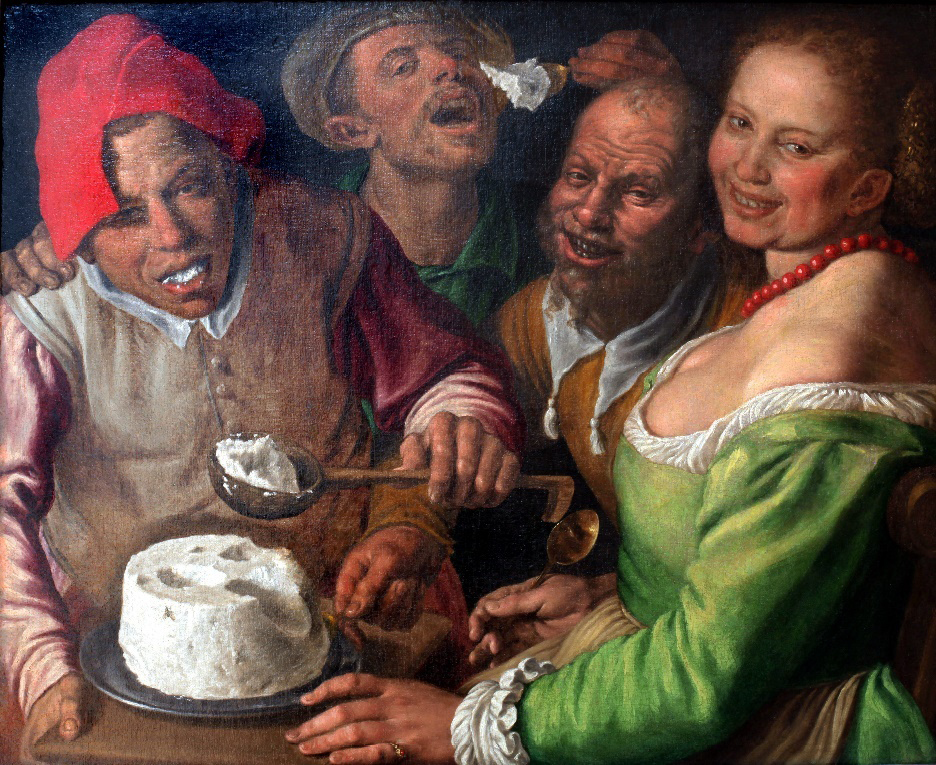
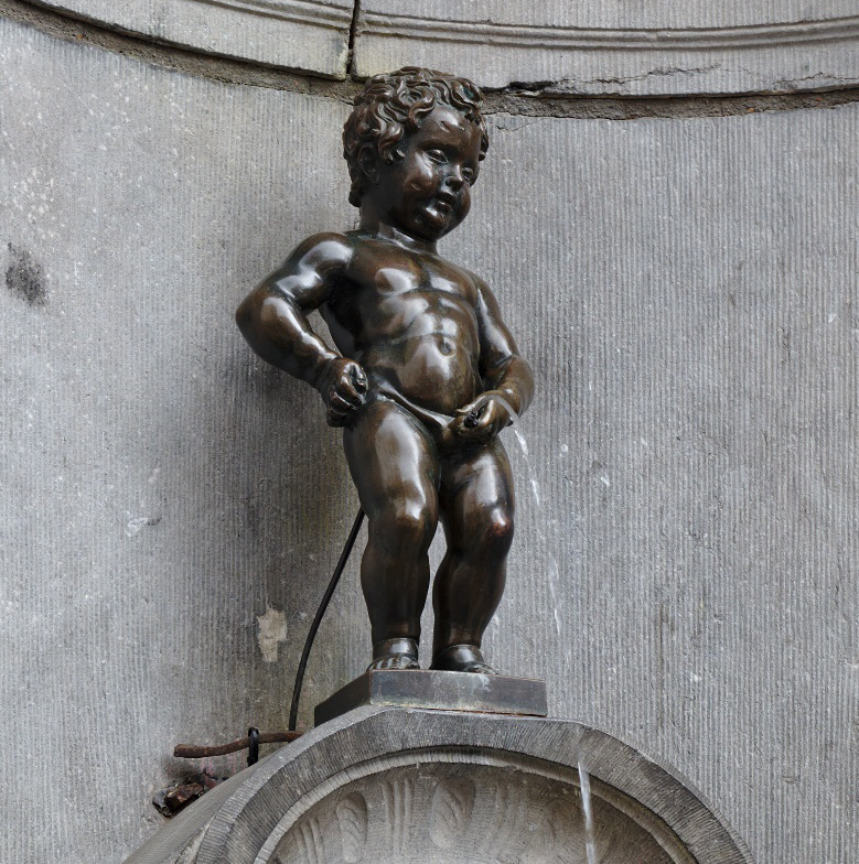
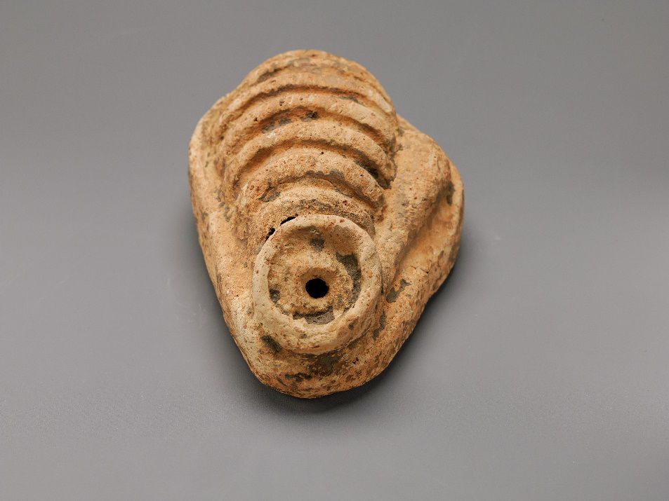
When triggering the lymphatic system, a painting such as “Portrait of Daniel Lambert of Leicester” by Benjamin Marshall (1806) may be useful to illustrate lymphedema (besides obesity). Useful artworks illustrating the respiratory system, are those of Piero della Francesca (hidden details on the coral pendant). Salvador Dalí’s installation of Mae West Room, particularly Mae West Lips, may be interesting to show for the oral cavity. Enhancing the concepts of “eating” or “swallowing” when talking about the esophagus or the stomach may be achieved by showing Vincenzo Campi’s artworks, for example, I Mangiatori di Ricotta (“The Ricotta Eaters”, c. 1585). Two curiosities of this nice painting are the artist’s self-portrait (dressed in red) and a skull design on the ricotta as a memento mori symbol (Figure 8). A curious way of enhancing the topic “intestines” and “defecation reflex” is by showing Giacomo Ceruti’s painting “The Dwarf” (Figure 4), asking the students not to focus on the first plane but on the background of the artwork. A tiny male character is observed in the typical position of defecation, in the middle of the street. Regarding the liver, the Bacchus paintings by Caravaggio show the artist himself with and without jaundice (a sign of hepatitis during and after healing). If talking, in turn, about the urinary system, Rembrandt’s etching prints on micturition are illustrated funnily, with a man and a woman “making water.” A famous, though tiny, landmark bronze fountain sculpture in central Brussels is the Manneken Pis depicting a little boy urinating into the fountain (Figure 9). Another example is “A Doctor Examining Urine,” an artwork by Trophime Bigot, the Candlelight Master, illustrating urine analysis. Triggers for the genital system are well represented by Leonardo da Vinci’s drawings as well as sculptures of female (uterus) and male (penis and scrotum) genital organs, especially as terracotta votive offerings coming from the Etruscan culture (Figure 10). Indeed, it was common in ancient Greek and Roman cultures to dedicate anatomical votive offerings to testify or acknowledge God’s power (Oberhelman, 2014). Finally, suggested works for beauty and aging may be used when discussing menopause (Figures 2-3). Table 3a shows a list of artworks associated with the lymphatic system and splanchnology.
| Topic | Period | Artist | Artwork | ||||||||||||||||
|---|---|---|---|---|---|---|---|---|---|---|---|---|---|---|---|---|---|---|---|
| Lymphatic system (lymphedema; obesity) | 1806 | Benjamin Marshall | Portrait of Daniel Lambert of Leicester | ||||||||||||||||
| Respiratory system (bronchial branching) | 1470-1485 | Piero della Francesca | Madonna of Senigallia and Brera Madonna (Montefeltro Altarpiece; details) | ||||||||||||||||
| Digestive system | |||||||||||||||||||
| Oral cavity | 1650 | Joos van Craesbeeck | The Temptation of Saint Anthony | ||||||||||||||||
| 1938 | Salvador Dalí | Mae West Lips Sofa (installation) | |||||||||||||||||
| Eating, swallowing; memento mori | c. 1585 | Vincenzo Campi | I Mangiatori di Ricotta (“The Ricotta Eaters”; with a self-portrait) | ||||||||||||||||
| Defecation reflex | 1720 | Giacomo Ceruti (“Il Pitocchetto”) | Il Nano (“The Dwarf”; detail) | ||||||||||||||||
| Jaundice (during illness) | 1594 | Caravaggio | Young Sick Bacchus (self-portrait) | ||||||||||||||||
| Jaundice (healing) | 1596 | Caravaggio | Bacchus (self-portrait) | ||||||||||||||||
| Urinary System | |||||||||||||||||||
| Micturition (child) | 1619; 1965* | Brussels landmark | Manneken Pis (“Little Pissing Man”); bronze fountain sculpture | ||||||||||||||||
| Micturition (man) | 1630 | Rembrandt | A Man Making Water (etching print) | ||||||||||||||||
| Micturition (woman) | 1631 | Rembrandt | A Woman Making Water (etching print) | ||||||||||||||||
| Urine analysis | 1630-1633 | Trophime Bigot (Candlelight Master) | A Doctor Examining Urine | ||||||||||||||||
| Genital system | |||||||||||||||||||
| Female genital system: uterus | 3rd-2nd century BCE | Etruscan culture | Votive terracotta uteri | ||||||||||||||||
| The foetus in the womb and other studies on reproduction | c. 1509-1516 | Leonardo da Vinci | Drawings | ||||||||||||||||
| Male genital system: penis and scrotum | 3rd-1st century BCE | Etruscan culture | Votive terracotta penis and scrotum | ||||||||||||||||
| 1506–1508 | Leonardo da Vinci | Weimar sheet (drawings) | |||||||||||||||||
| Coitus (anatomical studies) | 1490-1493 | Leonardo da Vinci | Sketch | ||||||||||||||||
| * This is a copy; the original one (1619) is kept at the Brussels City Museum. | |||||||||||||||||||
| Topic | Period | Artist | Artwork | ||||||||||||||||
|---|---|---|---|---|---|---|---|---|---|---|---|---|---|---|---|---|---|---|---|
| Normal gland | |||||||||||||||||||
| Young gland | 1484 | Sandro Botticelli | La Nascita di Venere (“The Birth of Venus”) | ||||||||||||||||
| Lactating gland | 1320-1325 | Ambrogio Lorenzetti | Madonna del Latte (“Madonna of Milk”) | ||||||||||||||||
| 1937 | Frida Kahlo | Mi Nana y Yo (“My Nurse and I”) | |||||||||||||||||
| Aging gland | 1507 | Albrecht Dürer | Avarice (Old Woman with a Purse) | ||||||||||||||||
| Abnormal gland | |||||||||||||||||||
| Abnormal anatomical position | 1487-1490 | Hans Memling | The Virgin Mary Nursing the Child Christ | ||||||||||||||||
| Breast cancer | |||||||||||||||||||
| 1969; 1972; 2014* | Nereo Costantino | Juliet statue (Juliet’s house, Verona) | |||||||||||||||||
| c. 1516 | Raffaello Sanzio | La Fornarina | |||||||||||||||||
| 1654 | Rembrandt | Bathsheba Holding King David’s Letter | |||||||||||||||||
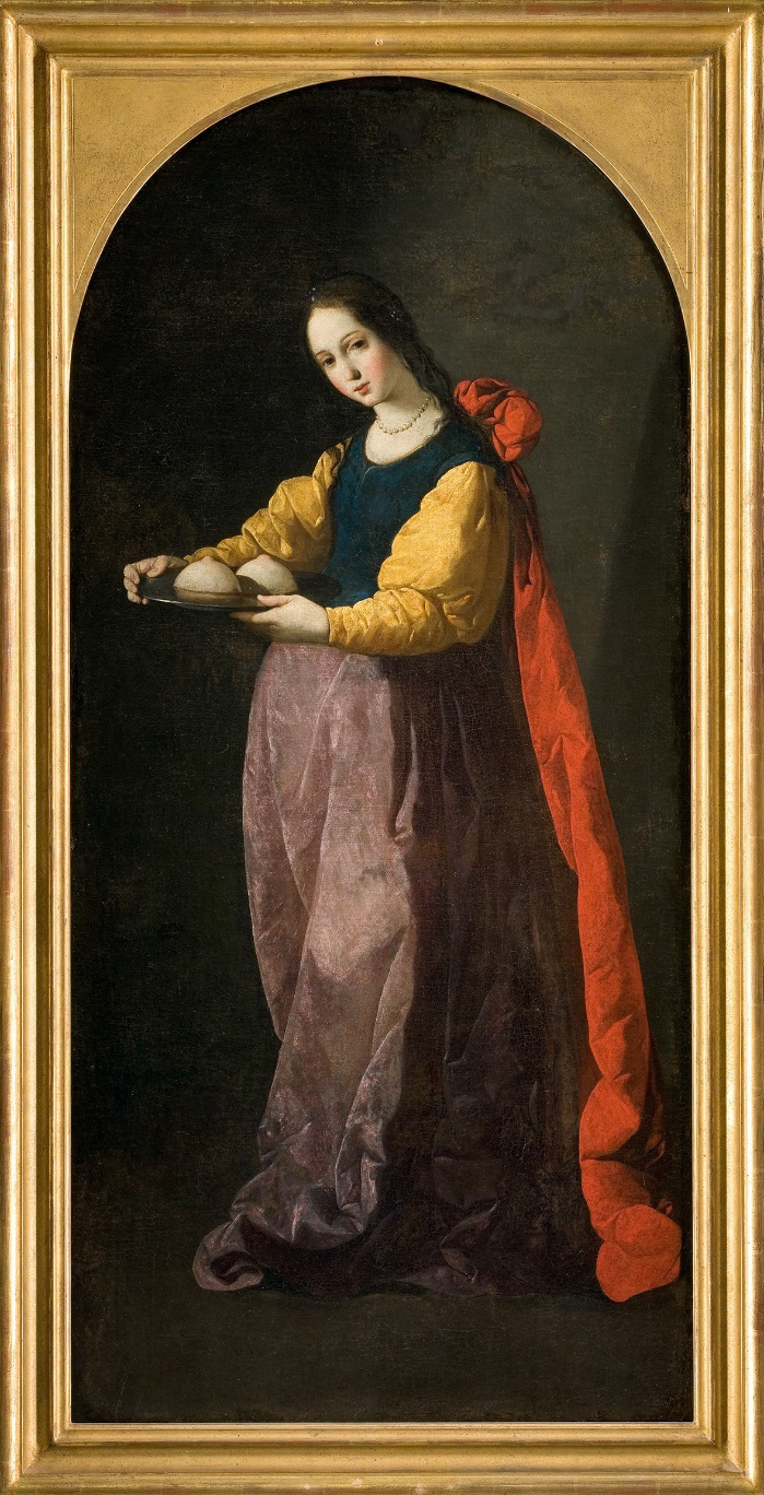
| Mastectomy | 1460-1470 | Piero della Francesca | Sant’Agata (“Saint Agatha”) | ||||||||||||||||
| 1630 | Francisco de Zurbarán | Santa Águeda (“Saint Agatha”) | |||||||||||||||||
| *Different copies of the statue have been made due to the continuous erosion caused by tourists rubbing Juliet’s breast. | |||||||||||||||||||
The mammary gland
The mammary gland is treated at various times according to the faculty we consider. In our case, it is traditionally linked to the female genital system: in other courses, in turn, it goes together with the integumentary system. The history of art provides examples of normal, active, and pathological mammary glands in young or aging women. The world’s most famous statue of Juliet in Verona (Italy) has been remade several times due to erosion caused by the tradition of rubbing her breasts for good luck. The erosion opens holes in the metal as cancer marks the women’s breasts. A mention of mastectomy may enrich the discussion on the personal life and human aspects of the women depicted. A good example of mastectomy is the martyrdom of Saint Agatha, like the ones illustrated in the artworks by Piero della Francesca and Francisco de Zurbarán (Figure 11). Table 3b shows a list of artworks associated with the mammary gland.
The nervous system
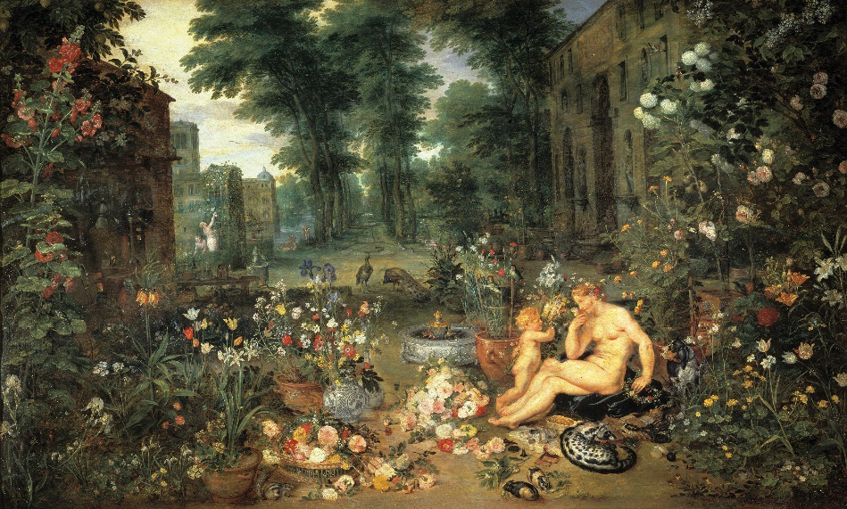
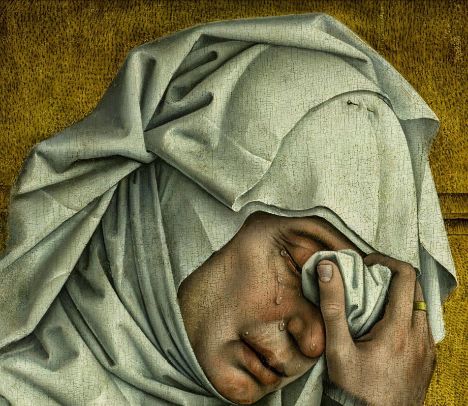
Some interesting artworks regarding pathologies of the nervous system, which allow a rich discussion, may be found in the last century (Radonjic, 2020), especially because with advancing medical technology many pathologies that were unknown or unidentified before, are now well described. Sometimes, we even know details on the clinical history of the figures illustrated in the artworks (i.e., Christine’s World by Andrew Wyeth, 1948). If talking, in turn, about the sense organs, Art offers a wide choice with allegorical paintings on the senses, especially the series done by Peter Paul Rubens together with Jan Brueghel the Elder during the 17th century (Figure 12). Delicate, realistic tears can be found in the painting “The Descent from the Cross” (“Deposition of Christ”, Figure 13) by one of the greatest Early Flemish artists (“Flemish Primitives”), Rogier van der Weyden, who lived in the 15th century. Table 4a shows a list of artworks associated with the nervous system.
| Topic | Period | Artist | Artwork |
|---|---|---|---|
| Nervous system | |||
| Poliomyelitis | 18th Egyptian Dynasty | Egyptian ancient art | The Doorkeeper (polio victim in an Egyptian stele) |
| Charcot-Marie-Tooth disease (hereditary motor and sensory neuropathy) | 1948 | Andrew Wyeth | Christina’s World |
| Sense Organs | |||
| Senses (sight, hearing, smell, taste, touch) | 1617-1618 | Peter Paul Rubens and Jan Brueghel the Elder | The Five Senses (allegorical series) |
| Hearing | 17th century | Jan van Kessel the Elder | Allegory of Hearing |
| Ear | |||
| 1889 | Vincent van Gogh | Self-Portrait with Bandaged Ear (self-mutilation) | |
| 20th century | Antonio Ligabue | Self-portraits series | |
| Ear pathology: cauliflower ear | late 4th–2nd century BCE | Greek, Hellenistic period | The Boxer at Rest (bronze statue) |
| Taste | 1645-1650 | Bartolomé Esteban Murillo | Children Eating Grapes and a Melon |
| Eyes | |||
| - | Natural, Monument Valley (Utah, USA) | The Eye of the Sun (rock) | |
| 5th century BCE or later | Classical Greek culture | Pair of stone eyes (designed for a statue) | |
| 12th century | Roman | Mosaic of Ecclesia Romana (polychrome mosaic, Old St. Peter’s Basilica apse) | |
| Eyes (tears) | Before 1443 | Rogier van der Weyden | The Descent from the Cross (detail Stabat Mater, Mary, left) |
| Red eyes | 1480-1500 | Dieric Bouts | Mater Dolorosa (Sorrowing Virgin) |
| 1928 | René Magritte | The False Mirror |
| Topic | Period | Artist | Artwork |
|---|---|---|---|
| Skin | |||
| Skin as a whole | 1562 | Marco d’Agrate | St. Bartholomew Flayed (Duomo of Milan; sculpture) |
| Epidermis (normal) | |||
| Infant (blush) | 1836 | Friedrich von Amerling | Portrait of Princess Maria Franziska von Liechtenstein Aged Two |
| Adult | c. 1460 | Rogier van der Weyden | Portrait of a Lady |
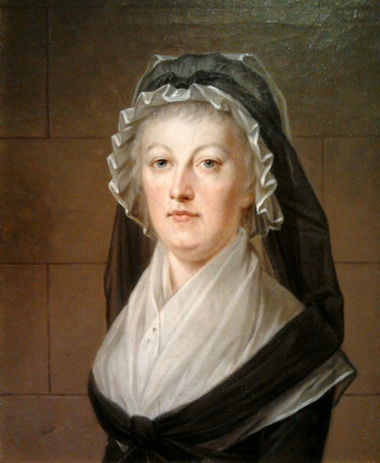
| Adult (pallor) | c. 1793 | Alexandre Kucharski | Marie Antoinette in the Temple Prison |
| Family | 1840 | Ferdinand Georg Waldmüller | Young Peasant Woman with Three Children at the Window |
| Old (wrinkles) | |||
| c. 1450 | Giorgio Schiavone (Juraj Culinovic) | Saint Bernardino of Siena | |
| Before 1768 | Christian Seybold | Portrait of an Old Woman with a Green Scarf | |
| Adnexa: hair | |||
| 1645 | Govern Flinck | Bearded Man with a Velvet Cap | |
| 1979 | Andrew Wyeth | Braids (The Helga Pictures series) | |
| Skin disorders | |||
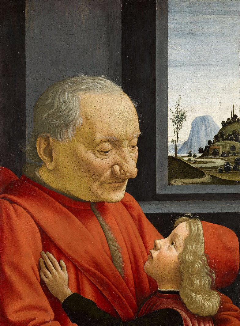
| Rhinophyma (complication of rosacea) | 1490 | Domenico Ghirlandaio | An Old Man and His Grandson |
| Alopecia | 1658 | Jan Vermeer | The Milkmaid |
| Hypertrichosis | c.1580 | Unknown | Portrait of Petrus Gonsalvus |
| Topic | Period | Artist | Artwork |
|---|---|---|---|
| Obesity, metabolic syndrome | Paleolithic (c. 25,000 -30,000 BCE) | Austria | Venus of Willendorf |
| c.1680 | Juan Carreño de Miranda | Eugenia Martínez Vallejo (La Monstrua Vestida) | |
| 20th-21st centuries | Fernando Botero | All his artworks (for example, Mujer Fumando Un Cigarrillo, “A Woman Smoking”, 1987, sculpture) | |
| Goiter, hypothyroidism | 1435-38 | Rogier van der Weyden | Virgin and Child in a Niche |
| 1460 | Andrea Mantegna | Virgin and Child | |
| 1512 | Michelangelo | The Creator Separating Light from Darkness (self-portrait); letters | |
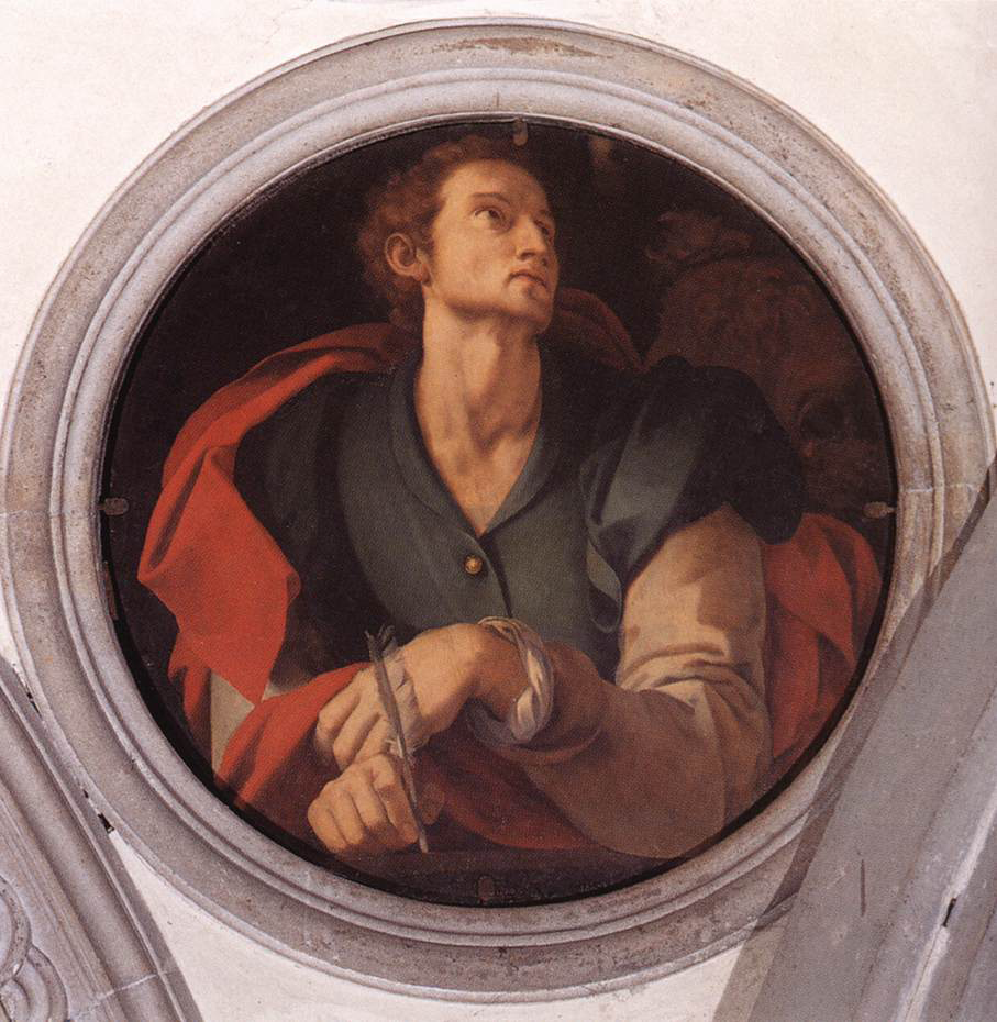
| c. 1525 | Jacopo Carucci (Pontormo) | San Luca (Saint Luke), decoration of the Capponi Chapel at Santa Felicita Church (Florence) | |
| 1612-1613 | Artemisia Gentileschi | Judith and Her Maidservant | |
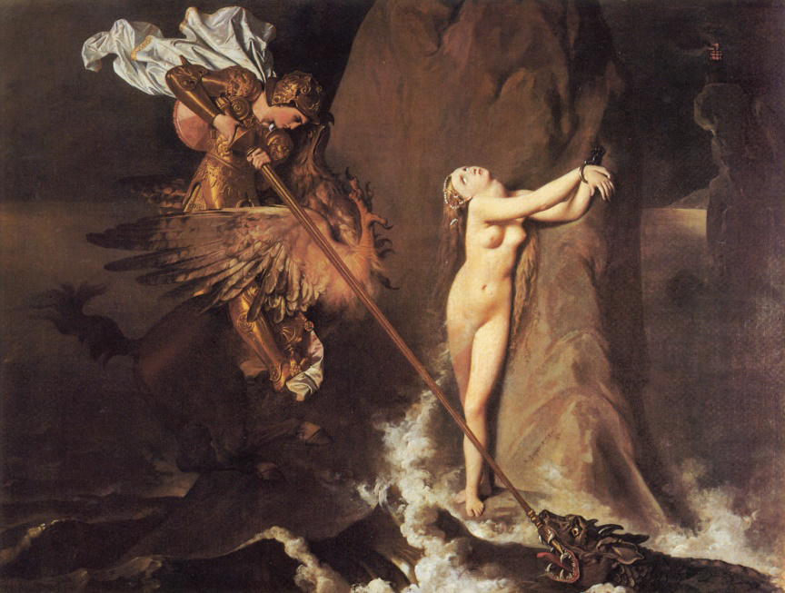
| 1819 | Jean-Auguste-Dominique Ingres | Roger Freeing Angelica | |
| Thyroglossal duct cyst | 1450-1463 | Piero della Francesca | Resurrection |
| 1463-65 | Piero della Francesca | Polyptych of the Misericordia | |
| Hyperthyroidism | 1499 | Albrecht Dürer | Self-portrait |
| Hirsutism | 1631 | José de Ribera | Magdalena Ventura with Her Husband and Son |
| Cushing’s syndrome (“moon face”) | 1517 | Hans Holbein the Younger | Adam and Eve |
| c. 1540 | Hans Holbein the Younger | Portrait of Henry VIII |
The endocrine system
Obesity and the related metabolic syndrome may be illustrated by choosing a Venus sculpture, like the Paleolithic Venus of Willendorf (Figure 14), or any of the characteristic artworks of the Colombian artist, Fernando Botero. Concerning the thyroid gland and its disorders, in turn, religious images show evident thyroid enlargements (Figures 15-16), i.e., hypothyroidism or a goiter (Traversari et al., 2017; Buta, 2021; Ashrafian, 2023, 2024). In that period, an enlarged or fat neck was considered a sign of beauty and prosperity (Sterpetti et al., 2015). Goiter is characterized by the loss or shallowing of the suprasternal notch recess or craniocervical neck flexion accentuating thyroid enlargement (Ashrafian, 2024). Interestingly, Michelangelo Buonarotti and Artemisia Gentileschi painted their self-portraits evidencing goiter. In addition, Artemisia Gentileschi’s famous painting “Judith and Her Maidservant” shows a goitrous neck (Christopoulou-Aletra et al., 2007). On the other hand, Piero della Francesca (1412-1492) painted two huge artworks showing himself with an evident lump or swelling in the middle of the anterior region of the neck, representing a thyroglossal duct cyst (Sterpetti et al., 2015).
Cushing’s syndrome is a pathology of the adrenal glands characterized by the so-called “moon face.” Two paintings of Hans Holbein the Younger, a German-Swiss artist, member of the Northern Renaissance style, and one of the greatest portraitists of the 16th century, may be good examples in this case. Table 4b shows a list of artworks associated with the endocrine system.
The integumentary system
When teaching this subject, one should first point to the characteristics of normal skin and its changes with aging. In this way, students get used to distinguishing skin physiological changes (blush, pallor, wrinkles). Alexander Kucharski’s painting “Marie Antoinette in the Temple Prison” (c. 1793) points out the French Queen’s pallor while imprisoned in the Tower of the Temple (Figure 17), before her execution by guillotine.
Abnormalities, like cyanosis or texture changes (i.e., rhinophyma, a complication of rosacea) may also be considered. Domenico Ghirlandaio shows a magnificent example of rhinophyma in his painting “An Old Man and His Grandson” (c. 1490, Figure 18). When talking about skin adnexa, in particular, the hair follicle, artworks showing subjects with thick beards, hypertrichosis, or alopecia, may be useful. As a curiosity, the first recorded case of hypertrichosis was described in 1580; it corresponded to Petrus Gonsalvus, a gentleman in the court of Henry II of France. On the other hand, examples of pseudoalopecia and true frontal alopecia may be observed in paintings from the 15th to the 17th centuries (Fernandez-Flores et al., 2020). A good example of how to illustrate alopecia is represented by Jan Vermeer’s famous painting “The Milkmaid” (1658). Table 4c shows a list of artworks associated with the integumentary system.
Artists’ disease
In the case of artists’ diseases, many are the examples to be suggested, which can be properly included along the whole human and clinical anatomy course program, as well as in ad hoc conferences or webinars. Besides their famous self-portraits, modern artists were also depicted in their daily lives through paintings or photographs. At times, their precious legacies of letters and poems provide insight into the struggles they faced. The self-portrait of a diseased Goya with his friend and physician, Dr. Arrieta (1820), well illustrates the bad painter’s conditions at that moment (note the painting’s background, which features various ghostly figures that suggest the artist’s hallucinations). Moreover, his work is divided into two main periods, light, and darkness, before and after his diseases.
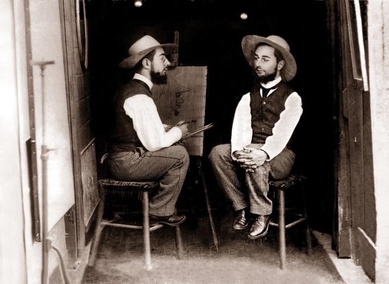
Henri Marie Raymond de Toulouse-Lautrec-Monfa, better known as Henri de Toulouse-Lautrec (1864–1901), had pycnodysostosis (Maroteaux-Lamy syndrome), a recessive autosomal dysplasia characterized by alteration of the bone matrix and bone fragility (he broke his legs at 13 and 14 years old); short stature (1,54 m), with an adult thorax and child inferior limbs. His skull was deformed, with a broad forehead. For the above reasons, he walked with great difficulty and suffered from recurrent pain (Markatos et al., 2018). He had to move with a cane and always wore a hat (Figure 19). Besides, he got syphilis and cerebral alcoholism. He died at 36 years old from a stroke. Curiously, his short stature helped him in his unique artistic style, the “cut-off” technique, subsequently adopted by other painters, where objects and figures appear cut off from the edge of the frame and from unusual angles.
Vincent van Gogh, in turn, was treated with digitalis by his physician for presumed neuropsychiatric diseases. This substance was considered in the 19th century as one of the treatments for epilepsy as well as for some psychiatric illnesses. One of the well-known side effects of systemic digitalis treatment is a disturbance in yellow-blue vision (xanthopsia). Due to digitalis-induced xanthopsia, van Gogh may have perceived the world with a yellow tint, which also resulted in the predominance of colored haloes around light sources in some of his famous works. Alternatively, the haloes in his works may be caused by glaucoma (Dahan and Shoenfeld, 2017). In an analogous way, when analyzing Claude Monet’s paintings (19th to 20th centuries), a change in color shades and focus of the water lilies series is noticed before and after cataracts surgery. Monet struggled to differentiate colors accurately and wrote about this to his friends. His letters provide a glimpse into the life of an artist who overcame such challenges to create masterpieces still revered today. Understanding his struggles can inspire us to appreciate his works on a deeper level.
Finally, two famous artists deserve attention. Firstly, Pierre-Auguste Renoir (1841-1919), secondly, Magdalena Carmen Frida Kahlo y Calderón, better known as Frida Kahlo (1907-1954). The former was a French artist, leader in the development of the Impressionist style. Around 1892, Renoir developed rheumatoid arthritis, worsened by cigarette smoking. He painted during the last twenty years of his life even after his rheumatoid arthritis severely limited his mobility. His hands were deformed and had also ankylosis of his right shoulder (Figure 20). The disease progressed in paralysis; as a consequence, the artist required a wheelchair in his last years. Nevertheless, Renoir remained able to grasp a brush, although he required an assistant to place it in his hand. The life of Renoir shows how disability cannot quell an artist’s desire to create. When asked about his plight the disabled artist gave the amiable reply of “the pain passes, but the beauty remains” (Gandhi, 2019). Frida Kahlo is also an outstanding example of how strongly a person reacts to tragedy and illness. Unconventional and vital, she was marked from birth by pain and illness. She was born with spina bifida. At 18 years old she had a terrible accident, leaving her with severe polytraumatism, including her spine. She needed thirty-two orthopedic surgeries, the necessity of lying in bed for years. Consequently, she was unable to have a child. Her parents arranged a mirror above her bed so she could see herself. Consequently, her artwork is full of self-portraits and symbols, representing her feelings on physical and sentimental pain. The latter can be read everywhere in her famous painting “The Broken Spine” (La Columna Rota, Figure 21) painted in 1944, shortly after she had spinal surgery to correct the ongoing consequences of the traffic accident. Her spine is replaced by an Ionic column, nails are placed all over her body, and a desertic, sterile background, represents her infertility. After the amputation of her leg, she wrote “Feet, what do I need you for when I have wings to fly?”
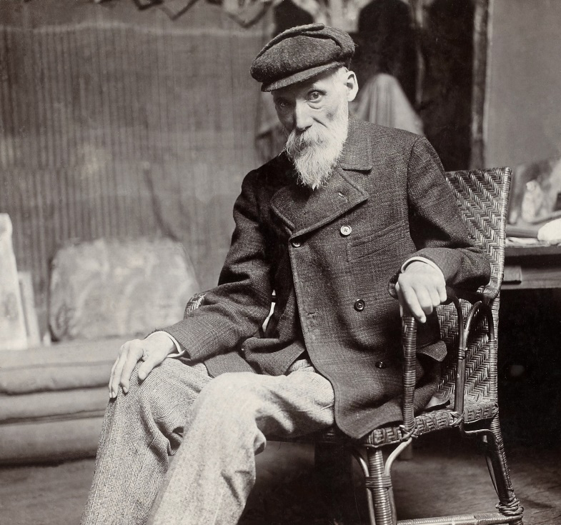
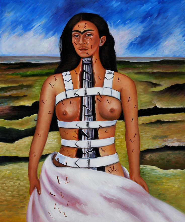
Table 5a shows a list of artworks associated with artists’ diseases.
| Artist | Period | Disease | Artwork |
|---|---|---|---|
| Piero della Francesca | 15th century | Thyroglossal duct cyst | Self-portraits (“Polyptych of the Misericordia” and “The Resurrection”) |
| Albrecht Dürer | 15th-16th centuries | Hyperthyroidism | Self-Portrait at Twenty-Eight |
| Michelangelo Buonarroti | 16th century | Goiter; hand osteoarthritis | Self-portraits, poems, and letters |
| Michelangelo Merisi “Caravaggio” | 16th-17th centuries | Syphilis; jaundice (acute hepatitis) | Self-portraits (Bacchus paintings during illness and after healing) |
| Francisco José de Goya y Lucientes | 18th-19th centuries | Encephalopathy caused by chronic lead poisoning (saturnism); labyrinthitis; deafness; hallucinations | A sharp difference in light and color is noticed in his artworks before and after his health problems; letters |
| Joseph Mallord William Turner | 18th-19th centuries | Color blindness and cataracts | A change in color shades of his artworks is noticed with the progress of his eye problems |
| Vincent van Gogh | 19th century | Alcoholism; xanthopsia; Mèniére syndrome; glaucoma; epilepsy | Self-portraits and letters |
| Henri de Toulouse-Lautrec | 19th century | Pycnodysostosis (genetic dwarfism); alcoholism and syphilis | Photographs |
| Pablo Picasso | 19-20th centuries | Dysmorphic mental disorder (body dysmorphic disorder) | Self-portraits |
| Claude Monet | 19th-20th centuries | Myopia and bilateral cataracts | A change in color shades and focus of the water lilies series is noticed before and after cataracts surgery; letters |
| Pierre-Auguste Renoir | 19th-20th centuries | Myopia, rheumatoid arthritis, and later paralysis | Self-portraits, letters, and photographs |
| Edgar Degas | 19th-20th centuries | Myopia and retinopathy | A change in color shades and in the focus of his paintings is noticed along the years; letters |
| Frida Kahlo | 20th century | Bifid spine; infertility; pain; polytraumatism; amputation; post-traumatic fibromyalgia | Self-portraits (La Columna Rota; “The Broken Column”, 1944), letters, photographs |
Students’ opinion
Class representatives from the first to fifth year of the medical undergraduate degree were asked to anonymously evaluate the use of artworks during Anatomy lessons, workshops, and conferences. Students may submit an adjective or sentence either by email or anonymously through chat. Tables 6-7 show a summary of their comments (for simplicity, comments are in alphabetical order).
| – Admirable | – Brilliant | – Concrete |
|---|---|---|
| – Creative | – Cutting-edge | – Different |
| – Engaging | – Fascinating | – Funny |
| – Innovative/very innovative | – Interesting/very interesting | – Original |
| – Pleasant | – Stimulating/very stimulating | – Unexpected |
| A cultural, interesting, and ludic way of reconstructing medicine along history. |
|---|
| A deep point of reflection on the concept of disease and medicine as elements influencing daily aspects of life, including artistic expression. |
| An enjoyable way to renew my art interest, especially if treating topics that we usually only see in textbooks. |
| Behind artworks, we have artists as well as models with their clinical history, strictly dependent on their own historical, social, cultural, and scientific period. Moreover, talented artists (from Monet to Michelangelo, from Egyptians to Kahlo) became “common” persons, suffering from the aches and diseases of our times. |
| Certain concepts are fixed easily and for a long time. |
| I have never thought that art could be so helpful. |
| It is helpful to develop lateral thinking to stimulate concentration during the lessons, especially at the beginning of the core curriculum. |
| It helps to apply anatomical knowledge in daily life and helps to think “out of the schemes”. |
| I have appreciated references to mental diseases and how these have an impact on art. |
| Anatomy, like philosophy and literature, brings me joy. They share the common denominator of getting to know oneself from different perspectives, all equally fascinating and intertwined. |
| Medicine is a fusion of sensitivity and technique. It is art more than art itself. |
| It is exciting to realize how medicine has influenced art. |
| Makes us reflect. |
| It offers an alternative perspective of artworks, a more medical one. It looks for and explains anatomical variations or pathologies, thus helping to fix these concepts immediately. |
| It opens the mind. |
| Perfect mix between art and medicine. |
| It shows me what it really means to have passion for a discipline. It is uncommon for a professor to share their own life, thoughts, and hobbies. These professors will remain in our memory as students. |
| It stimulates “photographic” memory, curiosity, and intuition. |
| The use of art as a tool to stimulate the clinical eye is advantageous in that daily life experiences may complement formal study from textbooks. |
| The use of artworks both during in-presence lessons and ad hoc webinars has allowed a pleasant moment of relaxation, in between so many university commitments. The university is too concentrated on the “lessons calendar” rather than on ludic or interactive activities, which may be able to improve a student’s day. |
Discussion
From the medical student’s point of view, Anatomy represents a hard and challenging discipline. It is a three-semester course given during the first two years of the medical degree curriculum. Therefore, teachers need to constantly innovate to keep both interest and concentration high. The use of artworks at any point of the program represents a new experience that helps to make anatomy teaching lighter and even funnier, without disregarding quality or contents. Good resolution images are easily found without problems of licensing and with no cost at all. It is always suggested to cite the details of each artwork (title, author, period) as well as the resources where they were obtained from. This work intends to be only a guide to whoever decides to start innovating teaching with artworks. Moreover, the suggested artworks herein contained may be changed and/or mixed with other types of artworks or even forms of Art, according to the personal preferences, the kind of audience and the type of intervention (undergraduate or postgraduate lesson; duration of the lesson, conference, or online seminar). It is always a “work in progress”, modelling the selection of artworks according to the main objectives of the lesson or talk.
Students actively and enthusiastically participated, and collaboratively communicated, both during in-presence lessons as well as during in-presence and online webinars. Guided by the teacher, they focused on distinctive features of the artworks shown, thus rendering learning enjoyable and meaningful. Discussions are rich and energetic, representing a unique opportunity to interact with faculty on a non-medical level. Students learned to disagree respectfully, appreciating other perspectives. Students learned to observe objectively, to navigate uncertainty, searching a collective and no single truth. It is highly recommended to enable anonymous chat during webinars, as it allows students to express their thoughts freely. When teaching, the teacher may encourage students, especially shy ones, to participate in class discussions or ask questions via email voluntarily. The feedback was positive, even if only part of the students replied, either directly or through their representatives.
Artistic creativity is one amazing feature of human uniqueness. Moreover, Art is a representation of human life experiences across different eras and times, intertwined with emotion and cultural expression. Artificial intelligence Art, in turn, lacks the human touch and is devoid of emotion and meaning.
According to the online Encyclopedia Britannica (2024), the term Art encompasses diverse media such as painting, sculpture, printmaking, drawing, decorative arts, photography, and installation. Art-based lessons or sessions tend to approach observation and pattern recognition, metaphorically, and symbolically. Artworks represent a powerful, easy-to-find resource to understand human body changes along life as well as the natural course of diseases. It is a useful teaching tool to refine visual skills and to build so-called visual literacy, in other words, the ability to reason anatomy, physiology, and pathophysiology from visual clues (Naghshineh et al., 2008; Bramstedt, 2016). Art has a positive impact on mindfulness, i.e., a state of mind that is open to new information, actively engaged in the present, aware of multiple perspectives (Langer, 2000). In a few words, students learn a broader conception of humanness and acquire a more complete “picture” of the subjects/patients.
A particular feature emerged after reading the students’ opinions. Medical humanities are essential during undergraduate studies to develop the student-person-future physician. The fact that a teacher talks about Art in a medical course, making them to notice details of an artwork never seen before, changes their perspective, stimulating critical and lateral thinking, against the rigidity of a single medical diagnosis. Seeing is defined not only as observation of physical signs and features, but also a process of understanding both person and context. Art opens the mind to different parallel solutions, all of them valid, if properly discussed. Michelangelo was once asked how he went about the sculpting process; his answer was: “I saw the angel in the marble and carved until I set him free”.
Finally, a crucial point to consider is “teaching the teachers”. There are many ways to form medical educators: seminars, courses, or webinars, which can be organized without much complexity. Sapienza University of Rome, for example, thanks to the Committee for Medical Education, organizes periodical seminars and courses addressed to medical teachers within the context of a tutorial on medical humanities.
Conclusions
An Art-based approach to teaching and training visual skills should be included continuously within the medical curriculum. It is our duty to stimulate a formal commitment with medical humanities in any biomedical degree, including both students and colleagues. The use of figurative art has a positive impact on students’ diagnostic skills, empathy, and mindfulness. Moreover, it favors critical and lateral thinking, as well as problem-solving, opening the mind to alternate ways of seeing. The natural consequence is to enhance students’ abilities to observe more deeply and to better understand real clinical situations, thus training more empathic (future) physicians.
Acknowledgements
Thanks are due to Prof. G. Familiari for the ideas given.
References
Ambrogi V. (2014) Piero della Francesca’s coral pendants: the hidden bronchial anatomy. Thorax. 69: 1161. https://doi.org/10.1136/thoraxjnl-2014-205344
Ashrafian H. (2023) Goiters in the renaissance era: multiple cases of thyroid autoimmunity and iodine deficiency. Best Pract Res Clin Endocrinol Metab. 37(2): 101748. https://doi.org/10.1016/j.beem.2023.101748. Epub 2023 Feb 15.
Ashrafian H. (2024) Goiter in the paintings by Rogier van der Weyden (1399-1464). J Endocrinol Invest. 47(2):483-484. https://doi.org/10.1007/s40618-023-02108-1. Epub 2023 May 12.
Barry D.S., Marzouk F., Chulak-Oglu K., Bennett D., Tierney P., O’Keeffe G.W. (2016) Anatomy education for the YouTube generation. Anat Sci Educ. 9: 90-96. https://doi.org/10.1002/ase.1550
Bramstedt K.A. (2016) The use of visual arts as a window to diagnosing medical pathologies. AMA J Ethics. 18(8): 843-854. https://doi.org/10.1001/journalofethics.2016.18.8.imhl1-1608
Britannica, The Editors of Encyclopaedia (2024). "Art". Encyclopaedia Britannica, 29 Mar. 2024, https://www.britannica.com/art/visual-arts. Accessed April 3, 2024.
Buta M.G. (2021) Genetic diseases in religious paintings. Medicine and Pharmacy Reports 94(3): 382-389. https://doi.org/10.15386%2Fmpr-1996
Christopoulou-Aletra H., Papavramidou N., Pozzilli P. (2007) Goitrous beauty in Artemisia Gentileschi’s Judit and her Maidservant. Thyroid. 17(1): 37-38. https://doi.org/10.1089/thy.2006.0223
Dahan S., Shoenfeld Y. (2017) A picture is worth a thousand words: Art and Medicine. IMAJ 19(12): 772-776.
Facebook https://www.facebook.com/arteanatomia/
Fernandez-Flores A., Marqués Serrano O., Serrano S., Fonseca E. (2020) True frontal alopecia in 17th-century paintings. Clinics in Dermatology 38: 574-579. https://doi.org/10.1016/j.clindermatol.2020.04.002
Friedlaender G.E., Friedlaender L.K. (2013) Art in science: enhancing observational skills. Clin Orthop Relat Res. 471: 2065-2067. https://doi.org/10.1007%2Fs11999-013-3000-0
Gandhi JS. (2019) The rheumatoid hands of Renoir. Am J Med. 132(5): 658-659. https://doi.org/10.1016/j.amjmed.2018.12.034. Epub 2019 Jan 16.
Google Arts & Culture https://artsandculture.google.com/
Heyn R. (2018) The art of observation, from visual artworks to medical diagnosis: initial experience at Sapienza University of Rome. In: Orefice C., Baños J.-E. Monographs 38. The role of humanities in the teaching of medical students; Dr. Antoni Esteve Foundation, Barcelona. Pp. 116-125.
Jasani S.K., Saks N.S. (2013) Utilizing visual art to enhance the clinical observation skills of medical students. Med Teach. 35(7): e1327-31. https://doi.org/10.3109/0142159X.2013.770131. Epub 2013 May 3.
Langer E.J. (2000) Mindful learning. Curr Dir Psychol Sci. 9: 220-223. https://doi.org/10.1111/1467-8721.00099
Marder L. (2020) Rembrandt's Self-Portraits. Retrieved from: ThoughtCo. Aug. 28, 2020, https://www.thoughtco.com/rembrandts-selfportraits-4153454 Accessed March 28, 2024.
Markatos K., Mavrogenis A.F., Karamanou M., Androutsos G. (2018) Pycnodysostosis: the disease of Henri de Toulouse-Lautrec. Eur J Orthop Surg Traumatol. 28(8): 1569-1572. https://doi.org/10.1007/s00590-018-2233-8. Epub 2018 May 24.
Miller A., Grohe M., Khoshbin S., Katz J.T. (2013) From the galleries to the clinic: applying art museum lessons to patient care. J Med Humanit. 34: 433-438. https://doi.org/10.1007/s10912-013-9250-8
Naghshineh S., Hafler J.P., Miller A.R., Blanco M.A., Lipsitz S.R., Dubroff R.P., Khoshbin S., Katz J.T. (2008) Formal art observation training improves medical students’ visual diagnostic skills. J Gen Intern Med. 23: 991-997. https://doi.org/10.1007%2Fs11606-008-0667-0
Oberhelman S.M. (2014) Anatomical votive reliefs as evidence for specialization at healing sanctuaries in the ancient Mediterranean world. AJH. 1(1): 47-62. https://doi.org/10.30958/ajh.1-1-4
Powley E., Higson R. (2013) The arts in medical education. A practical guide. Second edition. CRC Press, London, pp. 67-84.
Pyeritz R.E. (2021) Arachnodactyly represented in art. Am J Med Genet. 187C: 163-167. https://doi.org/10.1002/ajmg.c.31930. Epub 2021 May 22.
Radonjic A. (2020) The undiagnosed patient in Christina’s world. Am J Med. 133(2): 253-254. https://doi.org/10.1016/j.amjmed.2019.09.013. Epub 2019 Oct 14.
Srivastava A.A., Cohen S., Hailey D., Khoshbin S., Katz J.T., Gaske I.M. (2022) Training the eye, virtually: adapting an art in medicine curriculum for on-line learning. SN Soc Sci. (2022) 2:158. https://doi.org/10.1007/s43545-022-00442-4
Sterpetti A.V. (2019) Cardiovascular research by Leonardo da Vinci (1452-1519). Circ Res. 124(2): 189-191. https://doi.org/10.1161/CIRCRESAHA.118.314253
Sterpetti A.V., De Toma G., De Cesare A. (2015) Thyroid swelling in the art of the Italian Renaissance. Am J Surg. 210: 591-596. https://doi.org/10.1016/j.amjsurg.2015.01.027
Traversari M., Ballestrieri R., Galassi F.M. (2017) A likely cause of goiter in the Madonna col Bambino dormiente (1465/1470) by Andrea Mantegna (1431-1506). J Endocrinol Invest. 40: 237-238. https://doi.org/10.1007/s40618-016-0548-z
Wikimedia Commons https://w.wiki/Ns
YouTube https://www.youtube.com/channel/UCU1c-4GTyIXC8YH-1ZcicsA and https://www.youtube.com/c/RosemarieHeyn