https://doi.org/10.36253/ijae-15259
Anatomical study of an unusual variant of the great saphenous vein in cadaver: case report and nomenclature review
Section of Human Anatomy, Department of Anatomical, Histological, Forensic Medicine and Orthopedic Sciences, Sapienza University of Rome, Rome, Italy
§ These authors contributed equally and are listed in alphabetical order.
*Corresponding author. E-mail: guido.carpino@uniroma1.it
Abstract. During routine cadaveric dissection for undergraduate medical students, a variant of the great saphenous vein (GSV) was revealed. Its peculiarity resides in the relationship with the deep aponeurotic fascia in the thigh (i.e. fascia lata). Particularly, the GSV duplicated in two vessels at knee level: a “superficial” vein ran in the saphenous compartment (between superficial and deep aponeurotic fasciae), while the other deepened below the fascia lata. Given the clinical importance of the GSV and its frequent anatomical variations, this case report gave us the cue for reviewing the normal anatomy of GSV and for a critical revision of the nomenclature of its anatomical variations. GSV variants can be divided into two main patterns: i) the presence of a parallel accessory vein, and ii) vein duplication. The main features to distinguish these two variations reside in the caliber of the vein and its position with respect to the saphenous compartment. When compared to variants reported in scientific literature, our case seemed not to completely fit neither with the definition of a duplicated vessel or of an accessory vein. Remarkably, the presence of GSV variant may have key clinical implications. In particular, the relationship with fascial layers could influence the development of vein dilation and varicosity. Therefore, the understanding of GSV anatomy and its accurate study by ultrasound seem to be essential to support clinical and surgical practices relating to the superficial venous drainage of the lower limb.
Keywords: great saphenous vein, anatomical variant, fascia lata, duplication, accessory veins.
Contents
Normal anatomy of the great saphenous vein
Definition of GSV anatomical variations
The GSV variants and clinical implications
Case report
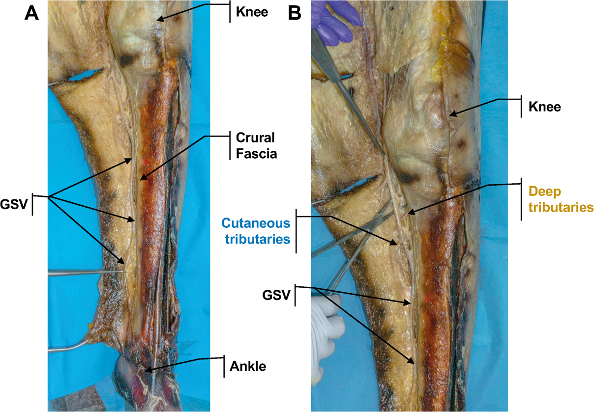
An anatomical variation of the great saphenous vein (GSV) was found in the left lower limb of a 108-year-old male cadaver during routine cadaveric dissection for undergraduate medical students. The left GSV had a normal origin from the dorsal venous arch of the foot, anterior to the medial malleolus and ascending the leg along its anteromedial surface, superficially to the crural fascia (Figure 1).
At the medial condyle of the tibia, a venous vessel was individuated posteromedially to the GSV and originating from the subcutaneous layer (Figure 2).
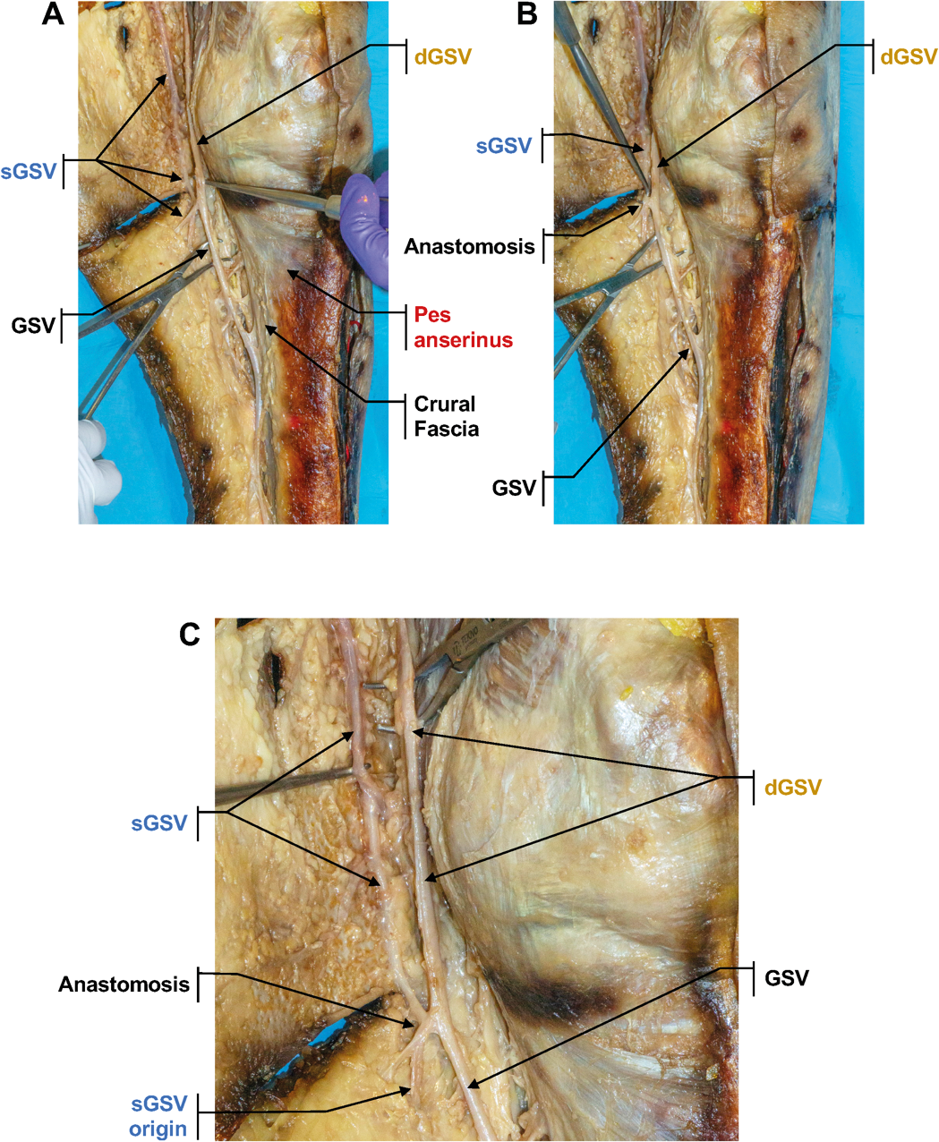
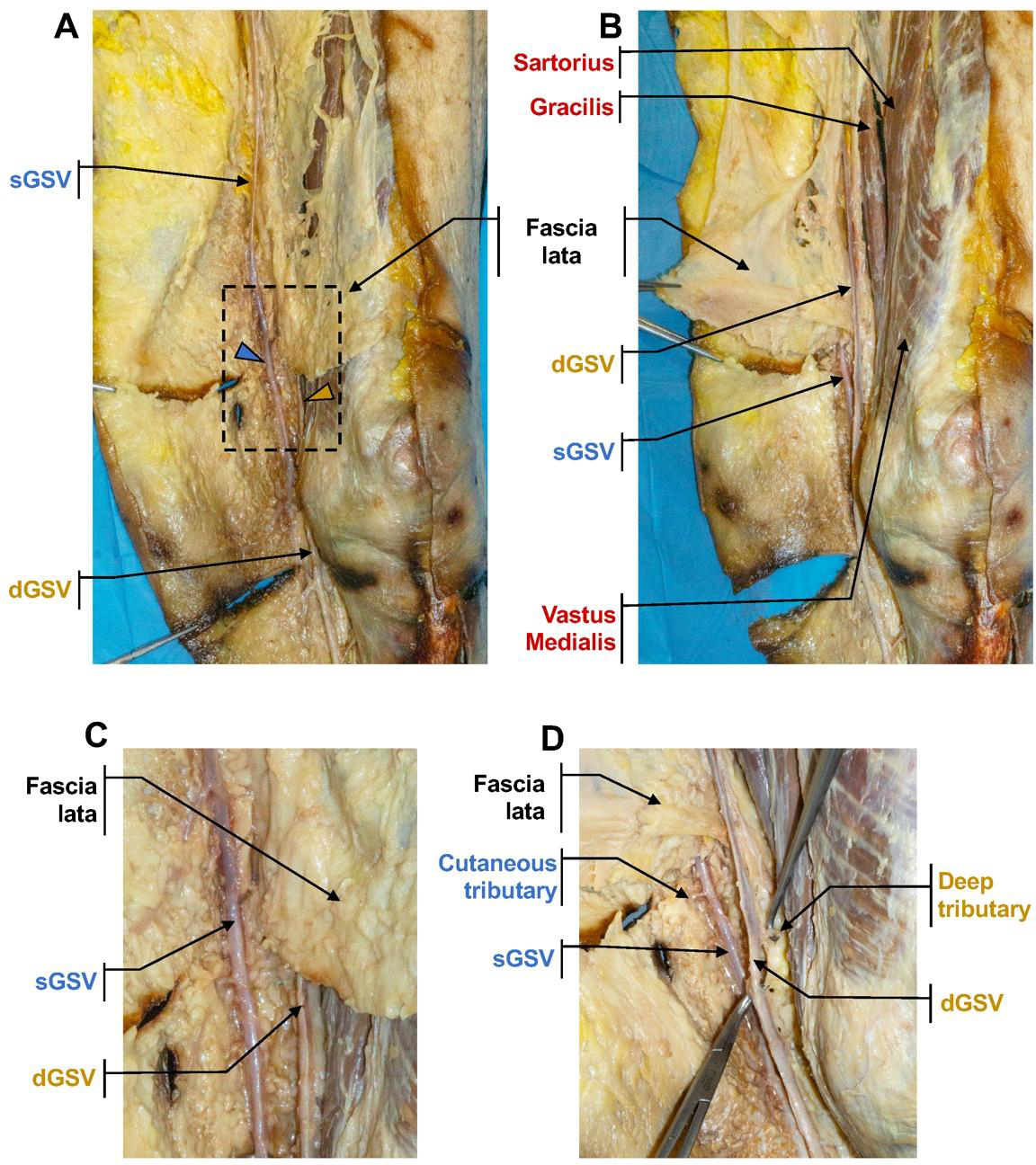
This vessel had the same diameter of the GSV, and an anastomosing branch connected the two vessels. After the anastomosis, the two veins ascended individually and parallelly to each other. The one initially individuated in the leg deepened below the fascia lata; the other ran superficially into the saphenous compartment (Figure 3). The two vessels kept the same diameter as they ascended in the thigh region. The right lower limb did not present the same variation.
Given the complexity of the examined variation, we introduced the following nomenclature for the two veins individuated in the thigh:
- –Superficial GSV (sGSV): the superficial venous vessel which lies in the saphenous compartment of the thigh and originates at the medial condyle of the tibia;
- –Deep GSV (dGSV): the continuation of the GSV in the thigh, running underneath the fascia lata.
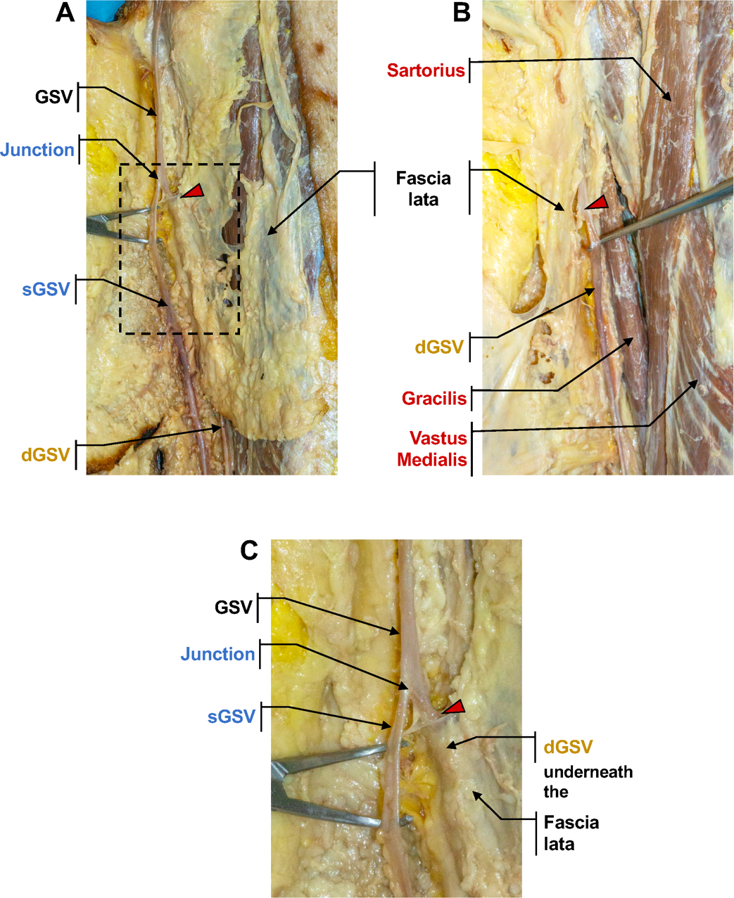
To expose the dGSV, the fascia lata was opened at the knee and partially removed during the dissection. In the thigh region, the fascia lata was cut on the midline and medially lifted in order to appreciate its relationship with the GSVs. At the upper one-third of the surface of the thigh, the dGSV pierced the fascia lata and joined the superficial vessel to form a single vein (Figure 4) which entered the saphenous hiatum, hence draining into the femoral vein. Notably, the dGSV received perforating veins from the deep veins of the limb, while the sGSV presented tributary branches from the cutaneous and subcutaneous layers (Figure 3).
Given the clinical importance of this vessel and its frequent anatomical variations, this case report gave us the cue for reviewing the normal anatomy of GSV and for a critical revision of the nomenclature of its anatomical variations.
Nomenclature review
Normal anatomy of the great saphenous vein
The great saphenous vein, also known as long saphenous vein, is a large blood vessel responsible for most of the superficial venous drainage of the lower limb (Caggiati et al. 2002). Its origin can be found superficially at the medial portion of the dorsal region of the foot about 2-3 cm to the front of the medial malleolus; there, the GSV arises as the continuation of the medial marginal vein of the foot and runs within the subcutaneous layer above the dorsal fascia of the foot (Becciolini et al. 2021). Once it passes the medial malleolus, the great saphenous vein proceeds (still subcutaneously) along the medial surface of the leg, accompanied by the tibial branch of the saphenous nerve (Meissner 2005). Then, the GSV continues its course running posteriorly to the medial condyle of the femur; here, it receives anastomotic branches from the small saphenous vein before the latter drains into the popliteal vein (Tuveri et al. 2009).
In the thigh, the GSV proceeds above the fascia lata, following the course of the sartorius muscle and collecting vessels of the superficial venous network. The GSV runs in the subcutaneous layer throughout the thigh. At the femoral triangle (3-4 cm below the inguinal ligament), it perforates the cribriform fascia to join the femoral vein (Souroullas et al. 2017).
Along its course, GSV is typically located within the so-called saphenous compartment, which is demarked by the superficial and aponeurotic deep fasciae (Chen and Prasad 2009). Moreover, it receives multiple tributaries from the deep venous system (i.e. the perforating veins), which are crucial in balancing blood flow during muscle contraction. Four crucial perforator groups have been identified: upper thigh (Hunterian), lower thigh (Dodd’s), at knee level (Boyd’s) and in the calf region (Cockett’s) (Cina et al. 2005).
Definition of GSV anatomical variations
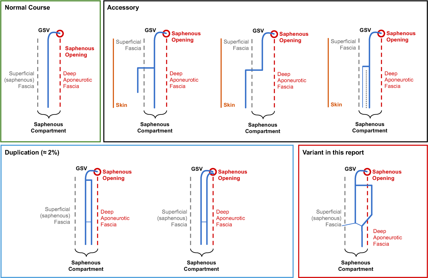
The GSV may present variations in terms of course, relations to the fascia, and possible duplications of the vessel (Black 2014; Chen and Prasad 2009; Ricci and Cavezzi 2002). Previous literature developed different classifications concerning the GSV’s variations (Chen and Prasad 2009). In summary, GSV variants could be divided into two main patterns: i) vein duplication and ii) the presence of a parallel accessory vein.
GSV duplication. Specific criteria must be met to classify a variation as a duplication. A duplication should present as two vessels that lie in the same plane, parallel to the skin and within the saphenous compartment, between the superficial fascia and the aponeurotic deep fasciae (i.e. crural fascia and the fascia lata, respectively in the leg and in the thigh) (Ricci and Caggiati 1999; Kumar et al. 2017). Moreover, the two GSVs should have similar caliber and should drain a common cutaneous territory (Kockaert et al. 2012). The two vessels can frequently present communicating branches and can have a complete course along the lower limb. Duplicated vessels can join together before draining into the femoral vein or, alternatively, can open separately into the femoral vein (Chen and Prasad 2009).
According to Chen and Prasad, duplication incidence is reported in 1% of cases (Chen and Prasad 2009). However, literature reports a range from 1% to 20% of duplication of the GSV. Kockaert M et al. indicated that authentic duplications of the GSV are less frequent than reported in literature, specifically 1.6% to 2% (Kockaert et al. 2012). Combining the retrospective duplex ultrasound and the prospective duplex ultrasound studies, Kockaert et al. indicated that true duplication of the GSV occurs in 1.8% of the duplex examinations of the GSV. GSV’s duplication is more frequent in male than female individuals, is more common unilaterally than bilaterally and in the thigh compared to the leg (Corrales et al. 2002).
Kumar et al. reported a unique variation of the GSV. In the cadaver they studied, the GSV followed its regular path in the lower two-thirds of the leg (Kumar et al. 2017); upon reaching the upper one-third of the medial aspect of the leg, it bifurcated into a thick anterior and a thinner posterior trunk that ascended parallel to each other. The two vessels merged together at the upper third of the thigh. Given the difference in size and caliber, the authors preferred to use the term “bifurcation” rather than “duplication”.
Parallel accessory vein. An accessory vein running in parallel with the GSV is usually smaller in size and does not drain the same cutaneous territory as the GSV (Cotton 1961; Caggiati 2000; Padavinangadi et al. 2015). Generally, the accessory vein runs superficially to the saphenous compartment. In some cases, the accessory vein can appear large and thick and may enter the saphenous compartment by piercing the superficial fascia (Chen and Prasad 2009). Schematically, an accessory vein could present with three most frequent patterns in the thigh (Chen and Prasad 2009; Ekin and Kurtul Yildiz 2017; Oguzkurt 2012):
i) A large subcutaneous tributary runs superficially to the saphenous compartment and pierces the superficial fascia to join the GSV at a variable level in the thigh.
ii) Proximally, the GSV lies within saphenous compartment and, distally, a large accessory vein is present in the subcutaneous layer with no other substantial vein visible in the saphenous compartment. The accessory vein pierces the saphenous fascia at a variable level in the thigh to join the GSV.
iii) An accessory saphenous vein runs together with the GSV below the superficial fascia into separate saphenous compartments; the two veins then join and lie within a single saphenous compartment before entering the saphenofemoral junction. In the latter situation, the accessory saphenous vein could be mistaken as a duplication of the GSV, especially at ultrasound examination.
In summary, in the presence of GSV variations and for an accurate classification, attention should be pointed at the caliber of the vein and its position with respect to superficial (saphenous) fascia (Caggiati 1999; Caggiati and Ricci 1997). The GSV runs in a deep position (so-called “saphenous compartment”) while accessory veins are in a superficial plane. Evidenced of this can be obtained by ultrasonography or computed tomography, and it can be considered a useful interventional and diagnostic indicator that can lead to the individuation of the saphenous vein from a large parallel accessory vein.
Echo-anatomy of the GSV
Duplex ultrasonography is a widely used tool to evaluate the venous system anatomy and determine possible variations especially in the clinical practice.
In the proximal thigh, the GSV can be individuated thanks to its typical appearance known as the “saphenous eye” or “Egyptian eye” in a transverse scan (Ricci and Caggiati 1999). This peculiar depict is due to its position within the saphenous compartment, demarked by the superficial and the aponeurotic deep fasciae.
Due to the presence of numerous tributaries, it may be difficult to identify the “saphenous eye” at the knee level. As a matter of fact, the “tibio-gastrocnemius angle” sign has been demonstrated to be a useful tool in the detection of the GSV in the knee area within the saphenous compartment (Oguzkurt 2012). The tibio-gastrocnemius angle is delimited by the tibia, the medial head of gastrocnemius muscle and the saphenous fascia. Of note, the GSV may be absent in this triangle in case of segmental hypoplasia of the GSV or if a segment of the GSV is located outside the saphenous compartment (Chen and Prasad 2009).
Moreover, another ultrasound marker for the GSV at the thigh level was described as the “E-point” (Ricci et al. 2017). This marker represents a very superficial position of the GSV which can be tracked in cross-section starting from the sapheno-femoral junction and optimally visualized where it crosses the adductor longus muscle, 3–5 cm below the junction.
The GSV variants and clinical implications
Varicose veins and valvular reflux. Varicose veins are twisted, dilated veins mostly located in the lower extremities (Jones and Carek 2008). Nearly 33% of the general population aged between 18 and 64 is affected by varicose veins (Aslam et al. 2022).
In the lower limb, the venous returns is sustained by three main factors: the muscle pump (Recek 2013; Uhl and Gillot 2015), venous valves (Goldman and Fronek 1989; Moore, Gohel, and Davies 2011), and perforating veins (Baliyan et al. 2016). Alterations in vein valves is pivotal in determining venous reflux and varicosity. The valvular dysfunction is presumed to be caused by a loss of elasticity in the vein wall, with incomplete valve closure and consequent valvular reflux (Clarke et al. 1992; Raetz, Wilson, and Collins 2019).
Venous reflux may occur along the course of the great saphenous vein. In this condition, the GSV often expands and enlarges its diameter: venous dilation may be variable based on the reflux entity and location along the GSV course (Kim et al. 2020; Thomson 1979). However, venous reflux and varices in the lower limb are not always associated to an incompetent or dilated GSV. It has been suggested that the presence of a large accessory vein or vein duplication could predispose to varicosity and reflux. Remarkably, the pathogenetic mechanisms leading to vein incompetence could be enhanced in accessory veins or duplications because these anatomical variants could have peculiar histological features (Caggiati 2000).
Hence, subjects with anatomical variants of the GSV may have a congenital predisposition to varicose vein disease based on relationships with fascial components. Accordingly, superficial and aponeurotic deep fasciae protect GSV within the saphenous compartment; differently, a parallel accessory vein lacks this fascial sheath, thus predisposing to vein incompetence (Ricci and Cavezzi 2002).
GSV’s clinical relevance in bypass surgery. The GSV represents main venous conduit used for patients undergoing coronary artery bypass grafting (Akca et al. 2020). Furthermore, the GSV is used in infrainguinal bypass surgery (Belvedere et al. 2020). In this context, in case of bypass for critical limb ischemia, the use of the GSV is reported to be superior compared to alternative autologous vein bypass (Arvela et al. 2012). It is interesting to note that the GSV can be also effectively and safely used as a temporary bypass in long and invasive surgical procedures.
In this light, surgeons must be aware of anatomical variants of the GSV, especially when considering duplications. The removal of GSVs may result in iatrogenic varicosity of the lower limb, particularly when accessory veins are present. As mentioned before, the accessory vessels often have a structural and histological predisposition to varicosity; also, the surgical removal of a duplicated GSV may increase the risk of venous reflux and the development of varices.
Conclusions
The reported anatomical variation of GSV is peculiar and seems to not completely fit with any conventional variations described above.
Similarly to GSV duplications, the two vessels (sGSV and dGSV, Figure 5 and 6) have the same caliber and ascend parallel to each another; however, they do not lie on the same plane and do not drain a common territory.
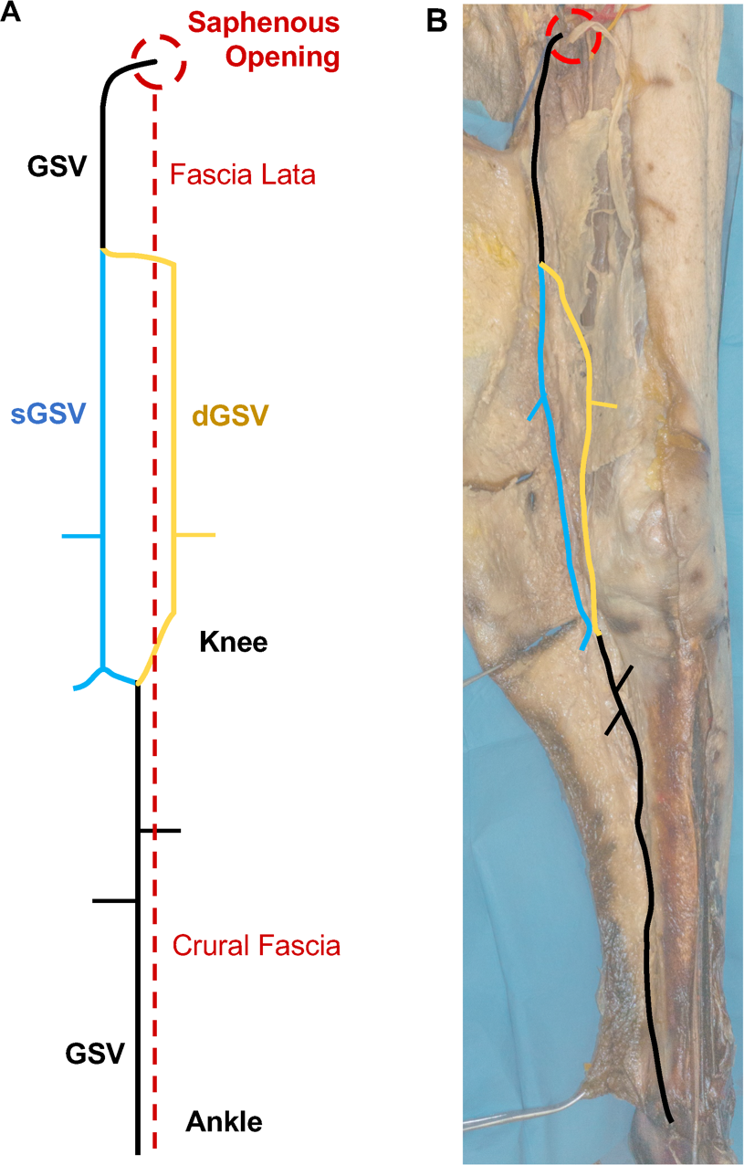
Uniquely, the superficial vessel (sGSV) resides in the saphenous compartment, while the deep one (dGSV) lies underneath the fascia lata. Moreover, they seem to have different drainage territories: sGSV receives tributaries from the subcutaneous layers of the thigh, whereas dGSV is reached by perforating (deep) veins. Finally, the two veins do not have the same origin. Based on these features, the observed variant does not fulfill the criteria for considering it as a duplication (Chen and Prasad 2009; Padavinangadi et al. 2015). Moreover, the sGSV cannot be defined as an accessory vein either. Generally, accessory veins have a smaller caliber compared to the GSV and do not run into the saphenous compartment (Ricci and Cavezzi 2002; Caggiati 2000).
Remarkably, the GSV has a conventional configuration in the leg, lying within the saphenous compartment and draining the superficial layers of this region. Just above the tibial tuberosity, the vessel pierced the aponeurotic deep fascia and continued as the dGSV. Parallel, at this level, the sGSV emerged from the subcutaneous layer and ascended within the saphenous compartment in the thigh. In the proximal thigh, the dGSV pierces the fascia lata and merged with the sGSV before draining into the femoral vein. Therefore, the peculiarity of this variation could affect its recognition at ultrasound examination. This could have an impact on the evaluation of eventual clinical implications; moreover, the actual incidence of the observed variation might be higher.
Although anatomical variants could predispose to venous reflux due to their relationship with fasciae (Ricci and Caggiati 1999; Thomson 1979), the subject did not show any varicosity; this is in agreement with the fact that both vessels are anatomically associated to an anatomical fascia, thus preventing excessive dilation.
In conclusion, we described an unusual anatomical variation of the GSV characterized by a peculiar relationship with the fasciae of the lower limb. Given the frequency and clinical implications of GSV variations, this case report furnished the opportunity to review the conventional anatomy of GSV and to summarize the most common variations in its course. An accurate ultrasound study of GSV is essential to support clinical and surgical practices relating to the superficial venous drainage of the lower limb.
Acknowledgements
The Authors wish to thank Prof. Alberto Caggiati for the critical feedback on the manuscript, and Luciano Sbraga for his ongoing support in the dissection activities.
The cadaver used in this study was donated to the Dissection Center for Human Anatomy, Department of Anatomical, Histological, Forensic Medicine and Orthopedic Sciences, Sapienza University of Rome, Rome, Italy. The Authors are grateful to those who expressed their will to donate their body for research and teaching purposes.
References
Akca F., Lam K.Y., Verberkmoes N., de Lathauwer I., Soliman-Hamad M., and van Straten B. (2020) Predictive value of great saphenous vein mapping prior to endoscopic harvesting in coronary artery bypass surgery. Interact Cardiovasc Thorac Surg. 31: 16-19.
Arvela E., Venermo M., Soderstrom M., Alback A., and Lepantalo M. (2012) Outcome of infrainguinal single-segment great saphenous vein bypass for critical limb ischemia is superior to alternative autologous vein bypass, especially in patients with high operative risk. Ann Vasc Surg. 26: 396-403.
Aslam M.R., Muhammad Asif H., Ahmad K., Jabbar S., Hayee A., Sagheer M.S., Rehman J.U., Khalid S., Hashmi A.S., Rajpoot S.R., and Sharif A. (2022) Global impact and contributing factors in varicose vein disease development. SAGE Open Med. 10: 20503121221118992.
Baliyan V., Tajmir S., Hedgire S.S., Ganguli S., and Prabhakar A.M. (2016) Lower extremity venous reflux. Cardiovasc Diagn Ther. 6: 533-43.
Becciolini M., Galletti S., Vallone G., Stella S.M., and Ricci V. (2021) Sonographic diagnosis of clinically unsuspected thrombosis of the medial marginal vein and dorsal arch of the foot. J Ultrasound. 24: 515-18.
Belvedere S., Gouil Q., Thompson C., and Solomon J. (2020) Computed Tomography Angiography in the Assessment of Great Saphenous Vein as Conduit for Infrainguinal Bypass Surgery. Vasc Endovascular Surg. 54: 313-18.
Black C.M. (2014) Anatomy and physiology of the lower-extremity deep and superficial veins. Tech Vasc Interv Radiol. 17: 68-73.
Caggiati A. (1999) The saphenous venous compartments. Surg Radiol Anat. 21: 29-34.
Caggiati A. (2000) Fascial relations and structure of the tributaries of the saphenous veins. Surg Radiol Anat. 22: 191-6.
Caggiati A., Bergan J.J., Gloviczki P., Jantet G., Wendell-Smith C.P., Partsch H., and International Interdisciplinary Consensus Committee on Venous Anatomical T. (2002) Nomenclature of the veins of the lower limbs: an international interdisciplinary consensus statement. J Vasc Surg. 36: 416-22.
Caggiati A., and Ricci S. (1997) The Long Saphenous Vein Compartment. Phlebology. 12: 107-11.
Chen S.S., and Prasad S.K. (2009) Long saphenous vein and its anatomical variations. Australas J Ultrasound Med. 12: 28-31.
Cina A., Pedicelli A., Di Stasi C., Porcelli A., Fiorentino A., Cina G., Rulli F., and Bonomo L. (2005) Color-Doppler sonography in chronic venous insufficiency: what the radiologist should know. Curr Probl Diagn Radiol. 34: 51-62.
Clarke G.H., Vasdekis S.N., Hobbs J.T., and Nicolaides A.N. (1992) Venous wall function in the pathogenesis of varicose veins. Surgery. 111: 402-8.
Corrales N.E., Irvine A., McGuinness C.L., Dourado R., and Burnand K.G. (2002) Incidence and pattern of long saphenous vein duplication and its possible implications for recurrence after varicose vein surgery. Br J Surg. 89: 323-6.
Cotton L.T. (1961) Varicose veins. Gross anatomy and development. Br J Surg. 48: 589-98.
Ekin E.E., and Kurtul Yildiz H. (2017) The importance of great saphenous vein types and segmental aplasia in venous reflux. J Clin Ultrasound. 45: 332-36.
Goldman M.P., and Fronek A. (1989) Anatomy and pathophysiology of varicose veins. J Dermatol Surg Oncol. 15: 138-45.
Jones R.H., and Carek P.J. (2008) Management of varicose veins. Am Fam Physician. 78: 1289-94.
Kim M.J., Park P.J., Koo B.H., Lee S.G., Byun G.Y., and Lee S.R. (2020) Association between venous reflux and diameter of great saphenous vein in lower thigh. J Vasc Surg Venous Lymphat Disord. 8: 100-05.
Kockaert M., de Roos K.P., van Dijk L., Nijsten T., and Neumann M. (2012) Duplication of the great saphenous vein: a definition problem and implications for therapy. Dermatol Surg. 38: 77-82.
Kumar N., Aithal A.P., Swamy R.S., Nayak S.B., Rao M.K.G., and Abhinitha P. (2017) Bifurcated Great Saphenous Vein: A Report on Its Therapeutic and Diagnostic Perspectives. J Cardiovasc Echogr. 27: 107-09.
Meissner M.H. (2005) Lower extremity venous anatomy. Semin Intervent Radiol. 22: 147-56.
Moore H.M., Gohel M., and Davies A.H. (2011) Number and location of venous valves within the popliteal and femoral veins: a review of the literature. J Anat. 219: 439-43.
Oguzkurt L. (2012) Ultrasonographic anatomy of the lower extremity superficial veins. Diagn Interv Radiol. 18: 423-30.
Padavinangadi A., Kumar N., Swamy R.S., Satheesha N.B., and Mohandas Rao K.G. (2015) Unilateral Double Great Saphenous Vein: A Clinically Significant Case Report. J Cardiovasc Echogr. 25: 116-18.
Raetz J., Wilson M., and Collins K. (2019) Varicose Veins: Diagnosis and Treatment. Am Fam Physician. 99: 682-88.
Recek C. (2013) Calf pump activity influencing venous hemodynamics in the lower extremity. Int J Angiol. 22: 23-30.
Ricci S., and Caggiati A. (1999) Echoanatomical Patterns of the Long Saphenous Vein in Patients with Primary Varices and in Healthy Subjects. Phlebology. 14: 54-58.
Ricci S., and Cavezzi A. (2002) Echo-Anatomy of Long Saphenous Vein in the Knee Region: Proposal for a Classification in Five Anatomical Patterns. Phlebology. 16: 111-16.
Ricci S., Moro L., Ferrini A., Rossi Bartoli I., and Antonelli Incalzi R. (2017) The E point: a new echographic great saphenous identification sign in specific anatomical variants and applications. Phlebology. 32: 120-24.
Souroullas P., Barnes R., Smith G., Nandhra S., Carradice D., and Chetter I. (2017) The classic saphenofemoral junction and its anatomical variations. Phlebology. 32: 172-78.
Thomson H. (1979) The surgical anatomy of the superficial and perforating veins of the lower limb. Ann R Coll Surg Engl. 61: 198-205.
Tuveri M., Borsezio V., Argiolas R., Medas F., and Tuveri A. (2009) Ultrasonographic venous anatomy at the popliteal fossa in relation to tibial nerve course in normal and varicose limbs. Chir Ital. 61: 171-7.
Uhl J.F., and Gillot C. (2015) Anatomy of the veno-muscular pumps of the lower limb. Phlebology. 30: 180-93.