https://doi.org/10.36253/ijae-14856
Transduodenal Surgical Ampullectomy: technical considerations
1 Department of Innovative Technologies in Medicine & Dentistry, University “G. d’Annunzio” Chieti-Pescara, Via dei Vestini, 31, 66100 Chieti, Italy
2 Unit of Surgical Oncology, Casa di Cura Luigi Pierangeli, Largo Luigi Pierangeli, Pescara, Italy
*Corresponding author: E-mail: paolo.panaccio@gmail.com
Abstract. The therapeutic approaches of ampullary tumours can be both surgical and endoscopic. Surgical options include transduodenal surgical ampullectomy, first described by Halsted in 1899 and the more radical pancreaticoduodenectomy introduced by Whipple in 1935. The endoscopic ampullectomy (papillectomy) is globally recognized as the first-line procedure for benign lesions. If endoscopic resection is not feasible and safe, a surgical approach has to be considered. Surgical treatment guarantees complete removal of the lesion nevertheless, it is burdened with a mortality rate of about 3% and high morbidity (around 20–40%) even in high-volume centers. For this reason in selected cases, the local excision technique seems to be an effective and less invasive alternative to more extensive surgery (pancreaticoduodenectomy) with consequent advantages in terms of lower morbidity and mortality. A pre-operative planning in a multidisciplinary board is mandatory prior to the procedure. Given that it remains a challenging procedure that requires particular surgical experience and operative skills offering good results in terms of morbidity and mortality and should be in the armamentarium of every pancreatic surgeon. The aim of this short research article is to offer to the lecturer a comprehensive view about the transduodenal ampullectomy in the treatment of localized ampullary diseases.
Keywords: Major Duodenal Papilla, Papilla of Vater, Transduodenal Surgical Ampullectomy, local excision of Ampulla Vateri tumours.
Contents
Indications for Transduodenal Surgical Ampullectomy (TSA)
Clinical outctome of Transduodenal Surgical Ampullectomy
Introduction
Knowledge of the anatomy of the Major Duodenal Papilla (MDP) or papilla of Vater and the relationship between Wirsung’s duct and distal common bile duct represents an important guideline for accurate diagnostic and therapeutic approach to a variety of biliopancreatic disease [1-3]. Tumours of the ampulla of Vater are relatively uncommon lesions of the digestive tract. Endoscopic papillectomy is considered as a gold standard in treatment of limited ampullary lesions. It is less extensive than transduodenal ampullectomy which involves excision of the entire ampulla together with small parts of the duodenal wall, bile duct, pancreatic duct and sometimes pancreatic parenchima, or pancreaticoduodenectomy (PD). The aim of this study is to report the main anatomical and embryological aspects of the ampulla of Vater and technical notes about transduodenal surgical ampullectomy (TSA) in the treatment of endoscopically non-resectable lesions of the ampulla.
Material and methods
Pre-operative evaluation
The purpose of pre-operative evaluation of ampullary lesions is to determine their malignant potential, resecability, and establish stage. Preoperative work-up included computed tomography (CT-scan) and/or magnetic resonance imaging (MRI), endoscopic ultrasound (EUS) with biopsy. Endoscopic retrograde cholangiopancreatography (ERCP) and intraductal ultrasonography (IDUS) were requested in case of jaundice. EUS has been described as a critical investigation for ampullary tumours, due to its ability to determine the extent of local invasion and identify lymph-nodes metastases [4,5].
Indications for Transduodenal Surgical Ampullectomy (TSA)
Notwithstanding, many authors have described specific indications or decision making algorithms based on their own clinical series, practices or experiences [6-9]. Briefly, according to our experience, suitable indications for surgical ampullectomy are: failure of endoscopic treatment (due to the presence of duodenal diverticulum or to a tumoral extension to pancreatic / duodenal /biliary wall), endoscopically unresectable bening periampullary tumors (low-grade dysplasia adenomas or high-grade/carcinoma in situ – pTis -), and selected cases of pT1 carcinoma with no lymph node involvement in patients with such comorbidities to preclude a highly complex surgical procedure such as PD. Generally accepted contraindications are T3 or T4, patients fit for PD, poorly differentiated or with nodal or distant metastases tumours. The role of TSA as a treatment for ampullary adenocarcinoma (AC) is now controversial due to the difficulty in assessment of lymph node involvement [10-12].
Surgical technique
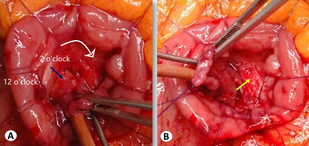
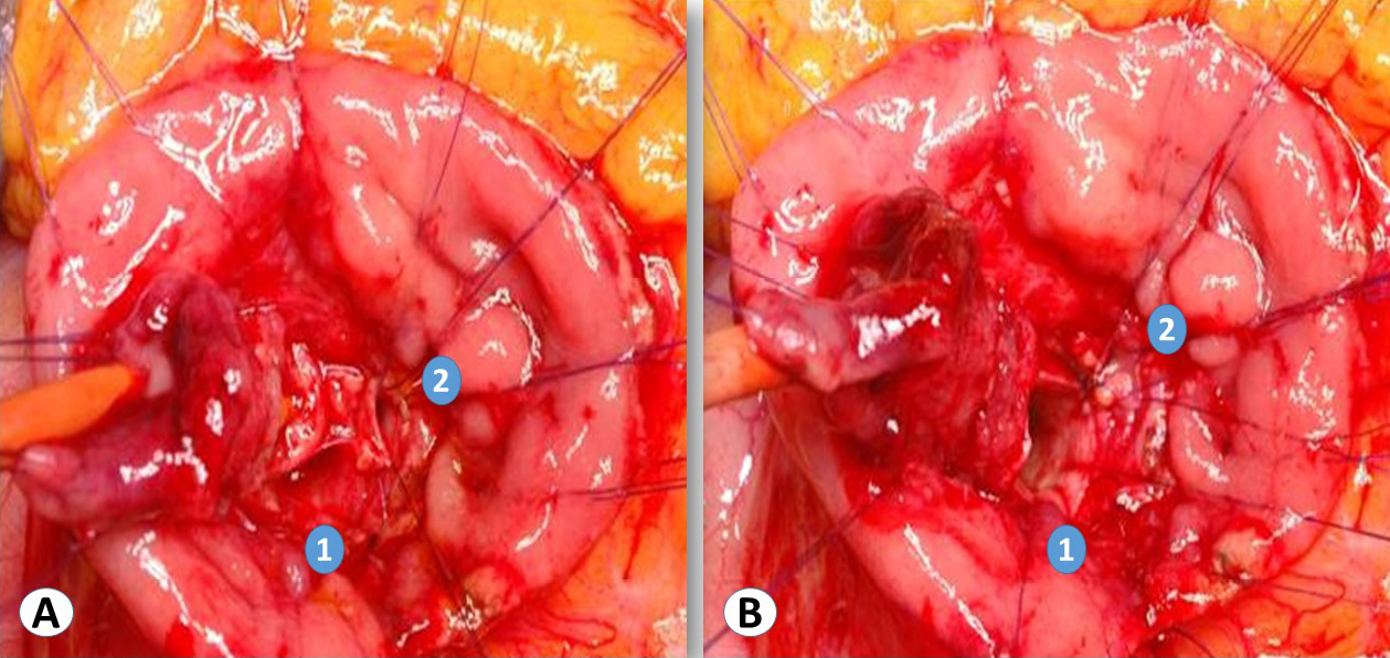
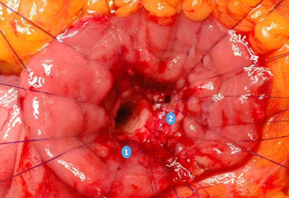
After a right subcostal incision, a complete exploration of the cavity was carried out. The procedure started by classical cholecystectomy. After identifying the duodenum, Cattel maneuver and subsequently Kocher’s maneuver was performed to allow complete exposure of the posterior duodenal wall. Bimanual palpation of the ampullary tumor complete this phase. A transverse duodenotomy of about 3–4 cm of the sec_ond duodenal portion was performed, on the antimesenteric side, facing the periampullary area. To separate the wall layers, submucosal injection of adrenaline (1:1000) was performed. A “lift sign” which would exclude an infiltration by the tumor of the deeper layers. The tumor mass was then transfixed with a stitch to pull it away and better dissect the ampulla of Vater. An optimal lateral traction of the ampulla with the tumor mass during the dissection is helpful to obtain tumor-free margins. Both Wirsung and bile ducts were then separately cannulated with a Fogarty catheter as shown in Figure 1. From a frontal view and considering the major axis of the duodenum, the papilla along with Wirsung and bile duct were usually located at around 2 o’clock. The dissection of the duodenal wall, reaching the muscular layer, was made in a circular clockwise manner starting at 11 o’clock. The dissection was continued clockwise until the whole papilla was completely excised and at least 1cm free-margin was obtained as shown in Figure 2. The pancreatic and bile ducts were therefore exposed and joined together with re-absorbable stitches (5.0 PDS) so that a common ostium was created and, finally, sutured with the duodenal wall. The final appearance was shown in Figure 3. Once removed, the whole papilla was sent for intraoperative frozen section analysis. If the pathological results did not meet the criteria of a potential curative local resection through TSA (for several reasons, such as infiltration of the muscular layer of the duodenal wall, extension of the lesion beyond the biliary and/or pancreatic duct and/or histology of ampullary invasive adenocarcinoma), a pancreaticoduodenectomy was considered. A nasoduodenal tube was placed and the duodenotomy closed transversely in a double layer with interrupted monofilament reabsorbable stitches (4.0 PDS). The procedure was concluded with double laminar drainage allocation. Lastly, some authors have proposed additional components to TSA: supraduodenal lymph nodes, anterior/posterior pancreatic head lymph nodes dissection in pT1 cancers, excision of the extrahepatic bile duct providing a reduction in biliary recurrences.
Results
Clinical outctome of Transduodenal Surgical Ampullectomy
No randomized clinical trial has been conducted comparing clinical outcomes in TSA, PD and endoscopic papillectomy. In recent systematic reviews and meta-analysis were reported the following results: R0 rate 96.4%, local recurrence rate 9.4%, complications rate 28.3%, mortality rate 0.9% [13,14].
Discussion
While endoscopic papillectomy remains the gold standard for benign lesions, transduodenal surgical ampullectomy (TSA) can be proposed in selected cases and when endoscopy fails. With limitations of current experience TSA is a feasible and effective. It represents and ideal therapeutic option in early ampullary lesions avoiding a demolitive procedure such as PD, with a better quality of life in a long-term follow up.
References
[1. Horiguchi SI, Kamisawa T. Major duodenal papilla and its normal anatomy. Dig Surg. 2010;27(2).
[2] Taliente F, Bianco G, Moschetta G, Franco A, Giovinazzo F, Agnes S, et al. From endoscopic resection to pancreatoduodenectomy: a narrative review of treatment modalities for the tumors of the ampulla of Vater. Chinese Clin Oncol [Internet]. 2022 Jun 1 [cited 2023 Apr 30];11(3). Available from: https://pubmed.ncbi.nlm.nih.gov/35818855/
[3] Dowdy GS, Waldron GW, Brown WG. Surgical Anatomy of the Pancreatobiliary Ductal System: Observations. Arch Surg. 1962;84(2).
[4] Ceppa EP, Burbridge RA, Rialon KL, Omotosho PA, Emick D, Jowell PS, et al. Endoscopic versus surgical ampullectomy: An algorithm to treat disease of the ampulla of vater. Ann Surg. 2013;
[5] Kandler J, Neuhaus H. How to Approach a Patient With Ampullary Lesion. Gastroenterology. 2018;155(6).
[6] Kobayashi A, Konishi M, Nakagohri T, Takahashi S, Kinoshita T. Therapeutic approach to tumors of the ampulla of Vater. Am J Surg. 2006;192(2).
[7] Aiura K, Shinoda M, Nishiyama R. Surgical technique for complete resection of the extrahepatic portion of the common bile duct and the ampulla of Vater for tumors of the ampulla of Vater. J Hepatobiliary Pancreat Sci. 2011;18(2).
[8] Scroggie DL, Mavroeidis VK. Surgical ampullectomy: A comprehensive review. World J Gastrointest Surg. 2021;13(11).
[9] Dubois M, Labgaa I, Dorta G, Halkic N. Endoscopic and surgical ampullectomy for non-invasive ampullary tumors: Short-term outcomes. Biosci Trends. 2016;10(6).
[10] Song J, Liu H, Li Z, Yang C, Sun Y, Wang C. Long-term prognosis of surgical treatment for early ampullary cancers and implications for local ampullectomy Hepato-biliary-pancreatic surgery. BMC Surg. 2015;15(1).
[11] Choi SB, Kim WB, Song TJ, Suh SO, Kim YC, Choi SY. Surgical outcomes and prognostic factors for ampulla of vater cancer. Scand J Surg. 2011;
[12] Aiura K, Hibi T, Fujisaki H, Kitago M, Tanabe M, Kawachi S, et al. Proposed indications for limited resection of early ampulla of Vater carcinoma: Clinico-histopathological criteria to confirm cure. J Hepatobiliary Pancreat Sci. 2012;19(6).
[13] Heise C, Ali EA, Hasenclever D, Auriemma F, Gulla A, Regner S, et al. Systematic review with meta‐analysis: Endoscopic and surgical resection for ampullary lesions. J Clin Med. 2020;9(11).
[14] Garg R, Thind K, Bhalla J, Simonson MT, Simons-Linares CR, Singh A, et al. Long-term recurrence after endoscopic versus surgical ampullectomy of sporadic ampullary adenomas: a systematic review and meta-analysis. Vol. 37, Surgical Endoscopy. 2023.